. 2004 Jul 19;200(2):267-72.
doi: 10.1084/jem.20040731.

The scavenger receptor MARCO is required for lung defense against pneumococcal pneumonia and inhaled particles

Affiliations

The scavenger receptor MARCO is required for lung defense against pneumococcal pneumonia and inhaled particles

Mohamed Arredouani et al. J Exp Med. .

Abstract

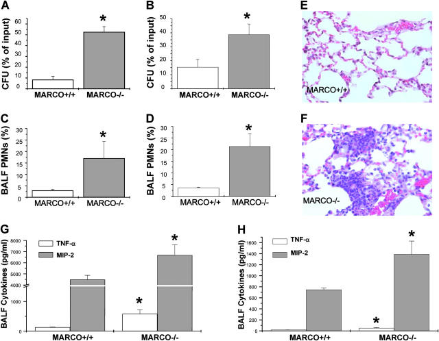
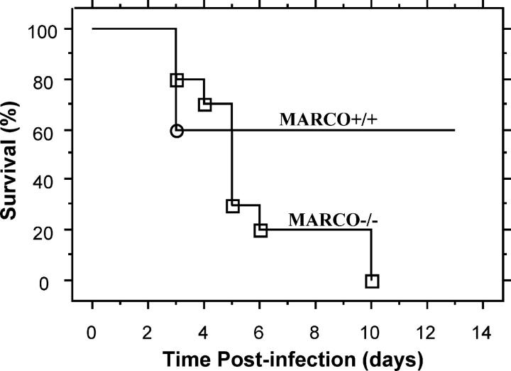
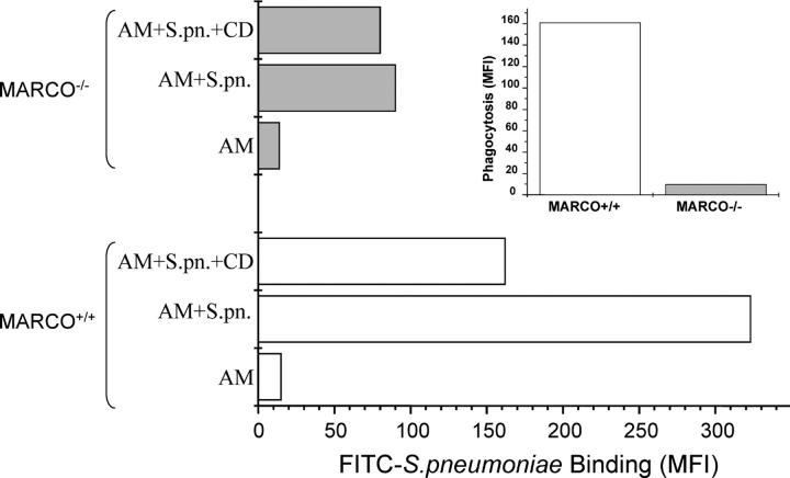
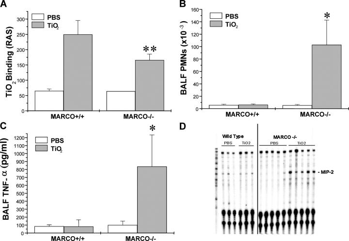
Alveolar macrophages (AMs) express the class A scavenger receptor macrophage receptor with collagenous structure (MARCO), but its role in vivo in lung defense against bacteria and environmental particles has not been studied. We used MARCO-deficient mice to directly test the in vivo role of AM MARCO in innate defense against pneumococcal infection and environmental particles. In a murine model of pneumococcal pneumonia, MARCO(-/-) mice displayed an impaired ability to clear bacteria from the lungs, increased pulmonary inflammation and cytokine release, and diminished survival. In vitro binding of Streptococcus pneumoniae and in vivo uptake of unopsonized particles by MARCO(-/-) AMs were dramatically impaired. MARCO(-/-) mice treated with the "inert" environmental particle TiO(2) showed enhanced inflammation and chemokine expression, indicating that MARCO-mediated clearance of inert particles by AMs prevents inflammatory responses otherwise initiated by other lung cells. Our findings point to an important role of MARCO in mounting an efficient and appropriately regulated innate immune response against inhaled particles and airborne pathogens.


Figures

Figure 1.
Figure 1.
(A and B) Pulmonary bacterial burden after infection with S. pneumoniae. MARCO+/+ and MARCO−/− (n = 7–9) were intranasally infected with 105 CFU of S. pneumoniae. Lung bacterial load was determined at 4 h (A) and 24 h (B) after infection. Data shown are representative of at least three separate experiments, and presented as mean ± SEM. *, P < 0.05 for −/− versus +/+. (C and D) Lung inflammation in response to S. pneumoniae infection. PMN recruitment into BALF of infected mice, harvested 4 h (C) and 24 h (D) after infection, presented as percentage of total cells in BALF. (E and F) Representative lung histology sections from infected mice that showed mostly normal histology in MARCO+/+ mice (E) and increased focal PMN infiltrates in MARCO−/− mice (F). (G and H) Infected mice (n = 4) BALF TNF-α and MIP-2 at 4 h (G) and 24 h (H) after infection as measured by ELISA. Data are presented as mean ± SEM. *, P < 0.05 for −/− versus +/+.
Figure 2.
Figure 2.
Absence of MARCO increases mortality after S. pneumoniae administration to mice. MARCO+/+ and MARCO−/− mice were intranasally inoculated with 2.2 × 10⁵ CFU of S. pneumoniae, and survival was monitored over a period of 13 d. Data are expressed as percentage of mice alive at each time point. Kaplan-Meier survival plots are shown for 10 animals per group. Mantel-Cox log-rank test showed that P = 0.0342. One experiment representative of two is shown.
Figure 3.
Figure 3.
Binding and phagocytosis of FITC-labeled S. pneumoniae (S.pn.) by MARCO+/+ and MARCO−/− AMs. AMs were harvested from untreated mice, and phagocytosis was measured as described in Materials and Methods. Fluorescence was measured on untreated cells (AM) and cells incubated with labeled bacteria in the absence (AM + S.pn.) or presence of cytochalasin D (AM + S.pn. + CD) to block internalization. The deduced phagocytosis rate as expressed in MFI units is shown in the inset. Data shown are representative of two experiments.
Figure 4.
Figure 4.
Lung inflammation in response to inhaled TiO2 particles. (A) MARCO+/+ and MARCO−/− AM binding of TiO2 as determined by right angle scatter (RAS) measurements of harvested BAL macrophages after intranasal instillation of particles to mice (n = 3 mice/group), **, P < 0.02 for TiO2 instilled MARCO−/− versus all other groups. (B) PMN recruitment into BALF of PBS and TiO2-treated mice, n > 9 mice/group. *, P < 0.05 for TiO2 instilled MARCO−/− mice versus all other groups. (C) BALF TNF-α as measured by ELISA, n > 3 mice/group. *, P < 0.05 for TiO2 instilled MARCO−/− mice versus all other groups. (D) Analysis of lung gene expression by RNase protection assay showing increased MIP-2 mRNA in lung samples from TiO2-treated MARCO−/− mice.

References

    1. Platt, N., R. Haworth, L. Darley, and S. Gordon. 2002. The many roles of the class A macrophage scavenger receptor. Int. Rev. Cytol. 212:1–40. - PubMed
    1. Nakamura, K., H. Funakoshi, F. Tokunaga, and T. Nakamura. 2001. Molecular cloning of a mouse scavenger receptor with C-type lectin (SRCL)(1), a novel member of the scavenger receptor family. Biochim. Biophys. Acta. 1522:53–58. - PubMed
    1. Pearson, A.M. 1996. Scavenger receptors in innate immunity. Curr. Opin. Immunol. 8:20–28. - PubMed
    1. Hampton, R.Y., D.T. Golenbock, M. Penman, M. Krieger, and C.R. Raetz. 1991. Recognition and plasma clearance of endotoxin by scavenger receptors. Nature. 352:342–344. - PubMed
    1. Suzuki, H., Y. Kurihara, M. Takeya, N. Kamada, M. Kataoka, K. Jishage, O. Ueda, H. Sakaguchi, T. Higashi, T. Suzuki, et al. 1997. A role for macrophage scavenger receptors in atherosclerosis and susceptibility to infection. Nature. 386:292–296. - PubMed

Publication types

MeSH terms