. 2004 Jul 20:2:58.
doi: 10.1186/1477-7827-2-58.

Endometrial glands as a source of nutrients, growth factors and cytokines during the first trimester of human pregnancy: a morphological and immunohistochemical study

Affiliations

Endometrial glands as a source of nutrients, growth factors and cytokines during the first trimester of human pregnancy: a morphological and immunohistochemical study

Joanne Hempstock et al. Reprod Biol Endocrinol. .

Abstract

Background: The maternal circulation to the human placenta is not fully established until 10-12 weeks of pregnancy. During the first trimester the intervillous space is filled by a clear fluid, in part derived from secretions from the endometrial glands via openings in the basal plate. The aim was to determine the activity of the glands throughout the first trimester, and to identify components of the secretions.

Methods: Samples of human decidua basalis from 5-14 weeks gestational age were examined by transmission electron microscopy and immunohistochemically. An archival collection of placenta-in-situ samples was also reviewed.

Results: The thickness of the endometrium beneath the implantation site reduced from approximately 5 mm at 6 weeks to 1 mm at 14 weeks of gestation. The glandular epithelium also transformed from tall columnar cells, packed with secretory organelles, to a low cuboidal layer over this period. The lumens of the glands were always filled with precipitated secretions, and communications with the intervillous space could be traced until at least 10 weeks. The glandular epithelium reacted strongly for leukaemia inhibitory factor, vascular endothelial growth factor, epidermal growth factor, transforming growth factor beta, alpha tocopherol transfer protein, MUC-1 and glycodelin, and weakly for lactoferrin. As gestation advanced uterine natural killer cells became closely approximated to the basal surface of the epithelium. These cells were also immunopositive for epidermal growth factor.

Conclusions: Morphologically the endometrial glands are best developed and most active during early human pregnancy. The glands gradually regress over the first trimester, but still communicate with the intervillous space until at least 10 weeks. Hence, they could provide an important source of nutrients, growth factors and cytokines for the feto-placental unit. The endometrium may therefore play a greater role in regulating placental growth and differentiation post-implantation than previously appreciated.


Figures

Figure 1
Figure 1
A) In the earliest specimen available, H710, the conceptus (C) can be seen embedded in the superficial endometrium overlying well-developed endometrial glands (G). M, myometrium. (Haematoxylin and eosin) Scale bar = 1.0 cm. B) The secretions within the lumens of the glands are heterogenous, being a mixture of carbohydrate-rich flocculent material (blue) and what appear to be lipid droplets (red). (Alcian blue and Neutral red) Scale bar = 100 μm.
Figure 2
Figure 2
Scattergram showing the relationship between endometrial thickness and gestational age.
Figure 3
Figure 3
Placenta-in-situ specimen (H1094) of 13.5 weeks gestational age showing the reduction in thickness of the endometrium (E) at this stage of pregnancy. The glands (G) have a more regular outline, but still contain precipitated secretions within their lumens. M, myometrium; IVS, intervillous space. (Haematoxylin and eosin) Scale bar = 1.0 mm.
Figure 4
Figure 4
A) Photomicrograph of a 1 μm resin section of 6 week decidua illustrating the columnar epithelium of the glands, their large apical projections and the heterogeneous nature of the secretions. (Methylene blue) Scale bar = 10 μm. B) At the ultrastructural level it can be seen that the cells possess large quantities of mitochondria and endoplasmic reticulum, and lipid droplets are abundant in the basal region. The cells are attached to a well-formed basal lamina (arrowed). Scale bar = 2 μm.
Figure 5
Figure 5
A) Photomicrograph of a 1 μm resin section of 10 week decidua. By now the epithelium is cuboidal in nature, although secretions are still present within the lumens. There appears to be an almost complete layer of additional cells (arrowed) beneath the basal lamina. Scale bar = 10 μm. B) At the ultrastructural level the cells appear more quiescent at this stage of gestation, although Golgi bodies and a few strands of rough endoplasmic reticulum remain. Scale bar = 1 μm.
Figure 6
Figure 6
Low power transmission electron micrograph of 10 week decidua demonstrating the heterogenous population of cells accumulated immediately beneath the epithelial basal lamina (arrowheads) at this stage of gestation. The smaller cells (arrowed) with large numbers of granules resemble uterine NK cells, whereas the larger more electron lucent cells (asterisks) resemble decidual cells. Scale bar = 5 μm.
Figure 7
Figure 7
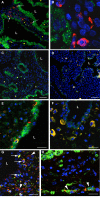
Confocal immunofluorescent images of decidua at 8 weeks (C, E, G, H) and 12 weeks (A, B, D, F) gestational age. In A) and B) the glandular epithelium has been immunolabelled for tocopherol transfer protein (green) and NK cells with CD56 (red). NK cells can be seen within the stroma between the glands, but also closely approximated (arrowed) to the basal lamina of the glandular epithelium. In C-F sections were immunolabelled for epidermal growth factor (EGF) (green) and CD56 (red). The epithelium reacts strongly at 8 weeks for EGF (C), but less so at 12 weeks (D). The NK cells lying beneath the glandular epithelium also react strongly for EGF (co-localisation yellow) (E and F). In G) and H) the sections were immunolabelled for human placental lactogen (red), and in G) for CD68 (green) and in H) for cytokeratin (green). Cells positive for both placental lactogen and CD68 (yellow) were considered to be macrophages, and were observed throughout the stroma but also closely approximated to the glandular epithelium (arrowed in G). Cells reacting only for placental lactogen, or for both placental lactogen and cytokeratin, were considered to be invading extravillous trophoblast cells (arrowheads in G and H), and were not found to be closely associated with the epithelium (E). Blue, DAPI; L, gland lumen. Scale bars C, D. = 60 μm and E - H = 30 μm.
Figure 8
Figure 8
Transmission electron micrographs of 15 week decidua illustrating A) the flattened nature of the glandular cells at this stage of gestation, and B) the accumulation of a flocculent osmiophilic material in their cytoplasm. L, gland lumen. Scale bars = 5 μm and 1 μm.
Figure 9
Figure 9
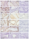
Photomicrographs of immunolabelled decidua at 6 weeks (A, D, G, J, M, P) and 12 weeks (B, E, H, K, N, Q) gestational age. The glandular epithelium reacted positively for LIF (A, B), VEGF (D, E), MUC-1 (G, H), alpha tocopherol transfer protein (J, K), TGFβ3 (M, N), and weakly for lactoferrin (P, Q). Negative controls; C, F, I, L, O and R. Scale bar = 200 μm.
Figure 10
Figure 10
Confocal photomicrograph of a frozen section of an 8 week villus A) immunolabelled for glycodelin (green) and cathepsin D (red) and B) under phase contrast. Vesicles labelled solely for glycodelin predominate in the apical region of the syncytiotrophoblast (S), and those for cathepsin D in the basal region. In the mid-zone the two labels co-localise (yellow) indicating that maternal proteins enter the trophoblast digestion pathway. IVS, intervillous space.

Similar articles

Cited by

References

    1. Hustin J, Schaaps JP. Echographic and anatomic studies of the maternotrophoblastic border during the first trimester of pregnancy. Am J Obstet Gynecol. 1987;157:162–168. - PubMed
    1. Jaffe R, Jauniaux E, Hustin J. Maternal circulation in the first-trimester human placenta-Myth or reality? Am J Obstet Gynecol. 1997;176:695–705. - PubMed
    1. Jauniaux E, Watson AL, Hempstock J, Bao Y-P, Skepper JN, Burton GJ. Onset of maternal arterial bloodflow and placental oxidative stress; a possible factor in human early pregnancy failure. Am J Pathol. 2000;157:2111–2122. - PMC - PubMed
    1. Kliman HJ. Uteroplacental blood flow. The story of decidulaisation, menstruation and trophoblast invasion. Am J Pathol. 2000;157:1759–1768. - PMC - PubMed
    1. Amoroso EC. Placentation. In: Parkes AS, editor. In Marshall's Physiology of Reproduction. 3. Vol. 2. London: Longmans Green and Co; 1952. pp. 127–311.

Publication types