. 2004 Aug 3;101(31):11245-50.
doi: 10.1073/pnas.0404297101. Epub 2004 Jul 20.

Central role for liver X receptor in insulin-mediated activation of Srebp-1c transcription and stimulation of fatty acid synthesis in liver

Affiliations

Central role for liver X receptor in insulin-mediated activation of Srebp-1c transcription and stimulation of fatty acid synthesis in liver

Guoxun Chen et al. Proc Natl Acad Sci U S A. .

Abstract

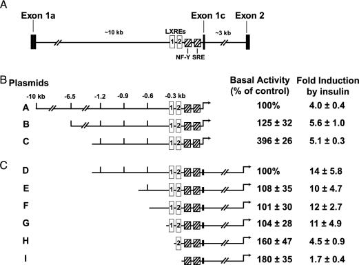
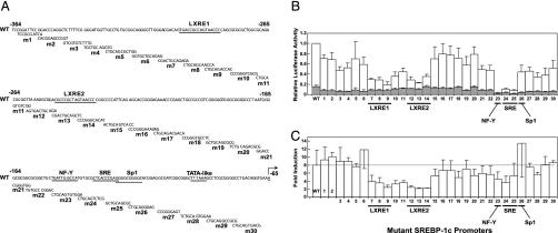
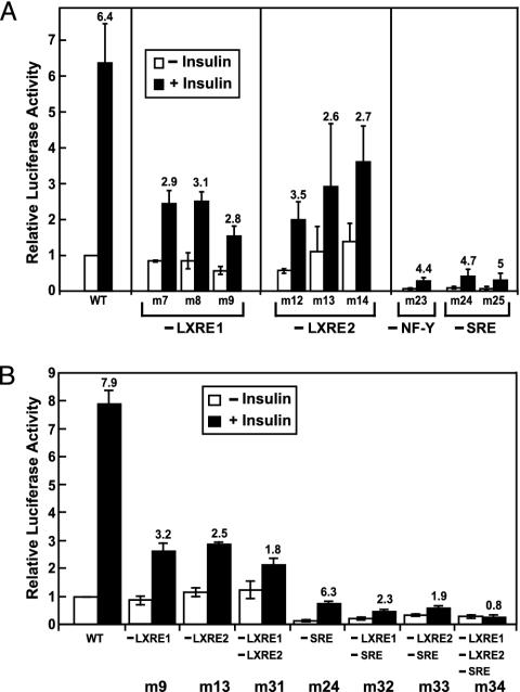
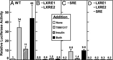
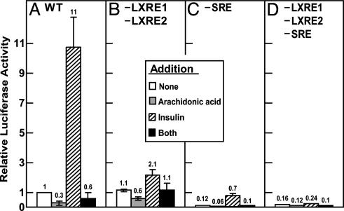
Transcription of the gene encoding sterol regulatory element-binding protein 1c (SREBP-1c) is known to be activated by insulin in the liver. The resultant SREBP-1c protein activates transcription of the genes required for fatty acid synthesis. Here, we use SREBP-1c promoter reporter constructs to dissect the mechanism of insulin activation in freshly isolated rat hepatocytes. The data show that a complete insulin response (increase of 6- to 11-fold) requires two binding sites for liver X receptors (LXRs), which are nuclear receptors that are activated by oxygenated sterols. Disruption of these binding sites did not lower basal transcription but severely reduced the response to insulin. In contrast, disruption of the closely linked binding sites for SREBPs and nuclear factor Y lowered basal transcription drastically but still permitted a 4- to 7-fold increase in response to insulin. Arachidonic acid, an inhibitor of LXR activation, blocked the response to insulin. We conclude that insulin activates the SREBP-1c promoter primarily by increasing the activity of LXRs, possibly through production of a ligand that activates LXRs or their heterodimerizing partner, the retinoid X receptor. Nuclear SREBPs and nuclear factor Y play permissive roles.


Figures

Fig. 1.
Fig. 1.
Activation of mouse SREBP-1c promoter in rat primary hepatocytes. (A) Schematic of partial mouse Srebp-1c gene, illustrating the upstream regulatory elements and the intron between exons 1c and 2. The proximal promoter regulatory region is shown with its cis-acting elements: two LXREs (open boxes) and one NF-Y and one SRE (hatched boxes). (B and C) Deletion analysis of SREBP-1c promoter region and its effects on basal and insulin-induced luciferase activities. The indicated plasmids were transfected into primary hepatocytes as described in Methods. Six hours after transfection, the medium was switched to medium B with or without 100 nM insulin and incubated for 21 h, after which the cells were harvested and assayed for dual luciferase activities as described in Methods. The 100% values for basal activity correspond to the normalized luciferase activity obtained with plasmid A (B) or plasmid D (C) in the absence of insulin. The fold induction in B and C was calculated as the ratio of normalized luciferase activity in the presence of insulin to that in the absence of insulin. Each value represents the mean ± SEM of three independent transfection experiments (each assayed in duplicate).
Fig. 2.
Fig. 2.
Nucleotide sequences (A), relative transcriptional activities (B), and fold induction (C) of WT and mutant mouse SREBP-1c promoters in primary rat hepatocytes. (A) The sequence of a portion of the normal SREBP-1c promoter is shown at the top and numbered according to a convention in which the A of the ATG initiation codon of mouse SREBP-1c is + 1. The putative binding sites for LXR (LXRE1 and LXRE2), NF-Y, SREBP (SRE), and Sp1 and the TATA-like sequence are underlined. The transcription initiation site is indicated by an arrow. Below the sequence of the WT promoter are shown 30 overlapping scramble mutations that are separately introduced into the DNA by site-directed oligonucleotide mutagenesis as described in Methods. The names of the mutant plasmids, m1–m30, are denoted on the left. The mutant scrambled sequence that was introduced is shown below the normal promoter sequence. (B) The height of each bar represents the relative luciferase activity of plasmid D (WT) or the indicated mutant plasmid (m1–m30) in transfected hepatocytes cultured in the absence (shaded bar) or presence (open bar) of insulin as described in Methods. The relative luciferase activity of the WT promoter (plasmid D) in the presence of insulin (mean of 12 independent experiments) was arbitrarily assigned a value of 1.0. Each bar for mutant promoters m1–m30 represents mean ± SEM of three to six independent transfections. (C) The data in B were replotted to show the fold induction of the various promoters by insulin. The fold induction was calculated as the ratio of normalized luciferase activity in the presence of insulin to that in the absence of insulin. The luciferase activity of each promoter in the absence of insulin was arbitrarily assigned a value of 1.0.
Fig. 3.
Fig. 3.
Mutations of LXRE1, LXRE2, and SRE in SREBP-1c promoter sequence reduce insulin-induced activation in primary rat hepatocytes. The indicated WT or scramble mutant plasmid was transfected into hepatocytes, incubated with or without 100 nM insulin, and assayed for dual luciferase activity as described in the legend to Fig. 1. Each value represents the mean ± SEM of three independent transfection experiments (each assayed in duplicate). In each experiment, all of the mutant plasmids were studied. The normalized luciferase activity of the WT promoter in the absence of insulin was arbitrarily assigned a value of 1.0. The numbers above the bars refer to the fold increase by insulin.
Fig. 4.
Fig. 4.
Maximal activation of SREBP-1c promoter by LXR agonist (T0901317) requires intact SRE as well as LXRE1 and LXRE2. The WT plasmid D (A) or the indicated mutant plasmid (B, plasmid m31; C, m24; D, m34) was transfected into primary rat hepatocytes as described in Fig. 1 and incubated in the absence or presence of 1 μM T0901317 or 100 nM insulin or both as indicated. After incubation for 21 h, the cells were harvested and assayed for dual luciferase activities. Relative luciferase activity was calculated as described in Fig. 3. The numbers above the bars refer to the fold increase relative to the WT value in the absence of addition. Each value represents the mean ± SEM of three independent transfection experiments (each assayed in duplicate).
Fig. 5.
Fig. 5.
Suppression of SREBP-1c promoter activity by arachidonic acid is mediated through the LXR elements. The WT plasmid D (A) or the indicated mutant plasmid (B, plasmid m31; C, m24; D, m34) was transfected into primary rat hepatocytes as described in Fig. 1 and incubated without or with 100 μM sodium arachidonate in the absence or presence of 100 nM insulin in medium B containing 0.1% (wt/vol) BSA. After incubation for 21 h, the cells were harvested and assayed for dual luciferase activities. Relative luciferase activity was calculated as described in Fig. 3. The numbers above the bars refer to the fold increase relative to the WT value in the absence of addition. Each value represents the mean ± SEM of three independent transfection experiments (each assayed in duplicate).

References

    1. McGarry, J. D. (1992) Science 258, 766–770. - PubMed
    1. Spiegelman, B. M. & Flier, J. S. (1996) Cell 87, 377–389. - PubMed
    1. Shimomura, I., Matsuda, M., Hammer, R. E., Bashmakov, Y., Brown, M. S. & Goldstein, J. L. (2000) Mol. Cell 6, 77–86. - PubMed
    1. Horton, J. D., Goldstein, J. L. & Brown, M. S. (2002) J. Clin. Invest. 109, 1125–1131. - PMC - PubMed
    1. Foufelle, F. & Ferre, P. (2002) Biochem. J. 366, 377–391. - PMC - PubMed

Publication types

MeSH terms