Pain encoding in the human forebrain: binary and analog exteroceptive channels
- PMID: 15269265
- PMCID: PMC6729871
- DOI: 10.1523/JNEUROSCI.1630-04.2004
Pain encoding in the human forebrain: binary and analog exteroceptive channels
Abstract
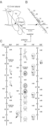
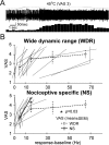
The neuronal system signaling pain has often been characterized as a labeled line consisting of neurons in the pain-signaling pathway to the brain [spinothalamic tract (STT)] that respond only to painful stimuli. It has been proposed recently that the STT contains a series of analog labeled lines, each signaling a different aspect of the internal state of the body (interoception) (e.g., visceral-cold-itch sensations). In this view, pain is the unpleasant emotion produced by disequilibrium of the internal state. We now show that stimulation of an STT receiving zone in awake humans (66 patients) produces two different responses. The first is a binary response signaling the presence of painful stimuli. The second is an analog response in which nonpainful and painful sensations are graded with intensity of the stimulus. Compared with the second pathway, the first was characterized by higher pain ratings and stimulus-evoked sensations covering more of the body surface (projected fields). Both painful responses to stimulation were described in terms usually applied to external stimuli (exteroception) rather than to internal or emotional phenomena, which were infrequently evoked by stimulation of either pathway. These results are consistent with those of functional imaging studies that have identified brain regions activated in a binary manner by the application of a specific, painful stimulus while increases in stimulus intensity do not produce increased activation. Such binary pain functions could be involved in pain-related alarm-alerting functions, which are independent of stimulus amplitude.
Figures


Similar articles
-
Neuroscience of the human thalamus related to acute pain and chronic "thalamic" pain.J Neurophysiol. 2024 Dec 1;132(6):1756-1778. doi: 10.1152/jn.00065.2024. Epub 2024 Oct 16. J Neurophysiol. 2024. PMID: 39412562 Review.
-
Psychophysics of CNS pain-related activity: binary and analog channels and memory encoding.Neuroscientist. 2006 Feb;12(1):29-42. doi: 10.1177/1073858405280553. Neuroscientist. 2006. PMID: 16394191 Review.
-
Thermal and pain sensations evoked by microstimulation in the area of human ventrocaudal nucleus.J Neurophysiol. 1993 Jul;70(1):200-12. doi: 10.1152/jn.1993.70.1.200. J Neurophysiol. 1993. PMID: 8360716
-
Thalamic stimulation-evoked sensations in chronic pain patients and in nonpain (movement disorder) patients.J Neurophysiol. 1996 Mar;75(3):1026-37. doi: 10.1152/jn.1996.75.3.1026. J Neurophysiol. 1996. PMID: 8867115
-
Responses of neurons in the region of human thalamic principal somatic sensory nucleus to mechanical and thermal stimuli graded into the painful range.J Comp Neurol. 1999 Aug 9;410(4):541-55. doi: 10.1002/(sici)1096-9861(19990809)410:4<541::aid-cne3>3.0.co;2-8. J Comp Neurol. 1999. PMID: 10398047
Cited by
-
Human Thalamic Somatosensory Nucleus (Ventral Caudal, Vc) as a Locus for Stimulation by INPUTS from Tactile, Noxious and Thermal Sensors on an Active Prosthesis.Sensors (Basel). 2017 May 24;17(6):1197. doi: 10.3390/s17061197. Sensors (Basel). 2017. PMID: 28538681 Free PMC article. Review.
-
Thalamic physiology of intentional essential tremor is more like cerebellar tremor than postural essential tremor.Brain Res. 2013 Sep 5;1529:188-99. doi: 10.1016/j.brainres.2013.07.011. Epub 2013 Jul 13. Brain Res. 2013. PMID: 23856324 Free PMC article.
-
Studies of properties of "Pain Networks" as predictors of targets of stimulation for treatment of pain.Front Integr Neurosci. 2011 Dec 5;5:80. doi: 10.3389/fnint.2011.00080. eCollection 2011. Front Integr Neurosci. 2011. PMID: 22164137 Free PMC article.
-
Neuroscience of the human thalamus related to acute pain and chronic "thalamic" pain.J Neurophysiol. 2024 Dec 1;132(6):1756-1778. doi: 10.1152/jn.00065.2024. Epub 2024 Oct 16. J Neurophysiol. 2024. PMID: 39412562 Review.
-
Attention to painful cutaneous laser stimuli evokes directed functional connectivity between activity recorded directly from human pain-related cortical structures.Pain. 2011 Mar;152(3):664-675. doi: 10.1016/j.pain.2010.12.016. Epub 2011 Jan 20. Pain. 2011. PMID: 21255929 Free PMC article.
References
-
- Apkarian AV, Shi T, Bruggemann J, Airapetian LR (2000) Segregation of nociceptive and non-nociceptive networks in the squirrel monkey somatosensory thalamus. J Neurophysiol 84: 484-494. - PubMed
-
- Becker DE, Yingling CD, Fein G (1993) Identification of pain, intensity, and P300 components in the pain evoked potential. Electroencephalogr Clin Neurophysiol 88: 290-301. - PubMed
-
- Blomqvist A, Zhang ET, Craig AD (2000) Cytoarchitectonic and immunohistochemical characterization of a specific pain and temperature relay, the posterior portion of the ventral medial nucleus, in the human thalamus. Brain 123: 601-619. - PubMed
-
- Bornhovd K, Quante M, Glauche V, Bromm B, Weiller C, Buchel C (2002) Painful stimuli evoke different stimulus-response functions in the amygdala, prefrontal, insula and somatosensory cortex: a single-trial fMRI study. Brain 125: 1326-1336. - PubMed
Publication types
MeSH terms
Grants and funding
LinkOut - more resources
Full Text Sources
Medical
Research Materials
