. 2004 Jul 21;24(29):6578-89.
doi: 10.1523/JNEUROSCI.1258-04.2004.

Modulation of synaptic plasticity by antimanic agents: the role of AMPA glutamate receptor subunit 1 synaptic expression

Affiliations

Modulation of synaptic plasticity by antimanic agents: the role of AMPA glutamate receptor subunit 1 synaptic expression

Jing Du et al. J Neurosci. .

Abstract

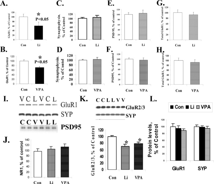
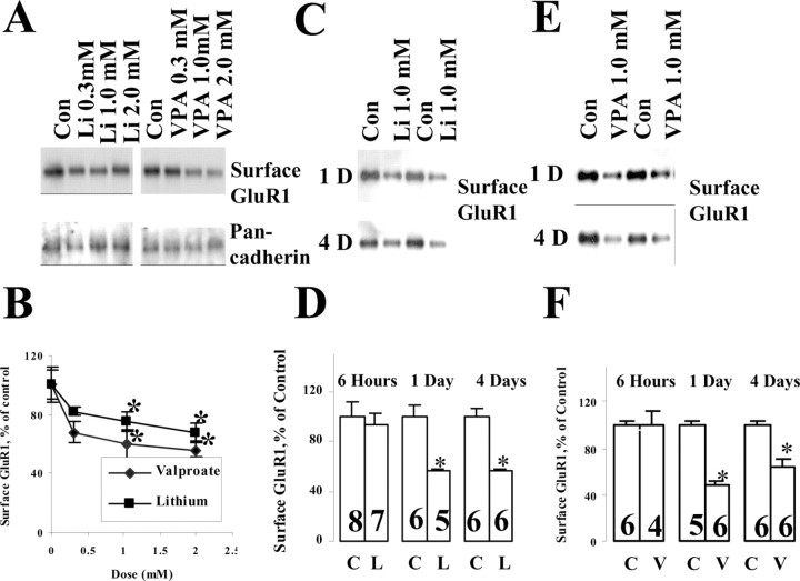
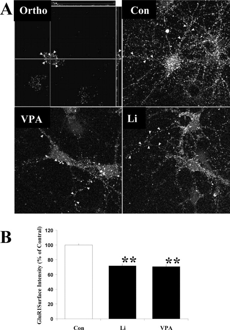
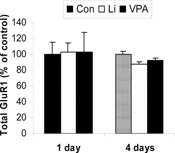
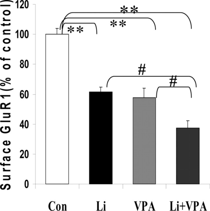
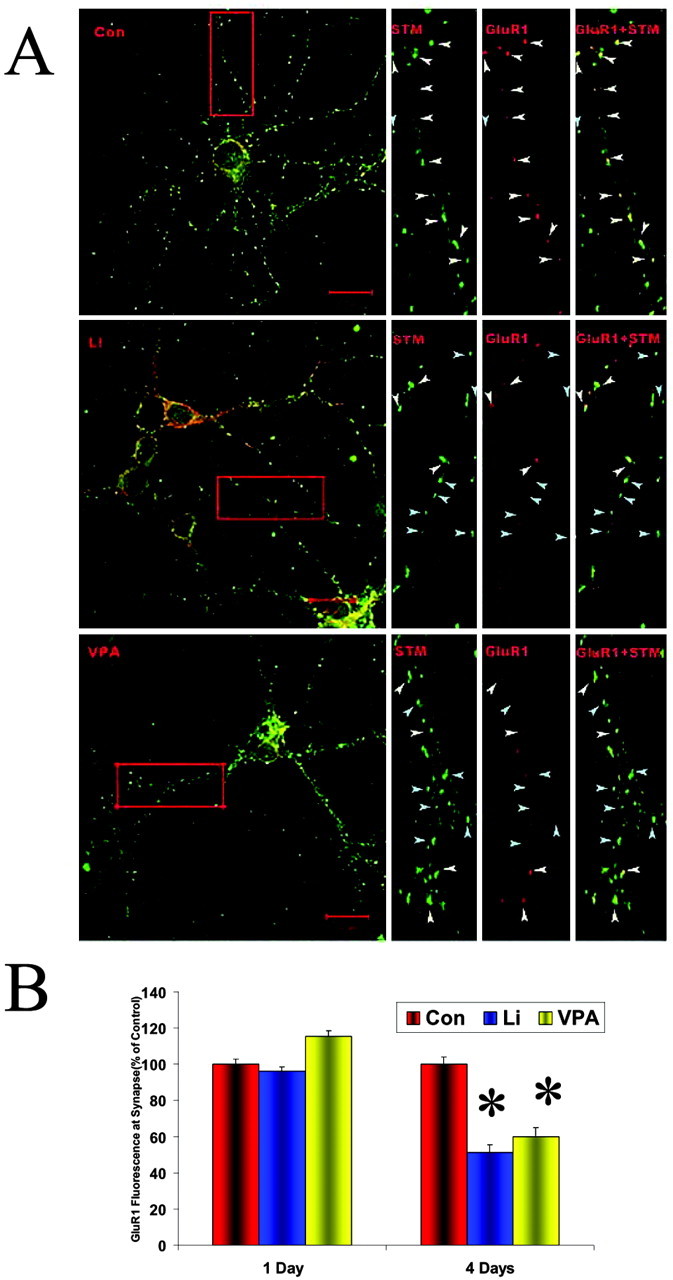
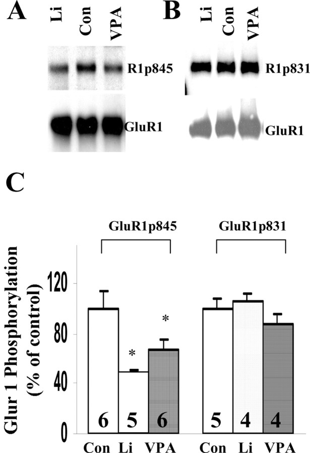
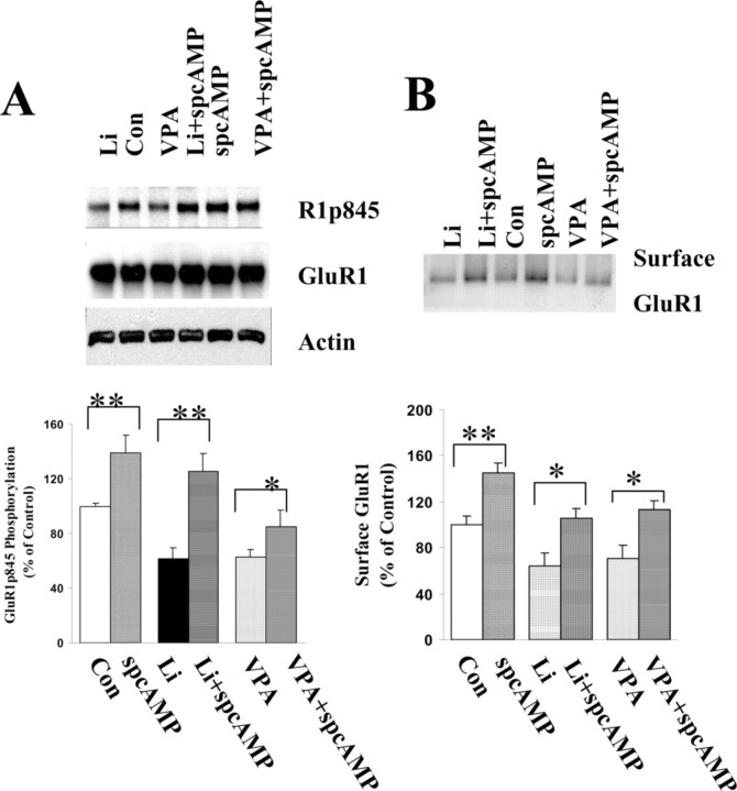
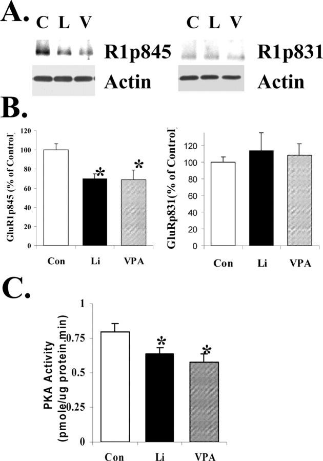
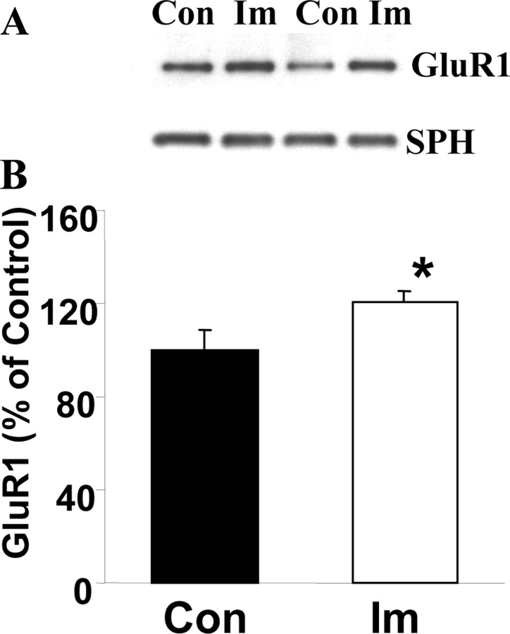
Increasing data suggest that impairments of cellular plasticity underlie the pathophysiology of bipolar disorder. In this context, it is noteworthy that AMPA glutamate receptor trafficking regulates synaptic plasticity, effects mediated by signaling cascades, which are targets for antimanic agents. The present studies were undertaken to determine whether two clinically effective, but structurally highly dissimilar, antimanic agents lithium and valproate regulate synaptic expression of AMPA receptor subunit glutamate receptor 1 (GluR1). Chronic (but not acute) treatment of rats with therapeutically relevant concentrations of lithium or valproate reduced hippocampal synaptosomal GluR1 levels. The reduction in synaptic GluR1 by lithium and valproate was attributable to a reduction of surface GluR1 distribution onto the neuronal membrane as demonstrated by three independent assays in cultured hippocampal neurons. Furthermore, these agents induced a decrease in GluR1 phosphorylation at a specific PKA site (GluR1p845), which is known to be critical for AMPA receptor insertion. Sp-cAMP treatment reversed the attenuation of phosphorylation by lithium and valproate and also brought GluR1 back to the surface, suggesting that phosphorylation of GluR1p845 is involved in the mechanism of GluR1 surface attenuation. In addition, GluR1p845 phosphorylation also was attenuated in hippocampus from lithium- or valproate-treated animals in vivo. In contrast, imipramine, an antidepressant that can trigger manic episodes, increased synaptic expression of GluR1 in hippocampus in vivo. These studies suggest that regulation of glutamatergically mediated synaptic plasticity may play a role in the treatment of bipolar disorder and raise the possibility that agents more directly affecting synaptic GluR1 may represent novel therapies for this devastating illness.


Figures

Figure 1.
Figure 1.
AMPA receptor subunit GluR1 was attenuated in synaptosomal preparation from long-term lithium- and valproate-treated animals. A, Quantification of GluR1 content in hippocampal synaptosomes from lithium-treated (n = 10) and control; (n = 12) animals (t test; p < 0.01). B, GluR1 protein content in hippocampal synaptosomes from valproate-treated (n = 11) and control (n = 12) animals (t test; p < 0.05). C, Quantification of synaptophysin protein in hippocampal synaptosomal preparation from lithium-treated (n = 5) and control (n = 6) animals. D, Synaptophysin protein levels in hippocampal synaptosomal preparation from valproate-treated (n = 8) and control (n = 7) animals. E, PSD95 protein levels in synaptosomal preparation from lithium-treated (n = 11) and control (n = 12) animals. F, PSD95 protein levels in synaptosomal preparation from valproate-treated (n = 12) and control (n = 12) animals. G, Total GluR1 expression levels in hippocampal tissue homogenates from lithium-treated (n = 9) and control (n = 9) animals. H, Total GluR1 expression levels in hippocampal tissue homogenates from valproate-treated (n = 5) and control (n = 9) animals. I, Samples of Western blot analysis of hippocampal synaptosomal preparation from lithium-treated (L) or valproate-treated (V) animals with anti-GluR1, anti-synaptophysin (SYP), or anti-PSD95 antibodies. J, NMDA receptor NR1 levels remained unchanged in synaptosomal preparation from lithium-treated (n = 10) or valproate-treated (n = 12) animals compared with control animals (n = 10). K, AMPA GluR2/3 receptors also were attenuated in hippocampal synaptosomal preparations from lithium- and valproate-treated animals (control, n = 7; lithium, n = 7; valproate, n = 6). ANOVA; *p < 0.05. L, GluR1 and synaptophysin (SYP) levels in synaptosomal preparation from short-term (5 d) lithiumtreated (n = 7), valproate-treated (n = 8), and control (n = 7) animals.
Figure 2.
Figure 2.
Surface GluR1 on cultured hippocampal neurons was decreased after lithium and valproate treatment within a therapeutic range in a dose- and time-dependent manner. Hippocampalprimary culture neurons were prepared from E18 Sprague Dawley embryos. After 10 d of culturing in B27 Neurobasal media, the neurons were treated with lithium or valproate with the dose indicated and for various durations. Surface proteins of the neurons were labeled with biotin, and then the cells were harvested with lysis buffer. Biotinylated surface proteins were precipitated by immobilized avidin and analyzed by Western blot analysis with anti-GluR1 or anti-pan-cadherin (surface protein control) antibodies. Data were analyzed by the Kodak Imaging System and pooled from three independent experiments. A, Dose dependency of lithium and valproate effect on surface GluR1 after 4 d of treatment. Con, Control. B, Dose-response curve for surface GluR1 expression after lithium and valproate treatment for 4 d. Pan-cadherin was used as a loading control (n = 2, n = 4; *p < 0.05). C, Lithium (1.0 mm) effects on surface GluR1 after 1 or 4 d of treatment. D, Quantification of the time course on lithium effect of GluR1 on neuronal surface (n = 3; n as indicated on the bars; t test; *p < 0.05). E, Valproate (1.0 mm) effect on GluR1 surface expression after 1 or 4 d of treatment. F, Quantification for the effect of valproate on GluR1 surface expression (n = 3; n as indicated on the bars; t test; *p < 0.05). C, Control; L, lithium; V, valproate.
Figure 3.
Figure 3.
A, Surface staining with anti-GluR1 N-terminal antibody after lithium and valproate treatment in cultured hippocampal neurons. Hippocampal neurons were cultured for 10 d, followed by treatment with Li (1.0 mm) or VPA (1.0 mm) for an additional 1 d. The surface of living neurons was labeled with anti-GluR1 (against N-terminal epitope) antibody and subsequently with Cy3-anti-rabbit IgG. Z-stack images were acquired with a 510-Meta confocal microscopy under exactly the same setup for different experimental groups. Three-dimensional images were reconstructed with 510-Meta software. The orthogonal picture (Ortho) indicates that GluR1 stainings are at the neuronal surface. B, Quantification of surface GluR1 in hippocampal neurons after lithium and valproate treatment. The mean fluorescent intensity on the longest dendrite was determined (n = 10 for each group; ANOVA; **p < 0.01). This experiment was repeated. Con, Control.
Figure 4.
Figure 4.
GluR1 total protein expression remained unchanged after lithium or valproate treatment in cultured hippocampal neurons. Hippocampal neurons were treated with lithium (1.0 mm) or valproate (1.0 mm) for 1 or 4 d. Equal amounts of proteins were applied for Western blot analysis by anti-GluR1 antibody. The loading of the protein was corrected by actin. Con, Control.
Figure 5.
Figure 5.
Treatment with both lithium and valproate had an additive effect on the surface GluR1 expression of hippocampal neurons. After being cultured for 10 d, hippocampal neurons were treated with lithium only, valproate only, and lithium plus valproate for 4 d. Surface GluR1 was determined by biotinylation assay. Samples were pooled from three independent experiments, with n of 9-12 (ANOVA; #,**p < 0.05). Con, Control.
Figure 6.
Figure 6.
Double immunostaining of GluR1 and synaptotagmin indicated that GluR1 was attenuated at synapses. A, Double immunostaining of GluR1 and synaptotagmin in hippocampal neurons treated with lithium or valproate for 4 d. After lithium (1.0 mm) or valproate (1.0 mm) treatment for 4 d, hippocampal neurons were double-stained with anti-GluR1 (red) and anti-synaptotagmin (STM; green) antibodies. Z-stack images were acquired by using a confocal microscope under the same setup for lithium-treated (Li) or valproate-treated (VPA) and untreated (Con) groups. Three-dimensional images were reconstructed by 510-Meta software. Portions of dendrite (red rectangle) were taken from the neurons for a close observation on their synapses. The GluR1-positive synapses are indicated with white arrows, and GluR1-negative synapses are indicated with blue arrows. B, Quantification of GluR1 (red) fluorescent intensity at the synapses. GluR1 fluorescent intensities were quantified at the individual synapses from the longest dendrite from seven neuronal images for each condition. The average length of the longest dendrite in each condition was not significantly different. In total, ∼150-190 synapses were measured for each condition (ANOVA; *p < 0.01). This experiment was repeated. Con, Control.
Figure 7.
Figure 7.
Phosphorylation of GluR1 at PKA site was attenuated significantly by lithium or valproate treatment in cultured hippocampal neurons. A, Phosphorylation of GluR1 at p845 (PKA site) after lithium (1.0 mm) or valproate (1.0 mm) treatment for 4 d in cultured hippocampal neurons. The membranes were stripped and reprobed with anti-GluR1 antibody. B, Phosphorylation of GluR1 at p831 (CaMKII site) after lithium (1.0 mm) or valproate (1.0 mm) treatment for 4 d. The same blot was stripped and reprobed with anti-GluR1 antibody. C, Quantification of GluR1 phosphorylation at p845 and p831 sites after lithium or valproate treatment for 4 d. Results were pooled from two to three independent experiments. The number for each condition is indicated on the bars (ANOVA; *p < 0.05). Con, Control.
Figure 8.
Figure 8.
Sp-cAMP enhanced GluR1 phosphorylation on the PKA site in lithium- or valproate-treated neurons and also resulted in an increase in GluR1 surface expression. Hippocampal neurons were cultured for 8 d in B27 plus Neurobasal medium and treated with lithium (1.0 mm) or valproate (1.0 mm) for an additional 4 d. Sp-cAMP (50 μm) was applied to the cultured hippocampal neurons for 30 min. The surface proteins were labeled with sulfo-NHS-LC-biotin, and the proteins were harvested with lysis buffer. Equal amounts of proteins were analyzed by Western blot with anti-phospho-GluR1p845 and anti-actin antibody. The blots were stripped and reprobed with anti-GluR1 antibody. A, Sp-cAMP significantly enhanced phosphorylation of GluR1 at the p845 site in all control, lithium-, and valproate-treated groups. B, Sp-cAMP treatment also reversed the effects of lithium and valproate on GluR1 surface expression. Data were pooled from two to three independent experiments (n = 5-8; t test; **p < 0.01 and *p < 0.05). Con, Control.
Figure 9.
Figure 9.
Both GluR1 phosphorylation at the PKA site and PKA autonomous activity were reduced in lithium- and valproate-treated animals in hippocampus. A, B, Phosphorylation of GluR1 in hippocampus from lithium (L) or valproate (V) chronically treated animals. C, Control. Rats were treated with lithium or valproate for 4 weeks; hippocampal tissues were isolated and homogenated. Equal amounts of homogenate proteins were separated by gel electrophoresis and analyzed with anti-GluR1p845, anti-GluR1p831, and anti-actin antibodies. The bands were analyzed by the Kodak Imaging System (n = 8 for all three groups; ANOVA; *p < 0.05). C, PKA activity in hippocampus from lithium or valproate chronically treated animals. Protein samples were prepared from one hippocampus of lithium- or valproate-treated animals. Equal amounts of proteins were assayed for PKA activity (n = 8 for all three groups; ANOVA; *p < 0.05).
Figure 10.
Figure 10.
Antidepressant imipramine (Im) induces an increase in synaptic GluR1 in vivo. Rats were treated with imipramine (10 mg/kg, twice daily, i.p.) for 4 weeks; hippocampal tissues were isolated, and synaptosome preparations were obtained by using a Ficoll gradient assay. A, B, Equal amounts of proteins were separated by gel electrophoresis and analyzed with anti-GluR1 antibodies and anti-synaptophysin antibodies. The bands were analyzed by the Kodak Imaging System (n = 8; t test; *p < 0.05). Con, Control.

References

    1. Backstrom P, Hyytia P (2003) Attenuation of cocaine-seeking behaviour by the AMPA/kainate receptor antagonist CNQX in rats. Psychopharmacology (Berl) 166: 69-76. - PubMed
    1. Burns LH, Everitt BJ, Kelley AE, Robbins TW (1994) Glutamate-dopamine interactions in the ventral striatum: role in locomotor activity and responding with conditioned reinforcement. Psychopharmacology (Berl) 115: 516-528. - PubMed
    1. Carlezon Jr WA, Nestler EJ (2002) Elevated levels of GluR1 in the midbrain: a trigger for sensitization to drugs of abuse? Trends Neurosci 25: 610-615. - PubMed
    1. Chao SZ, Ariano MA, Peterson DA, Wolf ME (2002) D1 dopamine receptor stimulation increases GluR1 surface expression in nucleus accumbens neurons. J Neurochem 83: 704-712. - PubMed
    1. Coyle JT, Duman RS (2003) Finding the intracellular signaling pathways affected by mood disorder treatments. Neuron 38: 157-160. - PubMed

Publication types

MeSH terms

LinkOut - more resources