. 2004 Jul 21;24(29):6590-9.
doi: 10.1523/JNEUROSCI.5747-03.2004.

Mood stabilizer valproate promotes ERK pathway-dependent cortical neuronal growth and neurogenesis

Affiliations

Mood stabilizer valproate promotes ERK pathway-dependent cortical neuronal growth and neurogenesis

Yanlei Hao et al. J Neurosci. .

Abstract

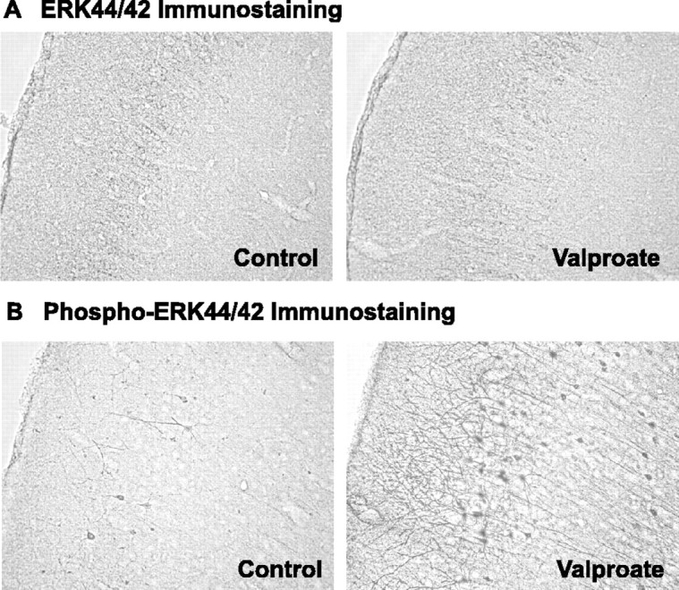
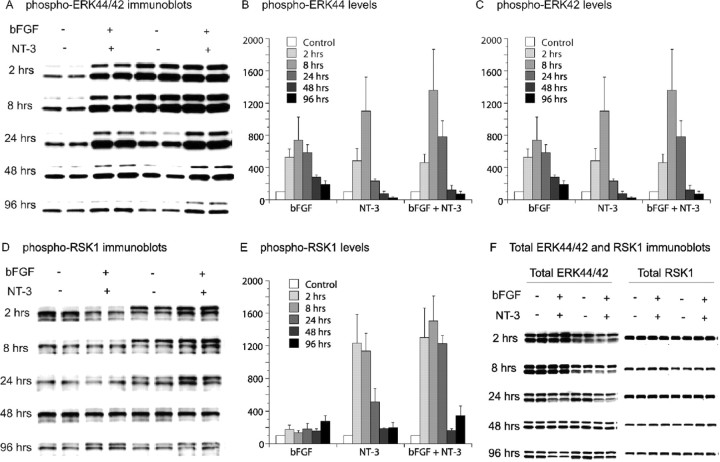
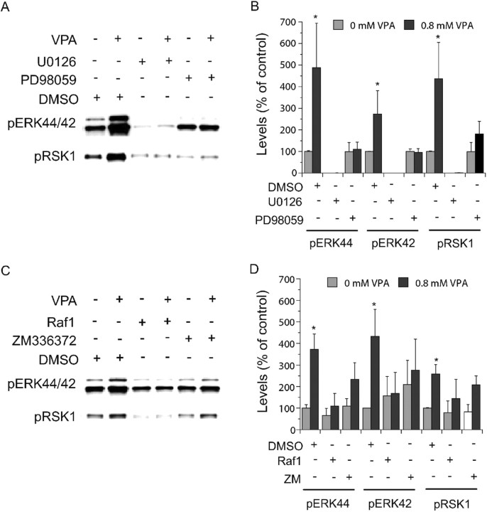
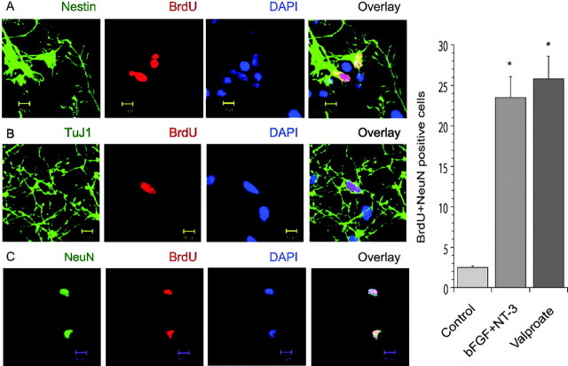
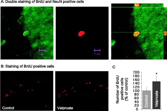
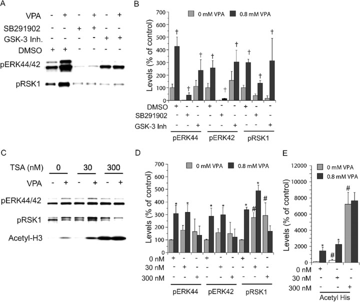
Manic-depressive illness has been conceptualized as a neurochemical illness. However, brain imaging and postmortem studies reveal gray-matter reductions, as well as neuronal and glial atrophy and loss in discrete brain regions of manic-depressive patients. The roles of such cerebral morphological deficits in the neuropathophysiology and therapeutic mechanisms of manic-depressive illness are unknown. Valproate (2-propylpentanoate) is a commonly used mood stabilizer. The ERK (extracellular signal-regulated kinase) pathway is used by neurotrophic factors to regulate neurogenesis, neurite outgrowth, and neuronal survival. We found that chronic treatment of rats with valproate increased levels of activated phospho-ERK44/42 in neurons of the anterior cingulate, a region in which we found valproate-induced increases in expression of an ERK pathway-regulated gene, bcl-2. Valproate time and concentration dependently increased activated phospho-ERK44/42 and phospho-RSK1 (ribosomal S6 kinase 1) levels in cultured cortical cells. These increases were attenuated by Raf and MEK (mitogen-activated protein kinase/ERK kinase) inhibitors. Although valproate affects the functions of GSK-3 (glycogen synthase kinase-3) and histone deacetylase (HDAC), its effects on the ERK pathway were not fully mimicked by selective inhibitors of GSK-3 or HDAC. Similar to neurotrophic factors, valproate enhanced ERK pathway-dependent cortical neuronal growth. Valproate also promoted neural stem cell proliferation-maturation (neurogenesis), demonstrated by bromodeoxyuridine (BrdU) incorporation and double staining of BrdU with nestin, Tuj1, or the neuronal nuclei marker NeuN (neuronal-specific nuclear protein). Chronic treatment with valproate enhanced neurogenesis in the dentate gyrus of the hippocampus. Together, these data demonstrate that valproate activates the ERK pathway and induces ERK pathway-mediated neurotrophic actions. This cascade of events provides a potential mechanism whereby mood stabilizers alleviate cerebral morphometric deficits associated with manic-depressive illness.


Figures

Figure 1.
Figure 1.
Increases in prefrontal cortical phospho-ERK44/42, but not total ERK44/42, immunoreactivities by chronic valproate treatment. Rats were fed regular or sodium valproate-supplemented (20 gm/kg) chows for 4 weeks, yielding a mean serum valproic acid concentration of 42 mg/l. Immunostaining of the brain slices was performed. Valproate treatment markedly increased intensities of phospho-ERK44/42 (B), but not of total ERK44/42 (A), in prefrontal cortical cells with neuronal characteristics. Two investigators, blind to the treatments, independently examined 10 sets of paired samples and reported clear increases in phospho-ERK44/42 staining intensities in eight sets.
Figure 2.
Figure 2.
Time-dependent activation of the ERK pathway by bFGF, NT-3, or in combination. Rat cortical cells isolated from E18 embryos were cultured in vitro for 8 d, at which time the culture reached confluence. The cells were then treated with bFGF and NT-3. Immunoblotting of phospho-ERK44/42, phospho-RSK1, total ERK44/42, and total RSK1 was conducted. bFGF (10 ng/ml), NT-3 (20 ng/ml), or a combination of both time dependently increased phospho-ERK44/42 (A-C) and phospho-RSK1 levels (D, E) but not total ERK44/42 and total RSK1 (F). Bar graphs (B, C, E) depict densitometric results representing mean ± SE of three or more sets of samples immunoblotted in duplicates as presented in the figure (A, D, F).
Figure 3.
Figure 3.
Concentration- and time-dependent activation of the ERK pathway and accumulation of acetylated histone-3 by valproate (VPA) in cortical cells. Rat cortical cells isolated from E18 embryos were cultured in vitro for 8 d, at which time the culture reached confluence. The cells were then treated with sodium valproate. Immunoblotting of phospho-ERK44/42, phospho-RSK1, total ERK44/42, total RSK1, or acetyl-H3 was conducted. Valproate concentration (48 hr) (A, B) and time (0.8 mm) (C, D) dependently increased phospho-ERK44/42, phospho-RSK1, and acetyl-H3 levels are shown. Bar graphs (B, D) depict densitometric results representing mean ± SE of three or more sets of samples immunoblotted in duplicates as presented in A and C. F(5, 18) = 9.974, p = 0.001 for phosho-ERK44; F(5,18) = 2.892, p = 0.0435 for phospho-ERK42; F(5,18) = 11.404, p < 0.0001 for phospho-RSK1; F(5,18) = 10.224, p < 0.0001 for acetyl-H3. *p < 0.05 compared with 8, 24, 48, 72, and 96 hr valproate-treated cells. #p < 0.05 compared with valproate-treated cells.
Figure 4.
Figure 4.
Involvement of ERK pathway components in valproate-(VPA) induced ERK pathway activation. E18, DIV 8 cortical cells obtained as described in Figures 2 and 3 were treated with valproate (0.8 mm) in the absence or presence of indicated inhibitors for 2 d. Immunoblotting was conducted as described in Figures 2 and 3. MEK inhibitors [PD98059 (50 μm) and U0126 (10 μm)] (A, B) and Raf inhibitors [Raf inhibitor I (1 μm) and ZM336372 (10 μm)] (C, D) attenuated valproate-induced increases in phospho-ERK44/42 and phospho-RSK1. U0126 and Raf1 inhibitor I attenuated basal phospho-ERK44/42 and phospho-RSK1 (A-D). Bar graphs (B, D) depict densitometric results representing mean ± SE from three or more sets of samples immunoblotted in duplicates on two gels as presented in A and C. *p < 0.05 compared with cells treated with DMSO alone.
Figure 5.
Figure 5.
Induction of neurite growth and cell reemergence by neurotrophic factors and valproate (VPA). For observation of cortical cell regeneration, gaps (indicated by arrows; bars indicate the edges of the gaps) were created by removing strips of cells from the middle of dishes or culture slides containing E18, DIV 8 confluent cortical cells as described in Figures 2 and 3. After 2 d, neurite growth and cell reemergence were more pronounced in cultures treated with bFGF (10 ng/ml), NT-3 (20 ng/ml), bFGF plus NT-3, or valproate (0.8 mm) (left) than controls. MEK inhibitor PD98059 (50 μm) blocked growth in valproate-treated and nontreated cultures (left). Similar results were also obtained in three or more sets of samples. Neurite lengths were traced in three sets of samples. Two day valproate (0.8 mm) treatment significantly increased neurite lengths in the gaps (right). *p < 0.05 compared with controls.
Figure 6.
Figure 6.
Induction of neurogenesis by neurotrophic factors and valproate in cortical cells. The cortical cell cultures were obtained and handled as described in Figure 5. To monitor cell proliferation, cultures were treated with BrdU (50 μg/ml) for 6 hr after creating gaps and treated with reagents as described in Figure 5 for 1, 2, or 5 d. The cultures were processed for staining of nuclei with DAPI (blue) and for staining of antigens of BrdU (red), nestin (green), TuJ1 (green), or NeuN (green). Images were obtained using a Zeiss LSM510 Meta multiphoton system. DAPI-positive cells, DAPI plus BrdU-positive cells, and cells undergoing mitosis were present (A, B), suggesting that cells in the gaps migrated from surrounding regions or were born de novo. BrdU plus nestin-(A), BrdU plus TuJ1-(B), or BrdU plus NeuN-(C) positive cells were present, suggesting the existence of neural stem cell neurogenesis. Valproate and the combination of bFGF and NT-3 significantly increased the numbers of both BrdU- and NeuN-positive cells, indicating that neurotrophic factor and valproate treatments promoted neurogenesis (right). *p < 0.05 compared with controls. Scale bars, 10 μm..
Figure 7.
Figure 7.
Induction of hippocampal neurogenesis by chronic valproate treatment in adult mice. Male C57BL/6 (25-30 gm) mice were treated first with a single injection of BrdU (300 mg/kg) and then fed valproate-containing chow (20 gm/kg) for 6 weeks. The potential effect of chronic valproate on hippocampal neurogenesis was investigated according to standard stereological techniques. Each treatment group contained six mice. Eight serial sections from each mouse were immunostained with antibodies against either BrdU (red) alone or BrdU and NeuN (green). The sections were examined using a Zeiss LSM510 Meta multiphoton system. Double staining of BrdU and NeuN revealed that the majority of the BrdU-positive cells in the dentate gyrus also stained positively for NeuN antibody (A). There were greater numbers of BrdU-positive cells in the section from the valproate-treated mouse (B). The difference in the numbers of BrdU-positive cells between the two groups was statistically significant (C). *p < 0.05 compared with controls. Scale bars, 10 μm.
Figure 8.
Figure 8.
Involvements of other signaling proteins in valproate-(VPA) induced ERK pathway activation. E18, DIV8 cortical cells obtained as described in Figures 2, 3, 4 were treated with valproate (0.8 mm) in the absence or presence of indicated inhibitors for 2d. Immunoblotting was conducted as described in Figures 2, 3, 4. p38 inhibitor [SB202190(2 μm)] lowered basal levels but not the magnitudes of valproate-induced increases in levels of phospho-ERK44/42 and phopspho-RSK1 (A, B). Selective GSK-3 inhibitor [GSK-3 inhibitor II (20 μm)] neither induced increases in basal levels of phospho-ERK44/42 and phospho-RSK1 nor altered valproate-induced increases in levels of phospho-ERK44/42 and phospho-RSK1 (A, B). Valproate and TSA induced increases in acetyl-H3 levels (C, E). The effects of valproate and TSA (at low concentration) on acetyl-H3 accumulation appeared to be additive (C, E). TSA alone appeared to elevate levels of phospho-ERK44/42 and phospho-RSK1, but the increases were not concentration dependent (C, D). A high concentration of TSA (300 nm) attenuated valproate-induced increases in phospho-ERK44/42 and phopspho-RSK1 levels (C, D). Bar graphs (B, D, E) depict densitometric results representing mean ± SE of three or more sets of samples immunoblotted in duplicates on two gels as presented in A and C. p < 0.05, with versus without VPA. *p < 0.05, with versus without VPA. #p < 0.05, TSA-nonVPA-treated cells versus nontreated cells.

References

    1. Abdel-Latif AA (2001) Cross talk between cyclic nucleotides and polyphosphoinositide hydrolysis, protein kinases, and contraction in smooth muscle. Exp Biol Med 226: 153-163. - PubMed
    1. Altar CA, Whitehead RE, Chen R, Wortwein G, Madsen TM (2003) Effects of electroconvulsive seizures and antidepressant drugs on brain-derived neurotrophic factor protein in rat brain. Biol Psychiatry 54: 703-709. - PubMed
    1. Altar CA, Laeng P, Jurata LW, Brockman JA, Lemire A, Bullard J, Bukhman YV, Young TA, Charles V, Palfreyman MG (2004) Electroconvulsive seizures regulate gene expression of distinct neurotrophic signaling pathways. J Neurosci 24: 2667-2677. - PMC - PubMed
    1. Andresen BT, Rizzo MA, Shome K, Romero G (2002) The role of phosphatidic acid in the regulation of the Ras/MEK/Erk signaling cascade. FEBS Lett 531: 65-68. - PubMed
    1. Apfel SC (2002) Is the therapeutic application of neurotrophic factors dead? Ann Neurol 51: 8-11. - PubMed

Publication types

MeSH terms

Substances