. 2004 Aug;2(8):E239.
doi: 10.1371/journal.pbio.0020239. Epub 2004 Jul 20.

Cellular and genetic analysis of wound healing in Drosophila larvae

Affiliations

Cellular and genetic analysis of wound healing in Drosophila larvae

Michael J Galko et al. PLoS Biol. 2004 Aug.

Abstract

To establish a genetic system to study postembryonic wound healing, we characterized epidermal wound healing in Drosophila larvae. Following puncture wounding, larvae begin to bleed but within an hour a plug forms in the wound gap. Over the next couple of hours the outer part of the plug melanizes to form a scab, and epidermal cells surrounding the plug orient toward it and then fuse to form a syncytium. Subsequently, more-peripheral cells orient toward and fuse with the central syncytium. During this time, the Jun N-terminal kinase (JNK) pathway is activated in a gradient emanating out from the wound, and the epidermal cells spread along or through the wound plug to reestablish a continuous epithelium and its basal lamina and apical cuticle lining. Inactivation of the JNK pathway inhibits epidermal spreading and reepithelialization but does not affect scab formation or other wound healing responses. Conversely, mutations that block scab formation, and a scabless wounding procedure, provide evidence that the scab stabilizes the wound site but is not required to initiate other wound responses. However, in the absence of a scab, the JNK pathway is hyperinduced, reepithelialization initiates but is not always completed, and a chronic wound ensues. The results demonstrate that the cellular responses of wound healing are under separate genetic control, and that the responses are coordinated by multiple signals emanating from the wound site, including a negative feedback signal between scab formation and the JNK pathway. Cell biological and molecular parallels to vertebrate wound healing lead us to speculate that wound healing is an ancient response that has diversified during evolution.


Conflict of interest statement

The authors have declared that no conflicts of interest exist.

Figures

Figure 1
Figure 1. Scab Formation and Resolution during Puncture Wound Healing
(A) Puncture wound assay. L3 larvae are punctured at the dorsal midline with a 100-μm diameter pin; they are then cultured and the healing wounds analyzed as shown. (B–E) Photomicrographs of heat-killed L3 larvae before wounding (B) and at the indicated times after wounding (C–E). Note larval growth during wound healing. Anterior is up. (F) L2 larva wounded as above and analyzed in L3, 60 h after wounding. Wounding in L2 allows visualization of late stages of wound healing without the complication of pupariation, which begins about 48 h after wounding in the standard L3 assay. (G) A mock-wounded L2 larva visualized 60 h after wounding. Note that it and the wounded larva (F) grew to a similar extent. (H–M) Close-up images of (B–G) showing unwounded cuticle (H and M) or wound sites (I–L, boxed regions in C–F) to show detail of scab. Micrographs are of living larvae taken shortly before the corresponding images of the whole heat-killed larvae above. Bar, 500 μm (for B–G), 50 μm (for H–M). (N) Timing of wound responses. Solid lines, time response was most often observed; dashed extensions to left, time response was occasionally observed; dashed extensions to right, time response was diminishing; BL, basal lamina.
Figure 3
Figure 3. Epidermal Cell Orientation and Fusion around Puncture Wounds
(A–E) w; UAS-GFP.nls/+; A58-Gal4/+ larvae that express GFP (green) in epidermal cell nuclei were mock-wounded (A) or puncture wounded (B–E), cultured for the indicated time, filleted open, fixed, and immunostained for Fasciclin III (red) to label the basolateral surface of the cells. (A) Pre-wounding. Dashed circle, size of the 100-μm pin used for wounding. (B) 2 h postwounding. Some cells at the wound margin have elongated and oriented toward the wound (arrowheads). (C) 8 h postwounding. Cells at the wound margin have begun fusing to form a syncytium. Note the syncytium with four nuclei that contains a partially degraded, radially-oriented membrane domain (arrow) and scattered puncta of Fasciclin III staining in the cytoplasm (arrowhead) that may be membrane breakdown intermediates. (D) 48 h postwounding. The central syncytium contains ten or more nuclei, some of which are located in extensions (arrowheads) that may represent recent fusions of peripheral cells with the central syncytium. Other peripheral cells have oriented toward the syncytium but not fused with it. (E) 60 h postwounding. A large syncytium with more than 30 nuclei. (F) 8 h postwounding. Larva was treated as above but immunostained for Coracle (red), a septate junction component. The central syncytium contains nine nuclei. Bar, 50 μm.
Figure 2
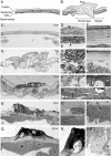
Figure 2. Ultrastructural Analysis of Puncture Wound Healing
(A) Schematic of unwounded epidermis showing the cell monolayer, its apical cuticle lining, and basal lamina. White ovals indicate nuclei. (B) Schematic of recently wounded epidermis showing a plug of cell debris in the wound gap. Cells and ruptured cuticle at the wound margin are shown. (C–S) TEM sections of unwounded (C–F) and wounded (G–S) larvae at the times indicated after wounding. Transverse sections through each wound site are shown (C, G, J, N, and Q) along with close-ups of the boxed regions at right. c, cuticle; d, debris; e, epidermis; ec, epicuticle; m, muscle; n, epidermal nucleus; p, plug; pc, procuticle; s, scab; t, trachea (C) Pre-wounding. The epidermis and cuticle are intact. (D) Apical surface of epidermal cell showing villi (arrowhead) that secrete cuticle. (E) Basal surface. Arrowhead, basal lamina. (F) Epidermal cuticle. The epicuticle (top three layers) overlies the striated procuticle layer. (G) 1 h postwounding. The epidermis and cuticle are discontinuous but the gap is filled with a plug (outlined by dashed line) of cell debris. The epidermis has partially separated from the cuticle beyond the wound margin (asterisks). (H) The plug contains highly vesiculated cell debris. (I) The epidermis separating (asterisk) from overlying cuticle appears vesiculated and is presumably necrotic. (J) 2 h postwounding. The outer portion of the plug has melanized to form an electron-dense scab (outlined by white dashed line). The epidermis and cuticle are still discontinuous. (K) Debris, including a necrotic trachea, in the plug. The plug is not bounded by a membrane or basal lamina. (L) Portion of scab showing melanized debris and trachea. (M) Close-up of a lamellipodium (bracket) extending into a plug at the outer edge of another 2-h wound. Note basal lamina (arrowhead) along the lamellipodium. (N) 8 h postwounding. The epidermis has migrated across the gap to reestablish continuity, and has secreted new cuticle beneath the scab. (O) A region of epidermal cell cytoplasm near wound plug debris contains vesiculated material (outlined by dashed line) that is probably phagocytosed debris. (P) The newly established epidermis under the wound has a continuous basal lamina (arrowhead) and apical villi (arrow) secreting cuticle. (Q) 24 h postwounding. The new cuticle underlying the scab is thicker and the scab is more electron dense. Four nuclei in close apposition are in a syncytium because there are no membranes separating them. (R) Portion of scab and old cuticle. Note that cuticle in contact with the scab is melanized. (S) Cytoplasmic extension (arrowhead) engulfing debris at the basal surface of the epidermis of another 24-h wound. Bar, 10 μm (C, G, J, N, and Q), 0.33 μm (D and E), 0.83 μm (F), 1 μm (H, M, and P), 2 μm (I and O), 1.67 μm (K and L), 4 μm (R), 1.25 μm (S).
Figure 4
Figure 4. Induction and Function of the JNK Pathway around Puncture Wounds
(A–C) Larvae carrying the JNK pathway reporter puc-lacZ, which expresses a nuclear β-galactosidase, were mock-wounded (A) or puncture wounded (B and C), and then cultured for the indicated times before staining with X-gal to visualize reporter activity (blue). There is little reporter activity in unwounded epidermis (A), but 4 h after wounding the reporter is expressed in a gradient emanating from the wound, with highest expression in the row of epidermal nuclei at the wound margin and decreasing levels in surrounding nuclei out to five cell diameters away. At 24 h (C), reporter expression has declined. (D–F) Larvae carrying the JNK pathway reporter msn-lacZ, treated as above. Wounding-induced reporter expression is seen out to seven cell diameters. (G–I) Larvae carrying msn-lacZ and A58-Gal4 and UAS-bskDN transgenes (to inactivate the JNK pathway in larval epidermis), treated as above. Reporter induction is inhibited, but the scab forms normally. (J and K) Larvae carrying msn-lacZ and either UAS-bskDN alone as control (J) or A58-Gal4 and UAS-bskDN transgenes (K), wounded as above and analyzed 24 h later by immunostaining for Fasciclin III and β-galactosidase. Reporter induction is inhibited in (K), but epidermal cells have oriented toward the wound, and although nuclear β-galactosidase staining is faint, careful inspection shows that the cells closest to the wound have fused to form a syncytium. Syncytium formation was confirmed using the A58-Gal4>UAS-GFP.nls marker. (L and M) Larvae carrying msn-lacZ and either UAS-bskDN alone as control (L) or A58-Gal4 and UAS-bskDN transgenes (M), wounded and analyzed 24 h later by TEM. Note that the epidermis in M has failed to spread across the wound gap and is still discontinuous (asterisks). No cuticle has been synthesized in the wound gap, but the cuticle flanking the wound appears thickened. Bar in (I), 100 μm (for [A–I]). Bar in (K), 50 μm (for [J and K]). Bar in (M), 5 μm (for [L and M]).
Figure 5
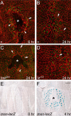
Figure 5. Cellular Responses and Genetic Requirements of Pinch Wound Healing
(A–D) Larvae carrying the msn-lacZ reporter and the indicated transgenes or mutations were pinched with a forceps to abrade a region of dorsal epidermis but leave the overlying cuticle intact. Wounded larvae were cultured for the indicated times and immunostained for Fasciclin III (red) and β-galactosidase (green). (A) 6 h after pinch wounding. Note the large epidermal gap (asterisk) at the wound site. Some cells at the wound margin have elongated and oriented toward the wound (arrowheads). Others have fused to form syncytia (arrow). (B) 24 h after pinch wounding. The epidermis has spread to close the gap. Note disorganization of epidermis and syncytia (arrows) at site of healed wound. (C) An A58-Gal4 and UAS-bskDN larva 24 h after pinch wounding. Epidermal spreading is inhibited and a large wound gap remains (asterisk). However, cells at the wound margin still orient toward the wound (arrowheads) and fuse to form syncytia (arrows). (D) A hemizygous lzr15 mutant larva 24 h after pinch wounding. lzr15 blocks crystal cell development and scab formation at puncture wounds (Figure 6), but no defects are observed in pinch wound healing. (E and F) Larvae carrying msn-lacZ reporter were mock-wounded (E) or pinch wounded (F), cultured for 4 h, and stained with X-gal (blue). Wounding induces reporter expression in a gradient extending out four cell diameters. The gap (asterisk) lacks a scab. Bar, 100 μM.
Figure 6
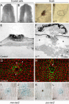
Figure 6. Effect of lz on Scab Formation and the Other Events of Puncture Wound Healing
(A and B) Posterior of lz+ (w1118) (A) and lzr15 mutant (B) L3 larvae. Larvae were heated so crystal cells appear as tiny black dots. No crystal cells are apparent in the lzr15 mutant. Bar, 200 μm. (C and D) Micrographs of control lz+ (w1118) (C) and lzr15 mutant (D) L3 larvae 4 h after puncture wounding. No scab is seen at the lzr15 wound site (encircled). Bar, 50 μm. (E and F) TEM sections through 24-h–old puncture wounds of a control lzr15/+ heterozygote (E) and a hemizygous lzr15 mutant larva (F), both carrying the msn-lacZ transgene. A consolidated, electron-dense scab has formed in the control larva (E), but only a diffuse plug with peripheral electron density is present at the lzr15 hemizygous wound (F). The electron density of the lzr15 plug might be due to residual melanization activity in the lzr15 mutant. Although reepithelialization is complete in the lzr15 mutant wound, the epidermis contains large vacuoles and abundant apical processes, and it is separated by a gap (asterisks) from the old cuticle and has not secreted new cuticle. Other 24-h lzr15 mutant wounds analyzed had necrotic or discontinuous epidermis at the wound site (not shown). Bar, 10 μm. (G and H) Fluorescence micrographs of 20-h puncture wounds in control (G) and lzr15 mutant (H) larvae carrying the msn-lacZ reporter that were treated as above and immunostained for Fasciclin III (red) and β-galactosidase (green). Epidermal cells at both control and lzr15 mutant wounds have fused to form syncytia (arrows), and cells in the control are oriented toward the wound site (arrowheads). The orientation response of epidermal cells in the lzr15 mutant is difficult to assess because cell borders out to six cell diameters away from the wound appear slack and wavy. Bar, 50 μm. (I–L) X-gal stains of 6-h–old puncture wounds of control lz+ (I and K) or lzr15 hemizygous mutant larvae (J and L) carrying either msn-lacZ (I and J) or puc-lacZ (K and L). Note the absence of scabs and the increase in reporter activity (blue) in lzr15. The basal level of reporter expression in unwounded epidermis was not increased in lzr15 (not shown). Bar, 50 μm.
Figure 7
Figure 7. Effect of lz on Survival Following Puncture Wounding
Control lz+ (w1118) and lzr15 mutant larvae were puncture wounded or mock-wounded and cultured for 4 or 24 h. The percentage of treated larvae alive and motile at each time is shown. Values are the average (± standard error of the mean) of three to six independent experiments with ten or more treated larvae per time point.
Figure 8
Figure 8. Model of the Cellular Events and Genetic Requirements of Larval Wound Healing
Puncture wounding disrupts the epidermis and overlying cuticle and triggers the three parallel series of events shown, each with distinct genetic requirements. Plug and scab formation stabilize the wound site, which promotes epidermal cell spreading and suppresses JNK activation, perhaps by a negative feedback mechanism (dashed line). The lz and Bc genes promote scab formation, presumably by promoting crystal cell development and the production and secretion of serum melanization factors by these cells. The spreading epidermal cells synthesize cuticle and basal lamina, and they clear wound site debris by phagocytosis. Pinch wounding disrupts the epidermis but not the overlying cuticle and triggers only the events shown in black. However, cuticle and basal lamina synthesis and phagocytosis have not been examined in pinch wounds and are only inferred to occur from the puncture wound studies. Wounding may induce additional signals (not indicated) that attract blood cells (plasmatocytes) and tracheal branches.

References

    1. Adachi-Yamada T, Nakamura M, Irie K, Tomoyasu Y, Sano Y, et al. p38 mitogen-activated protein kinase can be involved in transforming growth factor beta superfamily signal transduction in Drosophila wing morphogenesis. Mol Cell Biol. 1999;19:2322–2329. - PMC - PubMed
    1. Alster TS, Tanzi EL. Hypertrophic scars and keloids: Etiology and management. Am J Clin Dermatol. 2003;4:235–243. - PubMed
    1. Ashcroft GS, Yang X, Glick AB, Weinstein M, Letterio JL, et al. Mice lacking Smad3 show accelerated wound healing and an impaired local inflammatory response. Nat Cell Biol. 1999;1:260–266. - PubMed
    1. Ashida M, Brey PT. Role of the integument in insect defense: Pro-phenol oxidase cascade in the cuticular matrix. Proc Natl Acad Sci U S A. 1995;92:10698–10702. - PMC - PubMed
    1. Barwig B. Isolation and characterization of plasma coagulogen of the cockroach Leucophaea maderae (Blattaria) J Comp Physiol B. 1985;155:135–143.

Publication types

MeSH terms

Substances