. 2004 Aug;3(4):325-34.
doi: 10.1177/153303460400300402.

Combination of digital mammography with semi-automated 3D breast ultrasound

Affiliations

Combination of digital mammography with semi-automated 3D breast ultrasound

Ajay Kapur et al. Technol Cancer Res Treat. 2004 Aug.

Abstract

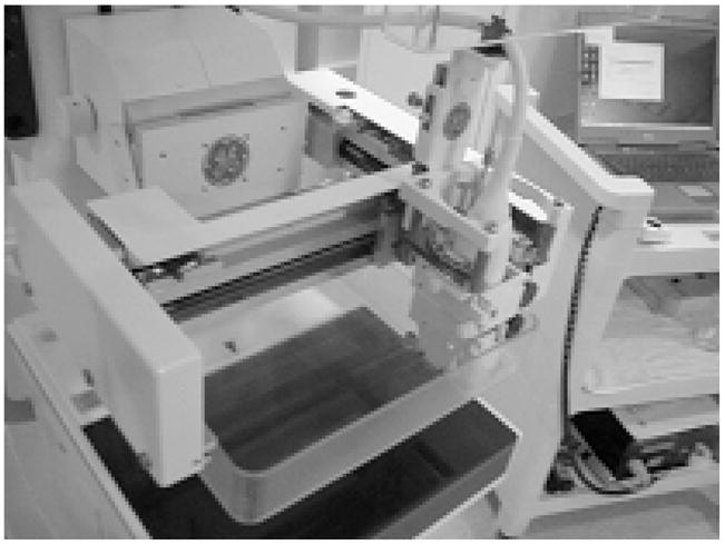
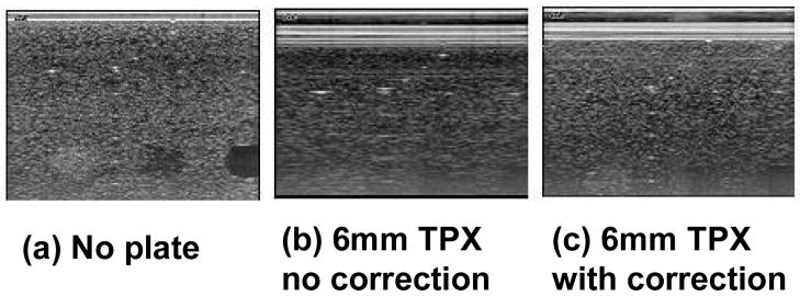
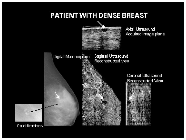
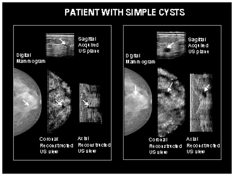
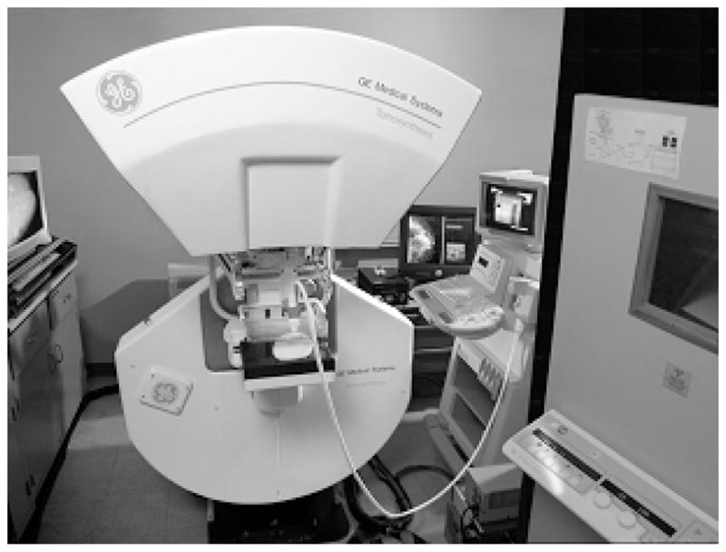
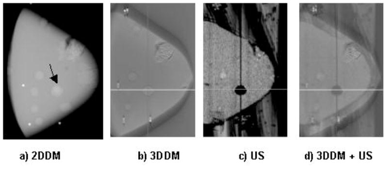
This paper describes work aimed at combining 3D ultrasound with full-field digital mammography via a semi-automatic prototype ultrasound scanning mechanism attached to the digital mammography system gantry. Initial efforts to obtain high x-ray and ultrasound image quality through a compression paddle are proving successful. Registration between the x-ray mammogram and ultrasound image volumes is quite promising when the breast is stably compressed. This prototype system takes advantage of many synergies between the co-registered digital mammography and pulse-echo ultrasound image data used for breast cancer detection and diagnosis. In addition, innovative combinations of advanced US and X-ray applications are being implemented and tested along with the basic modes. The basic and advanced applications are those that should provide relatively independent information about the breast tissues. Advanced applications include x-ray tomosynthesis, for 3D delineation of mammographic structures, and non-linear elasticity and 3D color flow imaging by ultrasound, for mechanical and physiological information unavailable from conventional, non-contrast x-ray and ultrasound imaging.


Figures

Figure 1
Figure 1
Dual-modality prototype compression paddle is a system to interface the 19×23cm2 full-field GE digital mammography x-ray detector on the Senographe 2000D FFDM system with the Logiq 9 ultrasound system using a M12L transducer array.
Figure 2
Figure 2
Prototype compression paddle mounted on the GE Senographe 2000D with an M12L ultrasound probe attached.
Figure 3
Figure 3
Correction for refraction effects. (a) RMI 404 GS phantom image acquired without any TPX plate at 9 MHz using an M12L probe. (b) The image is acquired using the same US settings with 6 mm of TPX placed on top of the phantom. (c) The imaging conditions are the same as (b), but refraction corrections were implemented in the beam forming code.
Figure 4
Figure 4
A benign circumscribed oval hypoechoic mass in a 36 year old patient with dense breast tissue, identified by white arrows, is seen on the acquired and reconstructed ultrasound planes. The mass was occult on the mammogram. Shown in the insert corresponding to the mammogram are benign calcifications not visible on the ultrasound images.
Figure 5
Figure 5
Two cysts seen with ultrasound are relatively occult on the corresponding mammogram. The cysts were similar in appearance on the hand-held direct contact scans as well as those obtained with the prototype system.
Figure 6
Figure 6
An early version of a combined tomosynthesis – ultrasound compression paddle system installed on a prototype tomosynthesis system developed at GE Global Research.
Figure 7
Figure 7
Phantom images of a modified CIRS triple modality biopsy phantom. Panel (a) is a 2D digital mammogram obtained on an early prototype 18×24 cm2 digital x-ray detector. Panel (b) shows a slice of the tomosynthesis (3DDM) image, which shows masses and a cluster of microcalcifications. Panel (c) shows a corresponding reconstructed ultrasound slice of the same phantom. Panel (d) is the same tomosynthesis slice merged with the reconstructed ultrasound slice, showing cyst-mass differentiation as well as microcalcifications.
Figure 8
Figure 8
B-scan (60 dB scale) and measured axial strain image (0–2.5% scale) of simulated intra-ductal carcinoma (5 mm radius) surrounded by glandular and fatty tissue

References

    1. Jackson VP. The role of US in breast imaging. Radiology. 1990;177:305–311. - PubMed
    1. Bassett LW, Kimme-Smith C. Breast sonography. AJR. 1991;156:449–455. - PubMed
    1. Jackson VP, Hendrick RE, Feig SA, Kopans DB. Imaging of the radiographically dense breast. Radiology. 1993;188:297–301. - PubMed
    1. Kolb TM, Lichy J, Newhouse JH. Occult cancer in women with dense breasts: detection with screening ultrasound – diagnostic yield and tumor characteristics. Radiology. 1998;207:191–199. - PubMed
    1. Berg W. Rationale for a trial of screening breast ultrasound. AJR. 2003;180:1225–1228. - PubMed

Publication types