. 2004 Aug;112(4):543-9.
doi: 10.1111/j.1365-2567.2004.01915.x.

Transferrin is required for early T-cell differentiation

Affiliations

Transferrin is required for early T-cell differentiation

M Fatima Macedo et al. Immunology. 2004 Aug.

Abstract

Transferrin, the major plasma iron carrier, mediates iron entry into cells through interaction with its receptor. Several in vitro studies have demonstrated that transferrin plays an essential role in lymphocyte division, a role attributed to its iron transport function. In the present study we used hypotransferrinaemic (Trf(hpx/hpx)) mice to investigate the possible involvement of transferrin in T lymphocyte differentiation in vivo. The absolute number of thymocytes was substantially reduced in Trf(hpx/hpx) mice, a result that could not be attributed to increased apoptosis. Moreover, the proportions of the four major thymic subpopulations were maintained and the percentage of dividing cells was not reduced. A leaky block in the differentiation of CD4(-) CD8(-) CD3(-) CD44(-) CD25(+) (TN3) into CD4(-) CD8(-) CD3(-) CD44(-) CD25(-) (TN4) cells was observed. In addition, a similar impairment of early thymocyte differentiation was observed in mice with reduced levels of transferrin receptor. The present study demonstrates, for the first time, that transferrin itself or a pathway triggered by the interaction of transferrin with its receptor is essential for normal early T-cell differentiation in vivo.


Figures
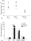
Figure 1
Figure 1
Reduced thymic cellularity in Tfrhpx/hpx mice with no major alterations in the four main thymic subpopulations.(a)Each dot represents the absolute thymocyte number from one mouse analysed. Data are shown for 16-week-old mice. Similar results were observed for 12-week-old mice.(b)Histograms show the percentage of TN, DP, CD8SP and CD4SP thymocytes for each genotype analysed. Genotypes differences are specified by black and white shading. Results show the mean ± SD values obtained from at least eight mice in each group. Data presented correspond to 16-week-old mice, similar results were observed for 12-week-old mice.
Figure 2
Figure 2
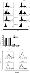
Thymocyte proliferation and apoptotic levels in Trfhpx/hpx mice. (a)Comparison of TN, DP, CD4SP and CD8SP thymocyte proliferation measured by BrdU incorporation, between Trfhpx/hpx and wild-type 17-week-old mice. Numbers in each panel indicate percentage of BrdU+ cells. (b) Histograms show the percentage of BrdU+ TN, DP, CD4SP and CD8SP thymocytes for Trfhpx/hpx and control mice (Trfhpx/+ and wild-type mice were used as controls). Genotypes differences are indicated by black and white colouring. Results show the mean ± SD values obtained from five mice in each group. Data presented correspond to 16–17-week-old mice. The significance of the differences between groups was tested by Student's t-test (*P = 0·03). (c)Annexin V staining of, CD4, CD8 and CD3 labelled, thymocytes from 16-week-old Trfhpx/hpx mice (black line n = 3) and control mice (grey line n = 4).
Figure 3
Figure 3
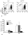
Trfhpx/hpx mice show a leaky block in the transition between the TN3 to TN4 stage of thymocyte differentiation. (a) TN thymocytes were analysed for the expression of CD44 and CD25. A representative example of a 12-week-old mice is shown. Similar results were observed for 16-week-old mice. Numbers in each panel indicate percentage of cells. (b) Similar numbers of TN1 thymocytes give rise to clearly less TN4 cells in the Trfhpx/hpx mice. Histograms show absolute numbers of TN1 and TN4 thymocytes. Genotypes differences are indicated by black and white shading. Results show mean ± SD values of: Trf+/+n = 3, Trfhpx/+n = 7, Trfhpx/hpxn = 6. Data presented correspond to 16-week-old mice, similar results were observed for 12-week-old mice. The significance of the differences between wild-type and Trfhpx/hpx mice was tested by Student's t-test (*P < 0·002).
Figure 4
Figure 4
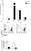
TrfR+/– mice show a leaky block in the transition between the TN3 to TN4 stage of thymocyte differentiation. (a)Histograms show the percentage of TN, DP, CD8SP and CD4SP thymocytes for TrfR+/– (n = 5) and control mice (n = 4). Genotypes differences are indicated by black and white colouring. Results show the mean ± SD values. (b)TN thymocytes were analysed for the expression of CD44 and CD25. A representative example of three independent experiments is shown. Numbers in each panel indicate percentage of cells. (c)Histograms show absolute numbers of TN1 and TN4 thymocytes for TrfR+/– (n = 4) and control mice (n = 3). Genotypes are specified by colour coding. Results show mean ± SD values. Data presented correspond to 16-week old-mice. The significance of the differences between groups was tested by Student's t-test (*P < 0·03; **P < 0·002).

References

    1. Kaplan J. Mechanisms of cellular iron acquisition: another iron in the fire. Cell. 2002;111:603–6. - PubMed
    1. Levy JE, Jin O, Fujiwara Y, Kuo F, Andrews NC. Transferrin receptor is necessary for development of erythrocytes and the nervous system. Nat Genet. 1999;21:396–9. - PubMed
    1. Tormey DC, Imrie RC, Mueller GC. Identification of transferrin as a lymphocyte growth promoter in human serum. Exp Cell Res. 1972;74:163–9. - PubMed
    1. Iscove NN, Melchers F. Complete replacement of serum by albumin, transferrin, and soybean lipid in cultures of lipopolysaccharide-reactive B lymphocytes. J Exp Med. 1978;147:923–33. - PMC - PubMed
    1. Brekelmans P, van Soest P, Voerman J, Platenburg PP, Leenen PJ, van Ewijk W. Transferrin receptor expression as a marker of immature cycling thymocytes in the mouse. Cell Immunol. 1994;159:331–9. - PubMed

Publication types

LinkOut - more resources