. 2004 Aug;72(8):4751-62.
doi: 10.1128/IAI.72.8.4751-4762.2004.

Interaction of Chlamydia trachomatis serovar L2 with the host autophagic pathway

Affiliations

Interaction of Chlamydia trachomatis serovar L2 with the host autophagic pathway

Hesham M Al-Younes et al. Infect Immun. 2004 Aug.

Abstract

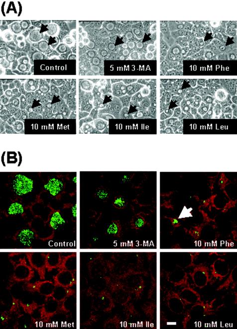
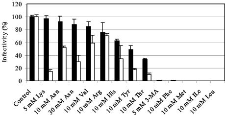
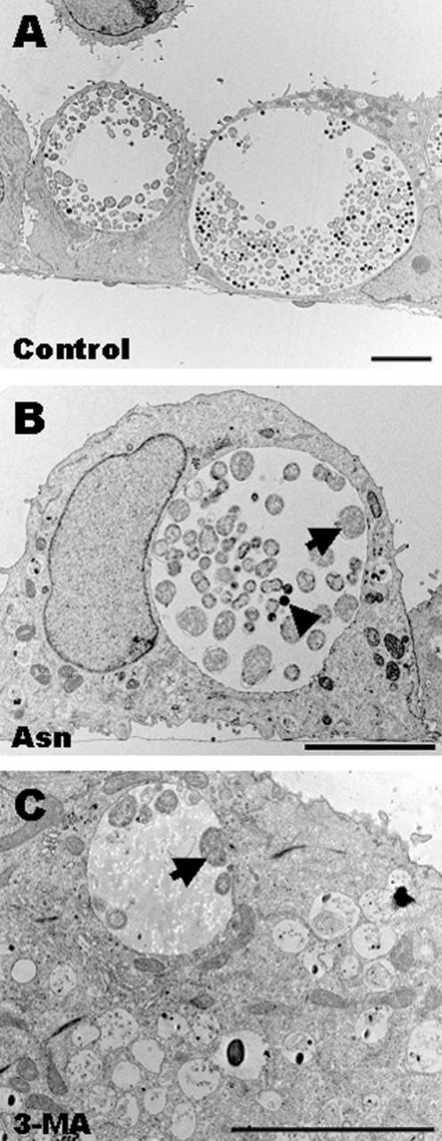
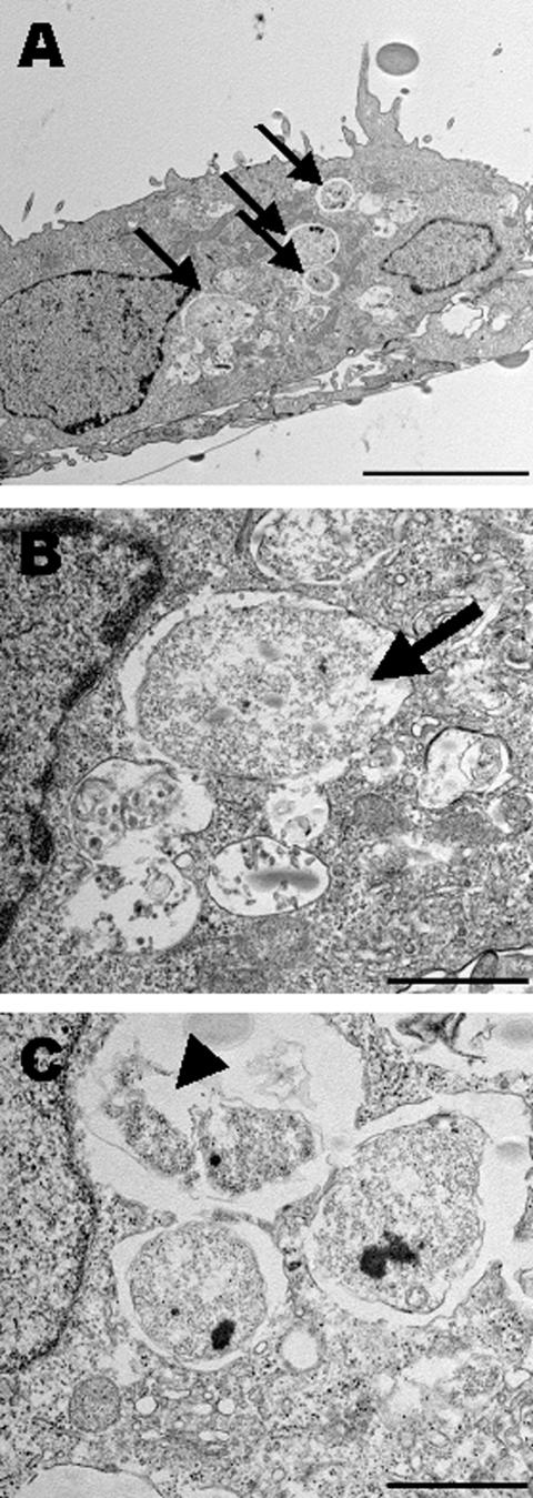
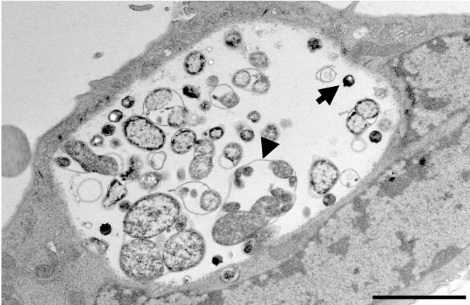
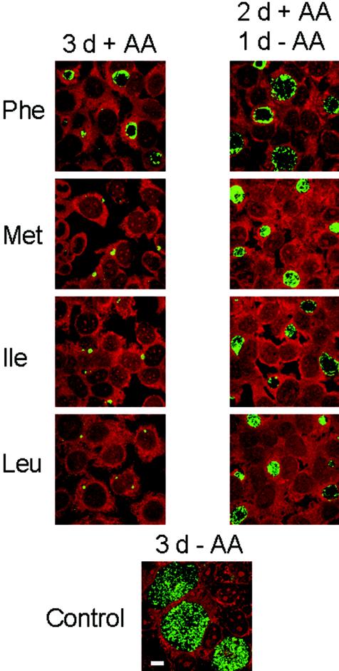
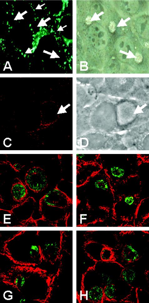
Chlamydiae are obligate intracellular pathogens that replicate within a membrane-bound compartment (the inclusion) and are associated with important human diseases, such as trachoma, pneumonia, and atherosclerosis. We have examined the interaction of the host autophagic pathway with Chlamydia trachomatis serovar L2 by using the specific autophagosomal stain monodansylcadaverine, antibodies to autophagosome-associated markers, and traditionally used autophagic inhibitors, particularly 3-methyladenine and amino acids. Chlamydial inclusions did not sequester monodansylcadaverine, suggesting absence of fusion with autophagosomes. Interestingly, exposure of cultures infected for 19 h to 3-methyladenine or single amino acids until the end of infection (44 h) caused various degrees of abnormalities in the inclusion maturation and in the progeny infectivity. Incubation of host cells with chemicals throughout the entire period of infection modulated the growth of Chlamydia even more dramatically. Remarkably, autophagosomal markers MAP-LC3 and calreticulin were redistributed to the inclusion of Chlamydia, a process that appears to be sensitive to 3-methyladenine and some amino acids. The present data indicate the lack of autophagosomal fusion with the inclusion because it was devoid of monodansylcadaverine and no distinct rim of autophagosomal protein-specific staining around the inclusion could be observed. However, high sensitivity of Chlamydia to conditions that could inhibit host autophagic pathway and the close association of MAP-LC3 and calreticulin with the inclusion membrane still suggest a potential role of host autophagy in the pathogenesis of Chlamydia.


Figures

FIG. 1.
FIG. 1.
Effect of excess individual amino acids and 3-MA on C. trachomatis L2 inclusion growth. (A) Cell monolayers were infected for 19 h and then exposed to IM containing exogenous individual amino acids or 3-MA. Microscopic examination was performed 44 h p.i., and phase-contrast images were obtained under the same magnification (×400). Inclusions are indicated by arrows. (B) Host cells were pretreated for 30 min with amino acids or 3-MA and then infected in the presence of respective chemicals until the end of the infection period (44 h). Cells were stained for Chlamydia (green) by using IMAGEN kit and examined by confocal microscopy. Bar, 10 μm.
FIG. 2.
FIG. 2.
Effect of excess amino acids and 3-MA on C. trachomatis L2 infectivity in vitro. Chemicals were added 19 h p.i. until the end of the infection course (44 h) (▪). In another set of experiments, chemicals were present in the cell monolayers 30 min before infection and also during infection (□). At 44 h p.i., cells were lysed and passaged onto fresh HEp-2 cells for determination of the infectious progeny yield. The percent infectivity is a reflection of the reduction in the titer (in IFU/milliliter) in infected cultures exposed to each chemical compared to the titer in control unexposed cell cultures. The results are expressed as the means of the percentage of infectivity ± the averages of the absolute deviations from the means for two different experiments.
FIG. 3.
FIG. 3.
C. trachomatis L2 inclusions in cell monolayers exposed to 3-MA or exogenous excess Asn, as viewed by EM. Host cells were pretreated with one of the chemicals for 30 min and then infected for 32 h in the presence of the respective chemical. (A) Control inclusions developed for 32 h in medium without additives contained normal forms of chlamydiae. (B) Inclusions developed in the presence of 30 mM Asn. Inclusions here are less mature and contain mostly RBs (arrow) compared to control inclusions (see panel A). Few EBs were observed (arrowhead). (C) Inclusions grown in the presence of 5 mM 3-MA contained low number of chlamydial forms, which were exclusively RBs (arrow). Bars, 5 μm.
FIG.4.
FIG.4.
EM examination of chlamydial inclusions grown under conditions of a high Met concentration. Host cells pretreated with 10 mM Met were infected for 48 h with C. trachomatis L2 at an MOI ∼10 in the presence of Met. (A) Ultrathin sections revealed that Met led to a complete growth arrest of inclusions. Moreover, multiple inclusions per infected host cell were detected (arrows). Most of the inclusions contained single typical RBs. (B and C) In addition, EM provided evidence of abnormalities in some of the formed RBs. Some RBs were partially degraded (arrow in panel B), whereas others were atypical and distorted (arrowhead in C). Bars, 5 μm (A) and 1 μm (B and C).
FIG. 5.
FIG. 5.
Morphology of C. trachomatis L2 inclusions developed in host cells exposed to 5 mM Lys 30 min before infection and until 48 h after infection. The major characteristic observed was the frequent presence of multiple RB-like forms that were enveloped, together with a single outer membrane. Notice the obvious loosening of the outer membrane (arrowhead). EB-like forms were also present (arrow). Bar, 2 μm.
FIG. 6.
FIG. 6.
Growth-inhibitory effects of excess Phe, Met, Ile, or Leu on C. trachomatis L2 can be simply reversed by additive removal. Left panels represent infected cell cultures that were continuously exposed to one of the additives for 72 h. Right panels represent infected cells that were incubated for 48 h with medium supplied with exogenous amino acids and for additional 24 h without additives in an attempt to restore the growth of inclusions. Replacement of medium generated an apparent recovery of inclusions (green). Bar, 10 μm.
FIG. 7.
FIG. 7.
Distribution of the autophagosome-associated markers in cell monolayers infected with C. trachomatis. (A and B) Fluorescence localization of the autofluorescent marker MDC in HEp-2 cells as visualized by conventional epifluorescence microscopy. MDC (small arrows) did not accumulate in chlamydial inclusions (large arrows). The corresponding phase-contrast image of panel A is shown in panel B. (C and D) MAP-LC3 distribution in cell cultures infected for 43 h. Confocal images revealed concentration of MAP-LC3-positive structures (red) adjacent to the inclusion (arrow). Panel D is a phase-contrast image of panel C. (E to H) Confocal images showing the distribution of calreticulin in cells infected for 43 h in the absence of additives (E) or in the presence of 1 mM Leu (F), Ile (G), or 3-MA (H). Chemicals were added at 19 h p.i. for additional 24 h. Cells were then fixed and stained with suitable antibodies.

Similar articles

Cited by

References

    1. Allan, I., and J. H. Pearce. 1983. Differential amino acid utilization by Chlamydia psittaci (strain guinea pig inclusion conjunctivitis) and its regulatory effect on chlamydial growth. J. Gen. Microbiol. 129:1991-2000. - PubMed
    1. Allan, I., T. P. Hatch, and J. H. Pearce. 1985. Influence of cysteine deprivation on chlamydial differentiation from reproductive to infective life-cycle forms. J. Gen. Microbiol. 131:3171-3177. - PubMed
    1. Al-Younes, H. M., T. Rudel, and T. F. Meyer. 1999. Characterization and intracellular trafficking pattern of vacuoles containing Chlamydia pneumoniae in human epithelial cells. Cell. Microbiol. 1:237-247. - PubMed
    1. Al-Younes, H. M., T. Rudel, V. Brinkmann, A. J. Szczepek, and T. F. Meyer. 2001. Low iron availability modulates the course of Chlamydia pneumoniae infection. Cell. Microbiol. 3:427-437. - PubMed
    1. Avruch, J., C. Belham, Q. Wenig, K. Hara, and K. Yonezawa. 2001. The p70 S6 kinase integrates nutrient and growth signals to control translational capacity. Prog. Mol. Subcell. Biol. 26:115-154. - PubMed

MeSH terms