

Genetic basis for the evolution of vertebrate mineralized tissue
- PMID: 15272073
- PMCID: PMC509207
- DOI: 10.1073/pnas.0404279101
Genetic basis for the evolution of vertebrate mineralized tissue
Abstract
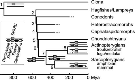
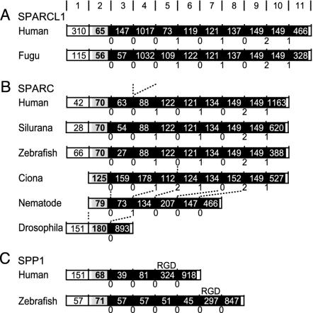
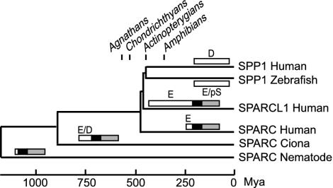
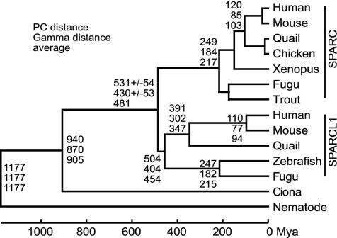
Mineralized tissue is vital to many characteristic adaptive phenotypes in vertebrates. Three primary tissues, enamel (enameloid), dentin, and bone, are found in the body armor of ancient agnathans and mammalian teeth, suggesting that these two organs are homologous. Mammalian enamel forms on enamel-specific proteins such as amelogenin, whereas dentin and bone form on collagen and many acidic proteins, such as SPP1, coordinately regulate their mineralization. We previously reported that genes for three major enamel matrix proteins, five proteins necessary for dentin and bone formation, and milk caseins and salivary proteins arose from a single ancestor by tandem gene duplications and form the secretory calcium-binding phosphoprotein (SCPP) family. Gene structure and protein characteristics show that SCPP genes arose from the 5' region of ancestral sparcl1 (SPARC-like 1). Phylogenetic analysis on SPARC and SPARCL1 suggests that the SCPP genes arose after the divergence of cartilaginous fish and bony fish, implying that early vertebrate mineralization did not use SCPPs and that SPARC may be critical for initial mineralization. Consistent with this inference, we identified SPP1 in a teleost genome but failed to find any genes orthologous to mammalian enamel proteins. Based on these observations, we suggest a scenario for the evolution of vertebrate tissue mineralization, in which body armor initially formed on dermal collagen, which acted as a reinforcement of dermis. We also suggest that mammalian enamel is distinct from fish enameloid. Their similar nature as a hard structural overlay on exoskeleton and teeth is because of convergent evolution.
Figures
References
-
- Ørvig, T. (1967) in Structure and Chemical Organization of Teeth, ed. Miles, A. E. W. (Academic, New York), Vol. I, pp. 45–110.
-
- Reif, W.-E. (1982) Evol. Biol. 15, 287–368.
-
- Reif, W.-E. (2001) Neues Jahrb. Geol. Palaontol. Abh. 219, 285–304. - PubMed
-
- Donoghue, P. C. & Sansom, I. J. (2002) Microsc. Res. Tech. 59, 352–372. - PubMed
-
- Smith, M. M. & Coates, M. I. (2001) in Major Events in Early Vertebrate Evolution, ed. Ahlberg, P. E. (Taylor and Francis, London), pp. 223–240.
Publication types
MeSH terms
Substances
Associated data
- Actions
- Actions
- Actions
- Actions
- Actions
- Actions
- Actions
- Actions
LinkOut - more resources
Full Text Sources
Other Literature Sources
Molecular Biology Databases
Research Materials
Miscellaneous
