Comparative Study
. 2005 Jan;18(1):1-10.
doi: 10.1038/modpathol.3800247.

Pulmonary pathology of severe acute respiratory syndrome in Toronto

Affiliations
Comparative Study

Pulmonary pathology of severe acute respiratory syndrome in Toronto

David M Hwang et al. Mod Pathol. 2005 Jan.

Abstract

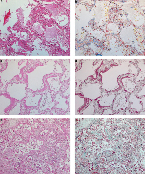
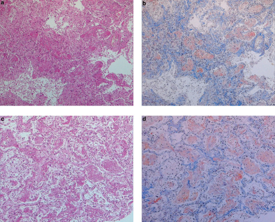
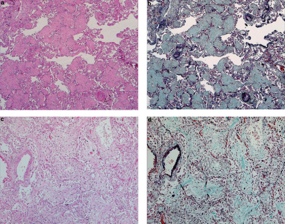
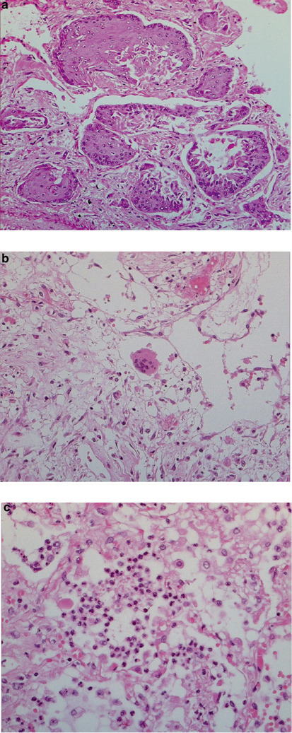
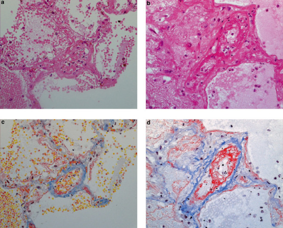
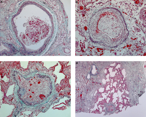
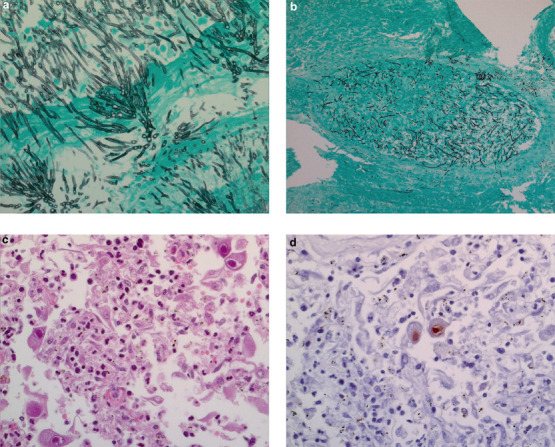
The severe acute respiratory syndrome (SARS) pandemic in Toronto resulted in a large number of autopsies on its victims. We describe the pulmonary pathology of patients who died in the 2003 Toronto outbreak. Autopsy material from the lungs of 20 patients who died between March and July 2003 were characterized by histology, molecular biology, and immunohistochemistry for cytokeratins, thyroid transcription factor-1, CD68, Epstein-Barr virus, cytomegalovirus, and human herpes simplex viruses. Matched controls were obtained from patients who died of other causes over the same interval. The mean duration of illness was 27 days (range 5-108 days). Post-mortem lung tissues from 19 of 20 patients with probable SARS were positive for SARS-associated coronavirus by RT-PCR. Histologically, all patients showed varying degrees of exudative and proliferative phase acute lung injury, evidenced in conventional and immunohistochemical stains by edema, inflammatory infiltrate, pneumocyte hyperplasia, fibrinous exudates, and organization. Eight of 20 patients showed predominantly a diffuse alveolar damage pattern of acute lung injury, six showed predominantly an acute fibrinous and organizing pneumonia pattern, and the remainder showed an admixture of the two patterns. Squamous metaplasia and scattered multinucleate giant cells were present in most cases. Vascular fibrin thrombi were a common finding and were often associated with pulmonary infarcts. Special stains demonstrated vascular endothelial damage of both small- and mid-sized pulmonary vessels. Two cases were complicated by invasive fungal disease consistent with Aspergillosis, and another by coinfection with cytomegalovirus. Our findings indicate that the lungs of patients who die of SARS are almost always positive for the SARS-associated coronavirus by RT-PCR, and may show features of both diffuse alveolar damage and acute fibrinous and organizing pneumonia patterns of acute injury. Cases of SARS may be complicated by coexistent infections and therapy-related lung injury.


Figures

Figure 1
Figure 1
Diffuse alveolar damage pattern of lung injury in SARS patients. (a) Early exudative phase diffuse alveolar damage showing vascular congestion, with interstitial and airspace edema and inflammatory cell infiltrates (H&E, original magnification × 200); (b) the same field showing fibrinous exudates by Martius scarlet blue stain (original magnification × 200); (c, d) exudative phase diffuse alveolar damage, with hyaline membranes (c, H&E, original magnification × 200; d, elastic trichrome, original magnification × 200); (e, f) organizing phase diffuse alveolar damage (e, H&E, original magnification × 100; f, elastic trichrome, original magnification × 100).
Figure 2
Figure 2
Acute fibrinous and organizing pneumonia pattern, acute exudative phase. Fibrinous exudates form ‘balls’ in alveolar spaces, rather than the hyaline membranes classically seen in diffuse alveolar damage pattern lung injury. (a, c) H&E; (b, d) Martius scarlet blue; all frames at × 100 original magnification.
Figure 3
Figure 3
Acute fibrinous and organizing pneumonia pattern, organizing phase. Fibromyxoid plugs in bronchioles and alveolar spaces, with minimal alveolar septal thickening. (a) H&E, original magnification × 40; (b) elastic trichrome (same field as (a)), original magnification × 40; (c) H&E, original magnification × 100; (d) elastic trichrome (same field as (c)), original magnification × 100.
Figure 4
Figure 4
Other histologic features of SARS in the lung. (a) Squamous metaplasia (H&E, original magnification × 200); (b) multinucleate giant cell (H&E, original magnification × 200); (c) focal acute bronchopneumonia (H&E, original magnification × 400).
Figure 5
Figure 5
Acute vascular/endothelial injury, with extravasation of fluid and blood cells. (a, b) H&E, original magnification × 400; (c, d) Martius scarlet blue, original magnification 400x, of the same fields in (a) and (b), respectively, accentuate intravascular and intra-alveolar fibrin.
Figure 6
Figure 6
Pulmonary thromboemboli in a SARS patient, duration of illness=52 days. (a) Nonocclusive organizing thromboembolus, original magnification × 100; (b) near-occlusive (original magnification × 200) and (c) occlusive organized thrombosis with recanalization (original magnification × 100); (d) pulmonary infarct associated with occluded vessel (original magnification × 25). All sections stained with elastic trichrome.
Figure 7
Figure 7
Coinfections in SARS patients. (a) Florid invasive fungal infection consistent with Aspergillosis in a 78-year-old (Gomori methenamine silver, original magnification × 400); (b) fungi consistent with Aspergillus spp, with angioinvasion (Gomori methenamine silver, original magnification × 100); (c) acute pneumonia, with cells containing intranuclear inclusions consistent with CMV (H&E, original magnification × 400), and staining positive for CMV by immunohistochemistry (d).

References

    1. Poutanen SM, Low DE, Henry B. Identification of severe acute respiratory syndrome in Canada. N Engl J Med. 2003;348:1995–2005. doi: 10.1056/NEJMoa030634. - DOI - PubMed
    1. Booth CM, Matukas LM, Tomlinson GA. Clinical features and short-term outcomes of 144 patients with SARS in the greater Toronto area. JAMA. 2003;289:2801–2809. doi: 10.1001/jama.289.21.JOC30885. - DOI - PubMed
    1. Tang P, Louie M, Richardson SE. Interpretation of diagnostic laboratory tests for severe acute respiratory syndrome: the Toronto experience. Can Med Assoc J. 2004;170:47–54. - PMC - PubMed
    1. Drosten C, Gunther S, Preiser W. Identification of a novel coronavirus in patients with severe acute respiratory syndrome. N Engl J Med. 2003;348:1967–1976. doi: 10.1056/NEJMoa030747. - DOI - PubMed
    1. Ksiazek TG, Erdman D, Goldsmith CS. A novel coronavirus associated with severe acute respiratory syndrome. N Engl J Med. 2003;348:1953–1966. doi: 10.1056/NEJMoa030781. - DOI - PubMed

Publication types

MeSH terms

LinkOut - more resources