

Dynamic movement of actin-like proteins within bacterial cells
- PMID: 15272301
- PMCID: PMC1299120
- DOI: 10.1038/sj.embor.7400209
Dynamic movement of actin-like proteins within bacterial cells
Abstract
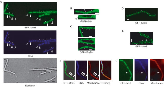
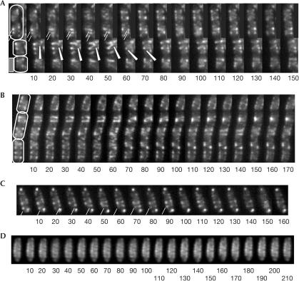
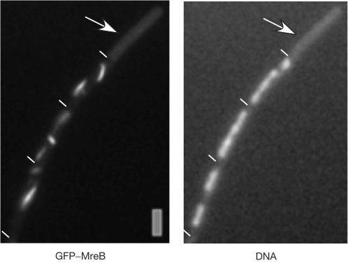
Actin proteins are present in pro- and eukaryotes, and have been shown to perform motor-like functions in eukaryotic cells in a variety of processes. Bacterial actin homologues are essential for cell viability and have been implicated in the formation of rod cell shape, as well as in segregation of plasmids and whole chromosomes. We have generated functional green fluorescent protein fusions of all three Bacillus subtilis actin-like proteins (MreB, Mbl and MreBH), and show that all three proteins form helical filaments underneath the cell membrane, the pattern of which is distinct for each protein. Time-lapse microscopy showed that the filaments are highly dynamic structures. A number of separate filaments of MreB and Mbl continuously move through the cell along helical tracks underneath the cell membrane. The speed of extension of the growing end of filaments is within the range of known actin polymerization (0.1 microm/s), generating a potential poleward or centreward pushing velocity at 0.24 microm/min for MreB or Mbl, respectively. During nutritional downshift and a block in topoisomerase IV activity, the filaments rapidly disintegrated, showing that movement occurs only in growing cells. Contrary to Mbl and MreBH filaments, MreB filaments were generally absent in cells lacking DNA, providing a further distinction between the three orthologues.
Figures
References
-
- Carballido-Lopez R, Errington J (2003) The bacterial cytoskeleton: in vivo dynamics of the actin-like protein Mbl of Bacillus subtilis. Dev Cell 4: 19–28 - PubMed
-
- Daniel RA, Errington J (2003) Control of cell morphogenesis in bacteria: two distinct ways to make a rodshaped cell. Cell 113: 767–776 - PubMed
-
- Defeu Soufo HJ, Graumann PL (2003) Actin-like proteins MreB and Mbl from Bacillus subtilis are required for bipolar positioning of replication origins. Curr Biol 13: 1916–1920 - PubMed
-
- Jones LJ, Carballido-Lopez R, Errington J (2001) Control of cell shape in bacteria: helical, actin-like filaments in Bacillus subtilis. Cell 104: 913–922 - PubMed
Publication types
MeSH terms
Substances
LinkOut - more resources
Full Text Sources
Other Literature Sources
Miscellaneous
