. 2004 Aug 4;23(15):3175-85.
doi: 10.1038/sj.emboj.7600325. Epub 2004 Jul 22.

CD95 ligand induces motility and invasiveness of apoptosis-resistant tumor cells

Affiliations

CD95 ligand induces motility and invasiveness of apoptosis-resistant tumor cells

Bryan C Barnhart et al. EMBO J. .

Abstract

The apoptosis-inducing death receptor CD95 (APO-1/Fas) controls the homeostasis of many tissues. Despite its apoptotic potential, most human tumors are refractory to the cytotoxic effects of CD95 ligand. We now show that CD95 stimulation of multiple apoptosis-resistant tumor cells by CD95 ligand induces increased motility and invasiveness, a response much less efficiently triggered by TNFalpha or TRAIL. Three signaling pathways resulting in activation of NF-kappaB, Erk1/2 and caspase-8 were found to be important to this novel activity of CD95. Gene chip analyses of a CD95-stimulated tumor cell line identified a number of potential survival genes and genes that are known to regulate increased motility and invasiveness of tumor cells to be induced. Among these genes, urokinase plasminogen activator was found to be required for the CD95 ligand-induced motility and invasiveness. Our data suggest that CD95L, which is found elevated in many human cancer patients, has tumorigenic activities on human cancer cells. This could become highly relevant during chemotherapy, which can cause upregulation of CD95 ligand by both tumor and nontumor cells.


Figures
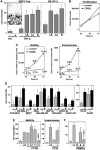
Figure 1
Figure 1
Stimulation of CD95 in CD95 apoptosis-resistant tumor cells induces invasiveness. (A) A 3-day in vitro invasiveness assay of MCF7(FB) and SK-OV-3 cells incubated with anti-APO-1 (αA), LzCD95L (Lz) or sCD95L (S).The inset shows fixed and stained cells that migrated through a Matrigel-coated membrane unstimulated (−) or stimulated (+) with anti-APO-1. (B) Cell proliferation in MCF7(FB) cells following stimulation with LzCD95L was determined by counting cells each day following trypsinization. (C) Increase in the total number of migrating (left) and invading (right) MCF7(FB) cells upon stimulation with LzCD95L. Percentages of cells compared to the total number of cells in the well are given. (D) Invasiveness assay of apoptosis-resistant cells (left panel) following stimulation with anti-APO-1 (asterisk), LzCD95L (underlined) or sCD95L. Two apoptosis-sensitive Type I tumor cell lines that are resistant to sCD95L also responded with increased invasiveness (right panel). Cell proliferation was controlled by MTS assay to ensure that the increase in cell number was not due to the increase in proliferation. Selected cell lines were also counted as described in (B). (E) Invasiveness (left) and motility (right) assay of MCF7(FB) cells incubated with anti-APO-1 (αA), CT26L or CT26 cells at the ratios 1:1, 3.3:1 (labeled as 3:1) and 6.7:1 (labeled as 7:1). The ratio of CT26 to MCF7(FB) cells in the invasiveness assays was 6.7:1. (F) PBMCs that had been activated for 16 h with PHA, followed by 6 days with IL-2 (A), or were not stimulated with either PHA or IL-2 (R) were cocultured with MCF7(FB) cells at a 6.7:1 ratio in an invasiveness assay. Where indicated, the PBMCs were preincubated with NOK-1. NOK-1 was maintained in the culture at 10 μg/ml.
Figure 2
Figure 2
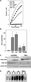
CD95-mediated activation of NF-κB contributes to in vitro invasiveness. (A) EMSA analysis of NF-κB activation of MCF7(FB) cells stimulated through CD95 or cells treated with TNFα or LzTRAIL. (B) EMSA analysis of NF-κB activity of cell extracts of MCF7(FB) cells stimulated with LzCD95L for 4 h and after pretreatment with BAY 11-7082 or either GST-TAT-IκBαmut or GST-TAT control (tat). (C) MCF7(FB) cells were treated with 2.5 μM BAY 11-7082 and subjected to invasiveness assay. The NF-κB inhibitor was maintained in the culture for the duration of the assay. (D) Invasiveness assay and (E) migration assay of MCF7(FB) cells incubated with GST-TAT-IκBαmut (mut) or GST-TAT control (t).
Figure 3
Figure 3
CD95L is more efficient than TNFα or TRAIL in inducing increased motility and invasiveness of MCF7(FB) cells. (A) CAT reporter gene analysis of MCF7(FB) cells transfected with an NF-κB promoter (κB) or mutated NF-κB promoter (mut) and treated with LzCD95L (Lz), anti-APO-1 (A), sCD95L or TNFα (for 4 h) and assayed over time. (B) A 3-day invasiveness assay of MCF7(FB) cells stimulated with 1 μg/ml LzCD95L, 1000 U/ml TNFα or 1 μg/ml LzTRAIL. (C) MCF7(FB) cells were incubated for the indicated times with LzCD95L or TNFα. Cell lysates were subjected to IKKβ immunoprecipitation followed by in vitro kinase assay (IKA) using GST-IκBα as the substrate. Phosphorylated IκBα was resolved and exposed to film. W, Western blot. (D) Jurkat cells deficient for RIP expression stimulated with either anti-APO-1/zVAD-fmk (A), LzCD95L/zVAD-fmk (Lz) or TNFα for 4 h were subjected to EMSA. P, parental Jurkat cells; R, RIP-deficient cells.
Figure 4
Figure 4
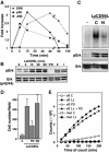
All three MAP kinase pathways are activated by LzCD95L in MCF7(FB) cells and activation of Erk is involved in CD95-induced invasiveness. (A) MCF7(FB) cells were stimulated with LzCD95L for indicated times and lysates of the cells were subjected to Western blot analysis for phosphorylation of indicated MAP kinases. Ratios of intensity of phosphorylated versus unphosphorylated kinase bands were determined by densitometry. (B) Kinetics of phosphorylation of p42/p44 Erk in LzCD95L-treated MCF7(FB) cells. As indicated (30 min stimulated), cells were preincubated with the following inhibitors: 20 μM PD98059 (PD), 40 μM zVAD (Z) or 40 μM zIETD (I). (C) MCF7(FB) cells were preincubated for 3 h with 140 μM of control peptide (C) or NEMO binding domain (NBD) peptide (N) and then stimulated for 2 h with LzCD95L. Nuclear extracts were purified and subjected to an EMSA (top panel). In parallel, cells were lysed and phosphorylation of Erk was analyzed by Western blotting (bottom panels). (D) Invasiveness assay of MCF7(FB) cells preincubated with PD98059 or p38 inhibitor SB203580. Effectiveness of the SB inhibitor was tested using phosphospecific antibodies (not shown). (E) CAT reporter assay of LzCD95L-stimulated MCF7(FB) cells following preincubation with PD98059.
Figure 5
Figure 5
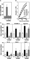
CD95-induced invasiveness requires caspase-8 activity. (A) Invasiveness assay of MCF7(FB) cells in the presence of zVAD-fmk (z) or zIETD-fmk (I). (B) κB-driven CAT reporter assay of MCF7(FB) cells preincubated with zVAD-fmk (z) or zIETD-fmk (I). (C) Invasiveness assay of NCI60 cells stimulated through CD95 in the presence of 20 μM PD98059 (PD) or 40 μM zIETD-fmk (I).
Figure 6
Figure 6
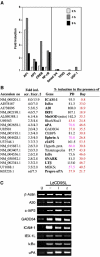
CD95 stimulation activates a transcriptional program. (A) MCF7(FB) cells were stimulated with LzCD95L as indicated and subjected to a Transignal DNA/Protein array to determine activation of transcription factors. The assay was performed three times and representative data are shown. Left, transcription factors that were activated in all three assays; right, transcription factors that were not significantly activated in any of the three assays. (B) MCF7(FB) cells were stimulated with anti-APO-1 and protein A for 8 h followed by isolation of RNA and gene chip analysis. The screen was performed twice (scr. 1 and scr. 2) using different MCF7(FB) clones. Only genes induced more than two-fold in both screens are shown. Prepro uPA could not be detected in the first screen due to a bad chip area. If a gene appeared more than once only the more highly induced signal is shown. Cells were treated with PD98059 and BAY 11-7082 in the second screen and percent induction of genes in the presence of these inhibitors is indicated. Genes that are inhibited more than 25% are shown in pink and more than 50% in red. (C) Semiquantitative RT–PCR of 8 h stimulated MCF-7(FB) cells treated with 40 μM zIETD-fmk (I) or zVAD-fmk (z) for the genes indicated. PCR products were subjected to agarose gel electrophoresis.
Figure 7
Figure 7
Urokinase plasminogen activator is a mediator of CD95-mediated invasiveness. Invasiveness (A) and motility (B) assay of MCF7(FB) cells incubated simultaneously with 1 μg/ml of LzCD95L and 20 μg/ml anti-uPA mAb or 20 μM uPA-STOP. The anti-uPA mAb was added each day. C, control; A, anti-APO-1; IgG, control IgG. The experiments shown are representative of three independent experiments. A 3-day MTS assay was performed to control for cell viability in the presence of these reagents, and no toxicity was observed (data not shown).

References

    1. Ahn JH, Park SM, Cho HS, Lee MS, Yoon JB, Vilcek J, Lee TH (2001) Non-apoptotic signaling pathways activated by soluble Fas ligand in serum-starved human fibroblasts. Mitogen-activated protein kinases and NF-kappaB-dependent gene expression. J Biol Chem 276: 47100–47106 - PubMed
    1. Alam A, Cohen LY, Aouad S, Sekaly RP (1999) Early activation of caspases during T lymphocyte stimulation results in selective substrate cleavage in nonapoptotic cells. J Exp Med 190: 1879–1890 - PMC - PubMed
    1. Alderson MR, Armitage RJ, Maraskovsky E, Tough TW, Roux E, Schooley K, Ramsdell F, Lynch DH (1993) Fas transduces activation signals in normal human T lymphocytes. J Exp Med 178: 2231–2235 - PMC - PubMed
    1. Algeciras-Schimnich A, Pietras EM, Barnhart BC, Legembre P, Vijayan S, Holbeck SL, Peter ME (2003) Two CD95 tumor classes with different sensitivities to antitumor drugs. Proc Natl Acad Sci USA 100: 11445–11450 - PMC - PubMed
    1. Aoki K, Kurooka M, Chen JJ, Petryniak J, Nabel EG, Nabel GJ (2001) Extracellular matrix interacts with soluble CD95L: retention and enhancement of cytotoxicity. Nat Immunol 2: 333–337 - PubMed

Publication types

MeSH terms