. 2004 Aug 18;23(16):3249-58.
doi: 10.1038/sj.emboj.7600329. Epub 2004 Jul 22.

Xenopus paraxial protocadherin has signaling functions and is involved in tissue separation

Affiliations

Xenopus paraxial protocadherin has signaling functions and is involved in tissue separation

Araceli Medina et al. EMBO J. .

Abstract

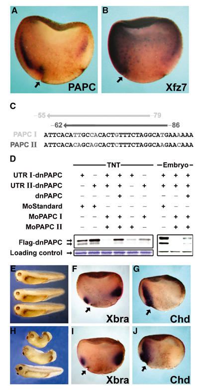
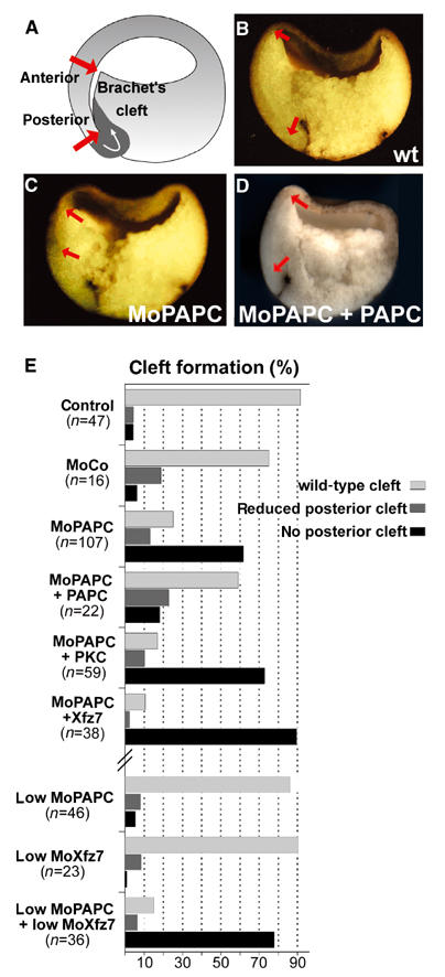
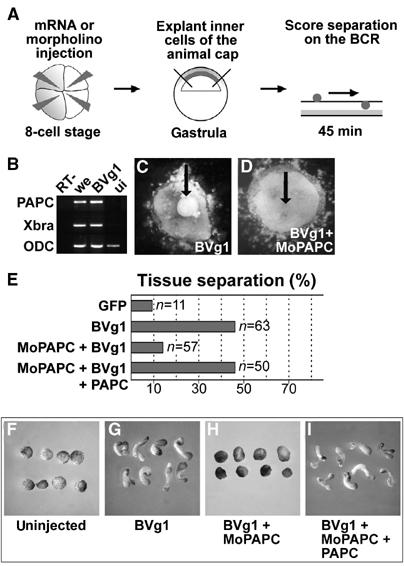
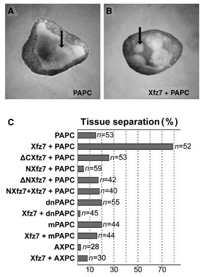
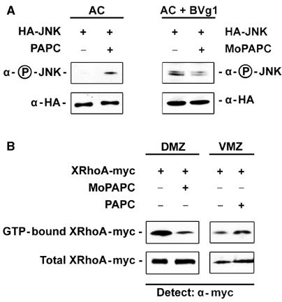
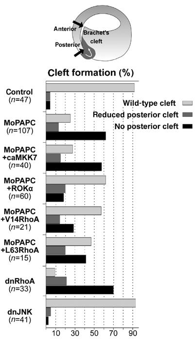
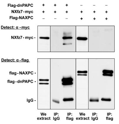
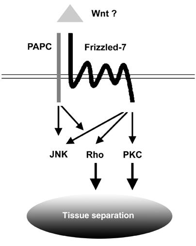
Protocadherins have homophilic adhesion properties and mediate selective cell-cell adhesion and cell sorting. Knockdown of paraxial protocadherin (PAPC) function in the Xenopus embryo impairs tissue separation, a process that regulates separation of cells of ectodermal and mesodermal origin during gastrulation. We show that PAPC can modulate the activity of the Rho GTPase and c-jun N-terminal kinase, two regulators of the cytoskeletal architecture and effectors of the planar cell polarity pathway. This novel signaling function of PAPC is essential for the regulation of tissue separation. In addition, PAPC can interact with the Xenopus Frizzled 7 receptor, and both proteins contribute to the development of separation behavior by activating Rho and protein kinase Calpha.


Figures

Figure 1
Figure 1
PAPC is expressed in the dorsal mesoderm and regulates morphogenesis during gastrulation. In situ hybridization of sagittally fractured gastrula-stage embryos showing, (A) expression of PAPC mRNA and (B) expression of Xfz7 mRNA. (C, D) Antisense morpholino oligonucleotides block translation of PAPC mRNA. (C) Morpholino oligonucleotides were designed to target the 5′UTRs of both PAPC alleles at the positions indicated. (D) MoPAPC I and II block translation of PAPC mRNA both in an in vitro coupled transcription/translation reaction (TNT) and in Xenopus embryos. (E–J) Knockdown of PAPC by microinjection of MoPAPC impairs morphogenesis without affecting mesoderm patterning. Xenopus embryos were injected at the 4–8 cell stage with a combination of both MoPAPC I and II (MoPAPC) into the DMZ. The embryos were analyzed by either in situ hybridization at early and midgastrula stages or were grown until the tailbud stage, at which time the phenotype was observed. (E) Uninjected embryos. (H) Embryos injected with 80 ng of MoPAPC exhibiting a range of phenotypic defects. Whole-mount in situ hybridizations for brachyury (Xbra; F, I) and chordin (chd; G, J) in control morpholino (MoCo)-injected embryos (F, G) and embryos injected with MoPAPC (I, J) are shown. Arrows indicate the dorsal blastopore lip.
Figure 2
Figure 2
Knockdown of PAPC by microinjection of MoPAPC perturbs formation of Brachet's cleft. MoPAPC (40 ng) was injected into two dorso-vegetal blastomeres at the eight-cell stage. Formation of Brachet's cleft was analyzed in embryos at stage 10.5 that were fractured sagittally through the dorsal midline. (A) Scheme describing the formation of Brachet's cleft at stage 10.5. The length of Brachet's cleft from the anterior to the posterior end, indicated by red arrows, was analyzed in control morpholino (MoCo) as well as MoPAPC-injected embryos. (B) Brachet's cleft develops normally in MoCo-injected embryos. (C) Development of posterior cleft is abolished in the MoPAPC-injected embryos. (D) Injection of 1 ng of PAPC mRNA rescues the effect of MoPAPC. (E) Summary of the experiments showing the effect of MoPAPC injection on formation of Brachet's cleft. Formation of posterior part of Brachet's cleft was impaired by knockdown of PAPC and the effect of MoPAPC was rescued by coexpression of 1 ng of PAPC mRNA. Injection of MoCo had no effect on Brachet's cleft formation. Microinjection of either 300 pg of Xfz7 or 500 pg of XPKCα mRNA did not compensate for the loss of PAPC function. PAPC and Xfz7 act synergistically to control Brachet's cleft formation. Injection of low doses of MoPAPC (20 ng) or MoXfz7 (40 ng) did not affect cleft formation. These morpholinos when injected together abolished the posterior part of Brachet's cleft.
Figure 3
Figure 3
PAPC function is necessary for BVg1-induced separation behavior and elongation of animal caps. (A) Scheme describing the blastocoel roof assay for analysis of separation behavior. (B) RT–PCR analysis of Xbra and PAPC expression in BVg1 mRNA (50 pg)-injected animal caps. (C) BVg1-injected animal cap cells show separation behavior. (D) Coinjection of 80 ng MoPAPC abolishes BVg1-induced separation behavior. (E) Compilation of the in vitro separation assays. (F–I) BVg1-induced elongation of animal caps requires PAPC function. (G) Injection of 50 pg of BVg1 mRNA induces elongation of animal caps. (H) MoPAPC (80 ng) injection abolishes BVg1-induced elongation of animal caps. (I) Injection of 800 pg of mRNA for PAPC rescues the effect of MoPAPC.
Figure 4
Figure 4
Xenopus PAPC and Xfz7 induce separation behavior in animal cap cells. (A) PAPC does not induce separation behavior when injected alone. (B) When Xenopus PAPC and Xfz7 are expressed together, they induce separation behavior of the animal cap cells. (C) Compilation of the experiments. RNAs for the following proteins were injected: Xenopus PAPC (400 pg), mPAPC (400 pg), dnPAPC (400 pg), Xfz7 (300 pg), ΔCXfz7 (300 pg), NXfz7 (300 pg), ΔNXfz7 (300 pg) and AXPC (600 pg).
Figure 5
Figure 5
PAPC activates JNK and RhoA. For the JNK activity assay, 200 pg of HA epitope-tagged JNK mRNA was injected either alone or in combination with 500 pg of PAPC mRNA into the animal blastomeres at the four-cell stage. HA-JNK was precipitated from extracts of 20 embryos at stage 11 and activation of JNK was monitored by its phosphorylation using a phosphospecific antibody. (A) JNK is activated by PAPC mRNA expression as measured by JNK phosphorylation. Equal amounts of immunoprecipitated HA-JNK were loaded in each lane. Injection of 100 pg of BVg1 mRNA activates JNK and injection of MoPAPC inhibits BVg1-induced activation of JNK. (B) PAPC expression enhances RhoA activity in the VMZ and PAPC function is required for RhoA activity in the DMZ. For gain of PAPC function assays, myc epitope-tagged RhoA DNA was injected into the VMZ either alone or in combination with 1 ng of PAPC mRNA. Activated RhoA was precipitated from 30 early gastrulae by RBD-GST, blotted onto a nitrocellulose membrane and detected using anti-myc Ab1 monoclonal antibody. Whole embryo extract was used as a loading control. Overexpression of PAPC causes activation of RhoA in the VMZ as monitored by precipitation of active RhoA by RBD-GST. Loss of PAPC function by MoPAPC (80 ng) injection greatly reduced RhoA activity in the DMZ.
Figure 6
Figure 6
PAPC-mediated regulation of Brachet's cleft formation requires RhoA. MoPAPC (40 ng) injection impairs formation of posterior Brachet's cleft. This effect of MoPAPC could be rescued by coinjection of Rho-associated protein kinase alpha mRNA (ROKα, 100 pg), constitutively active RhoA mRNA (V14RhoA, 50 pg), or by constitutively active human RhoA DNA (L63RhoA, 15 pg). Conversely, inhibition of RhoA signaling by injection of dominant-negative RhoA mRNA (N19RhoA, 1 ng) impaired posterior cleft formation. Activation of JNK signaling by microinjection of 200 pg of constitutively active MKK7 mRNA did not rescue the effect of MoPAPC and inhibition of JNK signaling by injection of 1 ng of dominant-negative JNK mRNA did not affect Brachet's cleft formation.
Figure 7
Figure 7
Xenopus PAPC and Fz7 interact. A flag epitope-tagged secreted extracellular domain of PAPC (flag-dnPAPC, 1 ng mRNA) or a flag epitope-tagged secreted extracellular domain of AXPC (flag-NAXPC, 1 ng mRNA) was injected with a myc epitope-tagged ectodomain of Fz7 (NXfz7-myc, 500 pg mRNA) into the animal blastomeres at the 2–4 cell stage. dnPAPC and NAXPC were immunoprecipitated at the gastrula stage using mouse anti-flag antibody and co-immunoprecipitated NXfz7 was detected using rabbit anti-myc antibody. NXfz7 and dnPAPC co-immunoprecipitated, indicating that they interact. The secreted ectodomain of Xfz7 does not precipitate with NAXPC, demonstrating the specificity of the PAPC and Fz7 interaction.
Figure 8
Figure 8
Xenopus Fz7 and PAPC regulate tissue separation. Xfz7 modulates PKCα activity and PAPC activates PCP signaling. The combined and balanced activities of PKCα and Rho are essential for tissue separation behavior in the dorsal mesoderm.

Similar articles

Cited by

References

    1. Alagramam KN, Murcia CL, Kwon HY, Pawlowski KS, Wright CG, Woychik RP (2001a) The mouse Ames waltzer hearing-loss mutant is caused by mutation of Pcdh15, a novel protocadherin gene. Nat Genet 27: 99–102 - PubMed
    1. Alagramam KN, Yuan H, Kuehn MH, Murcia CL, Wayne S, Srisailpathy CR, Lowry RB, Knaus R, Van Laer L, Bernier FP, Schwartz S, Lee C, Morton CC, Mullins RF, Ramesh A, Van Camp G, Hageman GS, Woychik RP, Smith RJ, Hageman GS (2001b) Mutations in the novel protocadherin PCDH15 cause Usher syndrome type 1F. Hum Mol Genet 10: 1709–1718 - PubMed
    1. Bradley RS, Espeseth A, Kintner C (1998) NF-protocadherin, a novel member of the cadherin superfamily, is required for Xenopus ectodermal differentiation. Curr Biol 8: 325–334 - PubMed
    1. Clark HF, Brentrup D, Schneitz K, Bieber A, Goodman C, Noll M (1995) Dachsous encodes a member of the cadherin superfamily that controls imaginal disc morphogenesis in Drosophila. Genes Dev 9: 1530–1542 - PubMed
    1. Das G, Reynolds-Kenneally J, Mlodzik M (2002) The atypical cadherin Flamingo links Frizzled and Notch signaling in planar polarity establishment in the Drosophila eye. Dev Cell 2: 655–666 - PubMed

Publication types