. 2004 Aug 3;101(31):11500-5.
doi: 10.1073/pnas.0404286101. Epub 2004 Jul 23.

Heterodimerization of type II phytochromes in Arabidopsis

Affiliations

Heterodimerization of type II phytochromes in Arabidopsis

Robert A Sharrock et al. Proc Natl Acad Sci U S A. .

Abstract

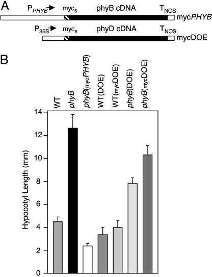
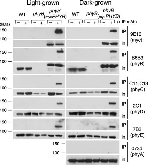
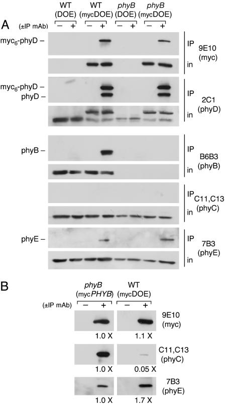
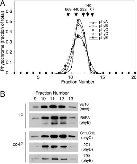
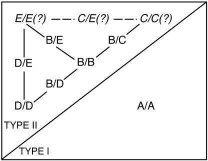
Coimmunoprecipitation of members of the phytochrome red/farred photoreceptor family from plant extracts has been used to analyze their heteromeric binding interactions. Phytochrome (phy)B or phyD apoproteins with six myc epitopes fused to their N termini are biologically active when expressed in Arabidopsis. Immunoprecipitation of either of these tagged proteins from seedling extracts coprecipitates additional type II phytochromes: six myc (myc6)-phyB coprecipitates phyC-phyE; and myc6-phyD coprecipitates phyB and phyE. No interaction of the epitope-tagged proteins with type I phyA was detected. Gel filtration chromatography shows that all five of the Arabidopsis phytochromes are present in seedlings as dimers, and that the heteromeric type II phytochrome complexes migrate at molecular masses characteristic of heterodimers. Similar levels of heterodimer formation are observed in extracts of dark-grown seedlings, where the phytochromes are cytosolic, and light-grown seedlings, where they are predominantly nuclear. These findings indicate that Arabidopsis, which until now has been thought to contain five homodimeric forms of phytochrome, in fact contains multiple species of both homodimeric and heterodimeric phytochromes. The conservation of the phytochrome family throughout angiosperms suggests that heterodimeric red/far-red receptors may be present in many flowering plants.


Figures

Fig. 1.
Fig. 1.
Activities of epitope-tagged phyB and phyD proteins. (A) Structures of the mycPHYB and mycDOE transgenes. myc6 epitopes were translationally fused to the PHYB and PHYD cDNA sequences. PPHYB and P35S represent the PHYB and cauliflower mosaic virus 35S promoter regions, and TNOS is the nopaline synthase terminator sequence. (B) Hypocotyl lengths of seedlings grown for 5 days under 20 μmol·s-1·m-2 R light.
Fig. 2.
Fig. 2.
Coimmunoprecipitation of phyC-phyE with the myc6-phyB protein from seedling extracts. Samples of extracts of light- and dark-grown seedlings of the WT, phyB mutant, and phyB(mycPHYB) lines were immunoprecipitated with the anti-myc 9E10 mAb or with 1 μg of nonimmune mouse IgG. Aliquots of the input extracted proteins and the immunoprecipitates were separated on 6% SDS gels, blotted, and probed with the indicated antibodies (in, input to IP reaction).
Fig. 3.
Fig. 3.
Binding interactions of type II phytochromes with the myc6-phyD protein. (A) Coimmunoprecipitation of phyB and phyE with the myc6-phyD protein from seedling extracts. Samples of extracts of light-grown seedlings of the WT and phyB mutant lines containing either the DOE or mycDOE transgenes were immunoprecipitated with the anti-myc 9E10 mAb or with nonimmune mouse IgG. Aliquots of the input extracted proteins and the immunoprecipitates were separated on 6% SDS gels, blotted, and probed with the indicated antibodies (in, input to IP reaction). (B) phyC coprecipitates more efficiently with phyB than with phyD. Extracts of dark-grown seedlings expressing similar levels of myc6-phyB or myc6-phyD were immunoprecipitated with the anti-myc 9E10 mAb or with nonimmune mouse IgG. Precipitated proteins were separated on 6% SDS gels, blotted, and probed with the indicated antibodies. Chemiluminescent immunoblot signals of the WT-(mycDOE) IPs relative to the phyB(mycPHYB) IPs were determined by densitometry.
Fig. 4.
Fig. 4.
Tissue-mixing experiment demonstrating in planta phytochrome interactions. Protein extracts were prepared from mixed light-grown seedlings in which phyB and myc6-phyD were expressed either in two different seedling populations (on the left) or in the same seedlings (on the right). Extracts were immunoprecipitated with the anti-myc 9E10 mAb or with nonimmune mouse IgG. Aliquots of the input extracted proteins and the immunoprecipitates were separated on 6% SDS gels, blotted, and probed with the indicated antibodies (in, input to IP reaction).
Fig. 5.
Fig. 5.
SEC of phytochrome dimers. Extracts of WT Arabidopsis and phyB- (mycPHYB) seedlings were prepared and separated on Superose 6. (A) Profiles of scanned immunoblots for each of the five Arabidopsis phytochromes from SEC of the WT extract. Column fractions were separated on 6% SDS gels, blotted, and probed with the phy-specific mAb. (B) Coimmunoprecipitation of phyC-phyE with myc6-phyB from individual fractions from SEC of an extract of the phyB(mycPHYB) line.
Fig. 6.
Fig. 6.
Dimerization interactions among the Arabidopsis phytochromes. Homo- and heterodimeric phytochromes that have been shown to be present in Arabidopsis seedlings are illustrated. Type II phytochrome dimer combinations that have not yet been investigated are shown in italics with question marks.

References

    1. Yamaguchi, R., Nakamura, M., Mochizuki, N., Kay, S. A. & Nagatani, A. (1999) J. Cell Biol. 145, 437-445. - PMC - PubMed
    1. Kircher, S., Gil, P., Kozma-Bognar, L., Fejes, E., Speth, V., Husselstein-Muller, T., Bauer, D., Adam, E., Schäfer, E. & Nagy, F. (2002) Plant Cell 14, 1541-1555. - PMC - PubMed
    1. Quail, P. H. (2002) Nat. Rev. Mol. Cell Biol. 3, 85-93. - PubMed
    1. Gyula, P., Schäfer, E. & Nagy, F. (2003) Curr. Opin. Plant Biol. 6, 446-452. - PubMed
    1. Sharrock, R. A. & Quail, P. H. (1989) Genes Dev. 3, 1745-1757. - PubMed

Publication types