. 2004 Aug;13(8):2059-77.
doi: 10.1110/ps.04637904.

Evolutionary constraints associated with functional specificity of the CMGC protein kinases MAPK, CDK, GSK, SRPK, DYRK, and CK2alpha

Affiliations

Evolutionary constraints associated with functional specificity of the CMGC protein kinases MAPK, CDK, GSK, SRPK, DYRK, and CK2alpha

Natarajan Kannan et al. Protein Sci. 2004 Aug.

Abstract

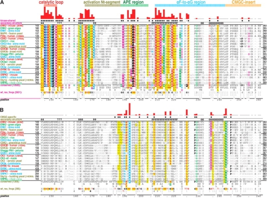
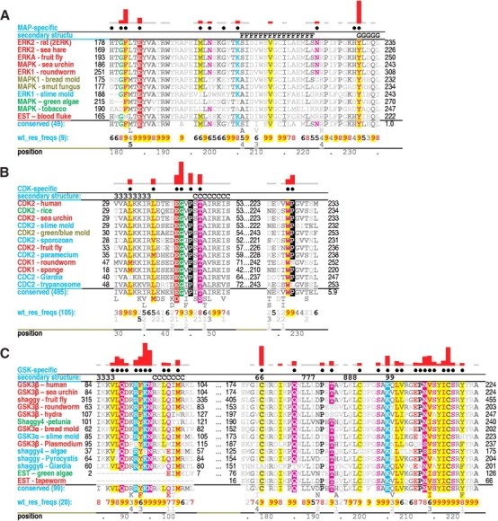
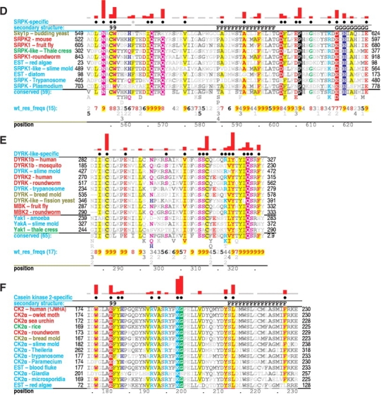
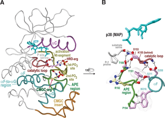
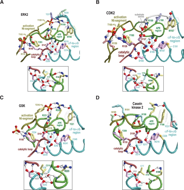
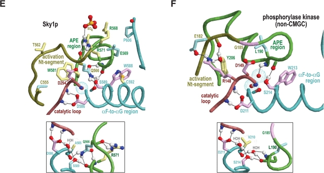
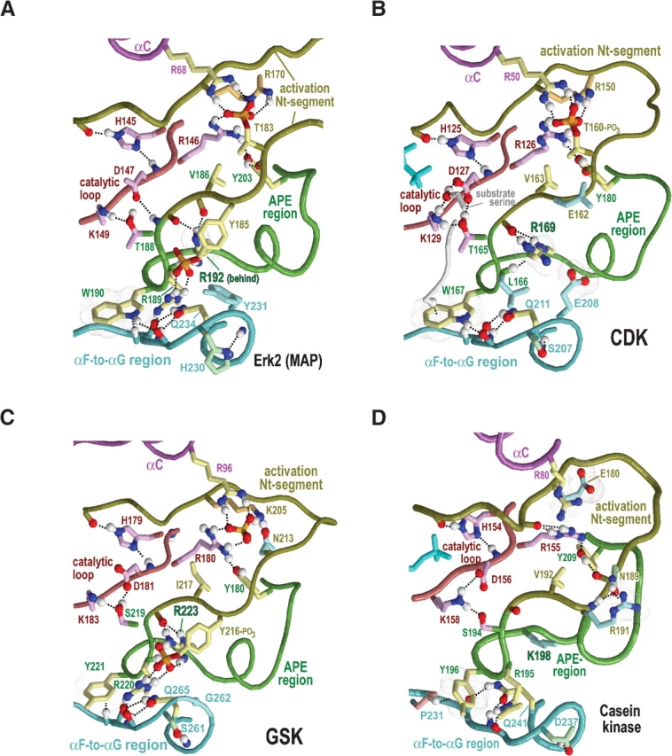
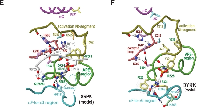
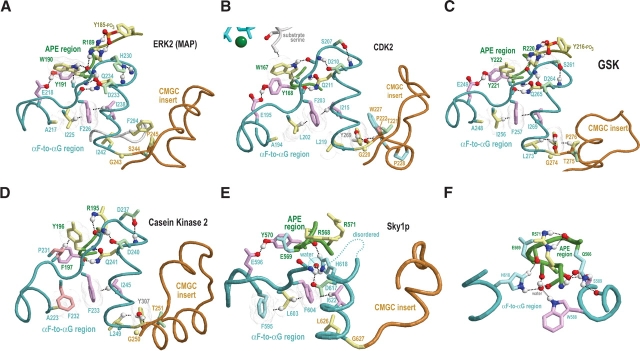
Amino acid residues associated with functional specificity of cyclin-dependent kinases (CDKs), mitogen-activated protein kinases (MAPKs), glycogen synthase kinases (GSKs), and CDK-like kinases (CLKs), which are collectively termed the CMGC group, were identified by categorizing and quantifying the selective constraints acting upon these proteins during evolution. Many constraints specific to CMGC kinases correspond to residues between the N-terminal end of the activation segment and a CMGC-conserved insert segment associated with coprotein binding. The strongest such constraint is imposed on a "CMGC-arginine" near the substrate phosphorylation site with a side chain that plays a role both in substrate recognition and in kinase activation. Two nearby buried waters, which are also present in non-CMGC kinases, typically position the main chain of this arginine relative to the catalytic loop. These and other CMGC-specific features suggest a structural linkage between coprotein binding, substrate recognition, and kinase activation. Constraints specific to individual subfamilies point to mechanisms for CMGC kinase specialization. Within casein kinase 2alpha (CK2alpha), for example, the binding of one of the buried waters appears prohibited by the side chain of a leucine that is highly conserved within CK2alpha and that, along with substitution of lysine for the CMGC-arginine, may contribute to the broad substrate specificity of CK2alpha by relaxing characteristically conserved, precise interactions near the active site. This leucine is replaced by a conserved isoleucine or valine in other CMGC kinases, thereby illustrating the potential functional significance of subtle amino acid substitutions. Analysis of other CMGC kinases similarly suggests candidate family-specific residues for experimental follow-up.


Figures

Figure 1.
Figure 1.
Contrast hierarchical alignments of various CMGC kinase families with representative sequences from distinct eukaryotic kingdoms for each family. The structural regions shown in Figures 3 ▶–6 ▶ and discussed in the text are indicated at the top. The histograms above the alignments plot the strength of the category-specific selective constraints imposed at each position (essentially using logarithmic scaling); these constraints also are indicated qualitatively through residue highlighting (with biochemically similar residues colored similarly). Dots below the histograms indicate residues assigned to that alignment’s category. Secondary structure is indicated directly above the aligned sequences, with β strands indicated by their number designations (i.e., 6–9 correspond to the β6–β9 strands, respectively) and helices by their letter designations (i.e., F and G correspond to the F-helix and the G-helix, respectively). The leftmost column of each alignment gives protein descriptions using the following color code to specify major eukaryotic taxa: metazoans, red; plants, green; fungi, dark yellow; and protozoans, cyan. See Materials and Methods for sequence identifiers. The foreground sequences (see text) are shown indirectly via the consensus patterns and corresponding weighted residue frequencies (wt_res_freqs) below the aligned sequences actually displayed. (Such sequence weighting adjusts for overrepresented families in the alignment.) Foreground alignment residue frequencies are indicated in integer tenths where, for example, a 5 indicates that the corresponding residue directly above it occurs in 50% to 60% of the weighted sequences. (A) Contrast hierarchical alignment of kinase-shared constraints. All protein kinases (10,583 sequences) comprise the foreground and all proteins, the background (see text for details). (B) Contrast hierarchical alignment of CMGC-specific constraints. CMGC kinases (1309 sequences) comprise the foreground and all protein kinases (i.e., the foreground set in A), the background sequences. Note that the alignment of SRPKs residues against P227 and G228 of the query (ERK2) is uncertain due to SRPK-specific insertions in this region; their structural locations are likewise uncertain due to disordering of this region in the Sky1p crystal structure. This may be related to SRPK-specific functions (see text).
Figure 2.
Figure 2.
Critical regions of contrast hierarchical alignments showing various family-specific constraints within CMGC kinases. CMGC kinases (operationally defined as protein kinases with either an arginine or a lysine at the CMGC-arginine position) constitute the background sequence set for each alignment. See legend to Figure 1 ▶ for a further description of alignment highlighting and notation. See Materials and Methods for sequence identifiers. For each alignment, the sequences displayed are from distinct phyla. See text for details. (A) MAP-specific constraints. (B) CDK-specific constraints. (C) GSK-specific constraints. (D) SRPK-specific constraints. (E) DYRK-specific constraints. (F) CK2-specific constraints.
Figure 2.
Figure 2.
Critical regions of contrast hierarchical alignments showing various family-specific constraints within CMGC kinases. CMGC kinases (operationally defined as protein kinases with either an arginine or a lysine at the CMGC-arginine position) constitute the background sequence set for each alignment. See legend to Figure 1 ▶ for a further description of alignment highlighting and notation. See Materials and Methods for sequence identifiers. For each alignment, the sequences displayed are from distinct phyla. See text for details. (A) MAP-specific constraints. (B) CDK-specific constraints. (C) GSK-specific constraints. (D) SRPK-specific constraints. (E) DYRK-specific constraints. (F) CK2-specific constraints.
Figure 3.
Figure 3.
Structural features of CMGC protein kinases discussed in the text. The structure of p38 MAP kinase (PDB 1cm8) is shown as a prototype of the CMGC group. Color scheme is as follows: Main-chain traces of key regions, colored as indicated at the top of Figure 1 ▶; main-chain traces of other regions are light gray; ATPs are cyan; phosphate moieties use the standard CPK color scheme; oxygen, nitrogen, and hydrogen atoms establishing hydrogen bonds are red, blue, and white, respectively; side chains of kinase-shared residues are pale magenta; and CMGC-specific residues are light yellow. Hydrogen bonds are depicted as dotted lines; CH–π interactions (Weiss et al. 2001) are depicted as dotted lines into dot clouds. (A) Key regions within the protein kinase N- and C-terminal domains. The locations of the αC, αF, and αG helices are indicated. (B) Close-up of regions that stabilize and interact with the CMGC-arginine (R192). Modeled substrate with a P + 1 proline is shown.
Figure 4.
Figure 4.
Structural features surrounding buried waters located between the APE region and the catalytic loop in CMGC and other protein kinases. In the main figures, the buried waters are shown as oxygens surrounded by turquoise dot clouds; in the inset, predicted hydrogen bonds formed by these waters are shown. Residue side chains and canonical glycine main chains are colored by functional categories as follows: kinase-shared, pale magenta; CMGC-specific, light yellow; intermediate between kinase-shared and CMGC-specific, light orange to pale red; family-specific, pale cyan; intermediate between CMGC-specific and family-specific, pale green; and subfamily-specific, light blue. Hydrogen bonding carbons are colored as their corresponding side chains, and van der Waals interactions are depicted as dot clouds. See the legend to Figure 3 ▶ for other bond representations and coloring conventions; see text for details. (A) Erk2 MAP kinase (PDB 2erk). (B) CDK2 (PDB 1qmz). (C) GSK (PDB 1gng). (D) CK2α (PDB 1lp4). In CK2α the water nearer the APE region is missing, presumably due to filling of its binding cavity by the side chain of L213, which is distinctively conserved within CK2α. (E) The SRPK-related Sky1p kinase (PDB 1how). The water nearer the APE region is replaced by the side chain of Q566, which occupies the conformationally strained position in SRPKs. Note that both the nitrogen and the oxygen of the glutamine side chain participate in hydrogen bonds that, in other protein kinases, are typically established by this water. (F) The (non-CMGC) phosphorylase kinase (PDB 2phk). As shown here, these buried waters also occur in non-CMGC protein kinases.
Figure 4.
Figure 4.
Structural features surrounding buried waters located between the APE region and the catalytic loop in CMGC and other protein kinases. In the main figures, the buried waters are shown as oxygens surrounded by turquoise dot clouds; in the inset, predicted hydrogen bonds formed by these waters are shown. Residue side chains and canonical glycine main chains are colored by functional categories as follows: kinase-shared, pale magenta; CMGC-specific, light yellow; intermediate between kinase-shared and CMGC-specific, light orange to pale red; family-specific, pale cyan; intermediate between CMGC-specific and family-specific, pale green; and subfamily-specific, light blue. Hydrogen bonding carbons are colored as their corresponding side chains, and van der Waals interactions are depicted as dot clouds. See the legend to Figure 3 ▶ for other bond representations and coloring conventions; see text for details. (A) Erk2 MAP kinase (PDB 2erk). (B) CDK2 (PDB 1qmz). (C) GSK (PDB 1gng). (D) CK2α (PDB 1lp4). In CK2α the water nearer the APE region is missing, presumably due to filling of its binding cavity by the side chain of L213, which is distinctively conserved within CK2α. (E) The SRPK-related Sky1p kinase (PDB 1how). The water nearer the APE region is replaced by the side chain of Q566, which occupies the conformationally strained position in SRPKs. Note that both the nitrogen and the oxygen of the glutamine side chain participate in hydrogen bonds that, in other protein kinases, are typically established by this water. (F) The (non-CMGC) phosphorylase kinase (PDB 2phk). As shown here, these buried waters also occur in non-CMGC protein kinases.
Figure 5.
Figure 5.
Category-specific structural features within and surrounding the activation segment. Electrostatic interactions are depicted as dot clouds. See the legends to Figures 3 ▶ and 4 ▶ for other bond representations and coloring conventions; see text for details. (A) ERK2 MAP kinase (PDB 2erk). (B) CDK2 (PDB 1qmz). (C) GSK (PDB 1gng). (D) CK2α (PDB 1ds5). (E) Model of substrate-bound SRPKs. This model is based on the non–substrate-bound form of the SRPK-related Sky1p protein (PDB 1how) and on CMGC-specific interactions revealed by our analysis. Residue numbering is based on the Sky1p structure. (F) Hypothetical model of DYRK (constructed as described in the text).
Figure 5.
Figure 5.
Category-specific structural features within and surrounding the activation segment. Electrostatic interactions are depicted as dot clouds. See the legends to Figures 3 ▶ and 4 ▶ for other bond representations and coloring conventions; see text for details. (A) ERK2 MAP kinase (PDB 2erk). (B) CDK2 (PDB 1qmz). (C) GSK (PDB 1gng). (D) CK2α (PDB 1ds5). (E) Model of substrate-bound SRPKs. This model is based on the non–substrate-bound form of the SRPK-related Sky1p protein (PDB 1how) and on CMGC-specific interactions revealed by our analysis. Residue numbering is based on the Sky1p structure. (F) Hypothetical model of DYRK (constructed as described in the text).
Figure 6.
Figure 6.
CMGC canonical structural features near the αF-to-αG region and the CMGC insert. See the legends to Figures 3 ▶ and 4 ▶ for bond representations and coloring conventions; see text for details and discussion. (A) ERK2 MAP kinase (PDB 2erk). (B) CDK2 (PDB 1qmz). (C) GSK (PDB 1gng). (D) CK2α (PDB 1lp4). (E) Sky1p (PDB 1how). (F) Sky1p structural features near a histidine (H618) that replaces the CMGC-glutamine residue within SRPKs.

Similar articles

Cited by

References

    1. Adams, J.A. 2003. Activation loop phosphorylation and catalysis in protein kinases: Is there functional evidence for the autoinhibitor model? Biochemistry 42 601–607. - PubMed
    1. Aubol, B.E., Chakrabarti, S., Ngo, J., Shaffer, J., Nolen, B., Fu, X.D., Ghosh, G., and Adams, J.A. 2003. Processive phosphorylation of alternative splicing factor/splicing factor 2. Proc. Natl. Acad. Sci. 100 12601–12606. - PMC - PubMed
    1. Battistutta, R., Sarno, S., De Moliner, E., Marin, O., Issinger, O.G., Zanotti, G., and Pinna, L.A. 2000. The crystal structure of the complex of Zea mays α subunit with a fragment of human β subunit provides the clue to the architecture of protein kinase CK2 holoenzyme. Eur. J. Biochem. 267 5184–5190. - PubMed
    1. Bax, B., Carter, P.S., Lewis, C., Guy, A.R., Bridges, A., Tanner, R., Pettman, G., Mannix, C., Culbert, A.A., Brown, M.J., et al. 2001. The structure of phosphorylated GSK-3β complexed with a peptide, FRATtide, that inhibits β-catenin phosphorylation. Structure 9 1143–1152. - PubMed
    1. Becker, W. and Joost, H.G. 1999. Structural and functional characteristics of DYRK, a novel subfamily of protein kinases with dual specificity. Prog. Nucleic Acid Res. Mol. Biol. 62 1–17. - PubMed

Publication types

Substances

LinkOut - more resources