. 2004 Sep;10(9):1380-7.
doi: 10.1261/rna.7460104. Epub 2004 Jul 23.

Solution structure of an RNA stem-loop derived from the 3' conserved region of eel LINE UnaL2

Affiliations

Solution structure of an RNA stem-loop derived from the 3' conserved region of eel LINE UnaL2

Seiki Baba et al. RNA. 2004 Sep.

Abstract

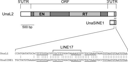
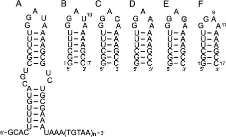
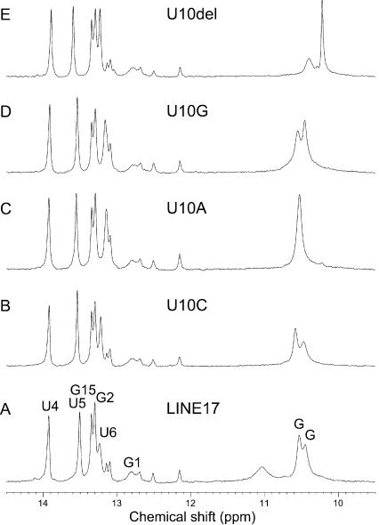
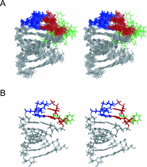
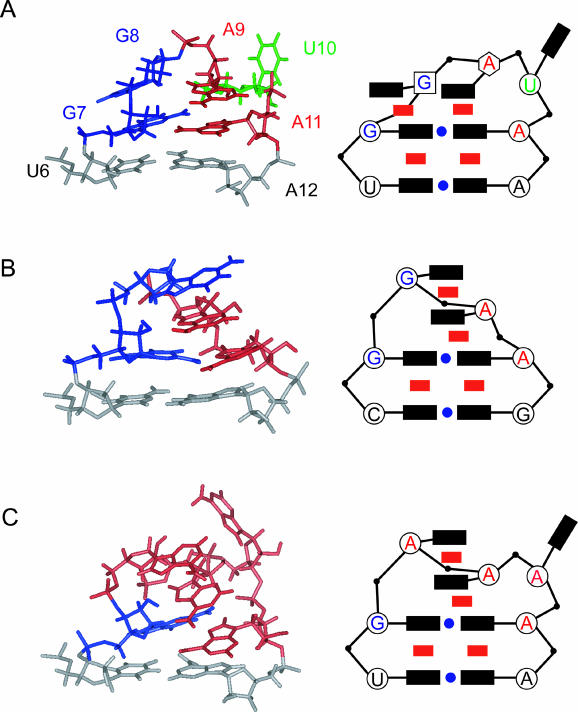
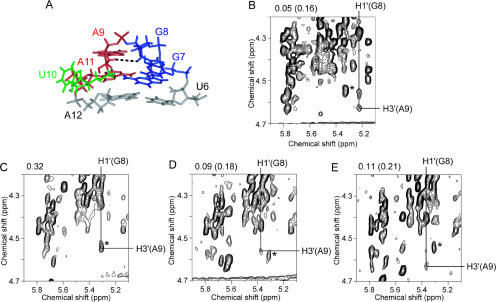
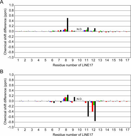
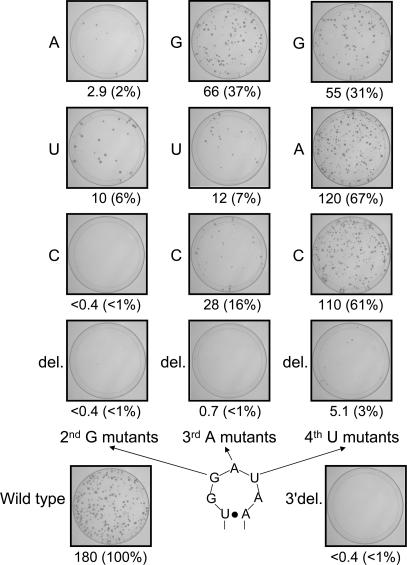
The eel long interspersed element (LINE) UnaL2 and its partner short interspersed element (SINE) share a conserved 3' tail containing a stem-loop that is critical for their retrotransposition. Presumably, the first step of retrotransposition is the recognition of their 3' tails by UnaL2-encoded reverse transcriptase. The solution structure of a 17-nucleotide RNA derived from the 3' tail of UnaL2 was determined by NMR. The GGAUA loop forms a specific structure in which the uridine is exposed to solvent with the third and fifth adenosines stacked. A sharp turn in the phosphodiester backbone occurs between the second guanosine and third adenosine. When the uridine is mutated (but not deleted), all mutants form the loop structure, indicating that the loop structure requires an exposed fourth residue. The retrotransposition assay in HeLa cells revealed that retrotransposition requires the second guanosine, although any nucleoside functions at the fourth position, suggesting that UnaL2 reverse transcriptase specifically recognizes the 5' side of the GGANA loop.


Figures

FIGURE 1.
FIGURE 1.
Schematic representation of full-length UnaL2 and UnaSINE1 from the eel and an alignment of their conserved 3′ tail regions. The single ORF comprises the shaded boxes, and the putative endonuclease (EN) and reverse transcriptase (RT) domains are indicated. The 5′ and 3′ untranslated regions (UTRs) are also indicated. The sequence alignment of the conserved 3′ tail is shown. The upper and lower stem regions are underlined by double lines and the region corresponding to LINE17 is indicated. The 3′ terminal repeats in UnaL2 and UnaSINE1 are underlined by a single line. The putative poly(A) signal in the UnaL2 sequence is indicated by a dotted line.
FIGURE 2.
FIGURE 2.
Secondary structure of the 3′ tail of UnaL2 and the 17-mer RNAs used in this study. (A) Secondary structure of the UnaL2 3′ tail that is putatively recognized by the UnaL2 reverse transcriptase. (B) The conserved RNA step-loop structure of the LINE17 3′ tail. (CF) Structures of U10C, U10A, U10A, and U10del, respectively.
FIGURE 3.
FIGURE 3.
Imino proton spectra of LINE17 and its mutants. (A) LINE17. (B) U10C. (C) U10A. (D) U10G. (E) U10del. Resonance assignments of LINE17 are shown.
FIGURE 4.
FIGURE 4.
Stereo view of the solution structures of LINE17. (A) The superposition of the final 20 structures. (B) The minimized average structure of LINE17. G, A, and U residues in the GGAUA loop of LINE17 are colored in blue, red, and green, respectively.
FIGURE 5.
FIGURE 5.
Comparison of the GGAUA, GGAA, and GAAAA loop structures and schematic representations. The following symbols are used in the schematic representation: black rectangle, base; red rectangle, stacking interaction; blue circle, hydrogen bonding interaction; open circle, C3′-endo ribose; open square, C2′-endo ribose; open hexagon, intermediate between C3′-endo and C2′-endo. (A) The GGAUA loop structure in LINE17 determined in this study. (B) The GGAA loop structure in Escherichia coli SRP RNA (Schmitz et al. 1999). (C) The GAAAA loop structure in boxB RNA in complex with the 36-mer N-terminal peptide of the N protein (N36) from bacteriophage λ (Schärpf et al. 2000).
FIGURE 6.
FIGURE 6.
The characteristic NOE (H1′ (G8)-H3′ (A9)) in the GGARA loop structures of LINE17 and mutants thereof. (A) The black dotted line indicates a NOE between H1′ (G8) and H3′ (A9) in the GGAUA loop of LINE17. (BE) NOESY spectra measured in D2O at 20°C with a mixing time of 200 msec. Resonance of the G8 H3′ proton is labeled as a horizontal line and that of the A9 H1′ proton is labeled as a vertical line. NOE cross peaks between H3′ (G8) and H1′ (A9) were observed. (B) LINE17. (C) U10C. (D) U10A. (E) U10G. The NOE signals due to H1′ (G7)-H3′ (G7) are indicated by asterisks. Note that the signal-to-noise ratio for LINE17 is higher than others because of higher sample concentration. The NOE volumes for H1′ (G8)-H3′ (A9) and H1′ (G7)-H3′ (G7) in parentheses relative to the average for H5–H6 of U4, U5, U6, and C16 were shown on the top of spectra. For U10C, the value for the overlapped peak is shown.
FIGURE 7.
FIGURE 7.
Chemical shift differences between LINE17 and the LINE17 mutants: blue, U10C; red, U10A; green, U10G; black, U10del. (A) Chemical shift differences of H8/H6 protons between LINE17 and each of the LINE17 mutants. (B) Chemical shift differences of the H1′ proton between LINE17 and each of the LINE17 mutants. The tenth residue is not shown because it is the mutated position in the LINE17 mutants.
FIGURE 8.
FIGURE 8.
Retrotransposition frequency (RF) in mutants of the stem–loop within the conserved 3′ tail region. RFs are calculated as described in Materials and Methods. RFs and relative value of RFs for mutants compared with the wild type are shown. Images show each 100-mm plate with G-418R colonies selected from ~2–4 × 10⁶ HygR cells. 3′ del indicates the UnaL2 mutant in which the 3′ conserved region is deleted.

References

    1. Allain, F.H. and Varani, G. 1995. Structure of the P1 helix from group I self-splicing introns. J. Mol. Biol. 250: 333–353. - PubMed
    1. Altona, C. and Sundaralingam, M. 1973. Conformational analysis of the sugar ring in nucleosides and nucleotides. Improved method for the interpretation of proton magnetic resonance coupling constants. J. Am. Chem. Soc. 95: 2333–2344. - PubMed
    1. Boeke, J.D. and Devine, S.E. 1998. Yeast retrotransposons: Finding a nice quiet neighborhood. Cell 93: 1087–1089. - PubMed
    1. Britten, R.J., Baron, W.F., Stout, D.B., and Davidson, E.H. 1988. Sources and evolution of human Alu repeated sequences. Proc. Natl. Acad. Sci. 85: 4770–4774. - PMC - PubMed
    1. Deininger, P.L. and Batzer, M.A. 1993. Evolution of retroposons. Evolutionary Biol. 27: 157–196.

Publication types

Associated data

LinkOut - more resources