Comparative Study
. 2004 Aug;165(2):523-31.
doi: 10.1016/S0002-9440(10)63317-2.

Active caspase-6 and caspase-6-cleaved tau in neuropil threads, neuritic plaques, and neurofibrillary tangles of Alzheimer's disease

Affiliations
Comparative Study

Active caspase-6 and caspase-6-cleaved tau in neuropil threads, neuritic plaques, and neurofibrillary tangles of Alzheimer's disease

Huishan Guo et al. Am J Pathol. 2004 Aug.

Abstract

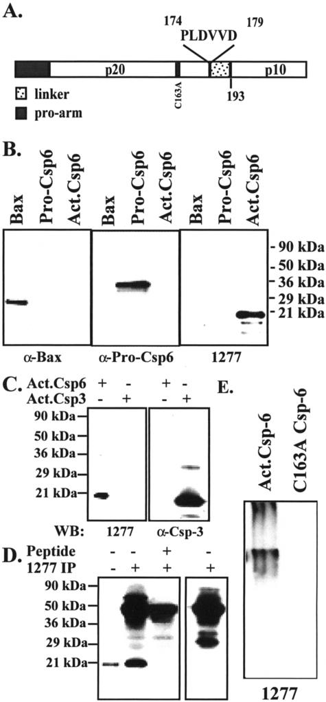
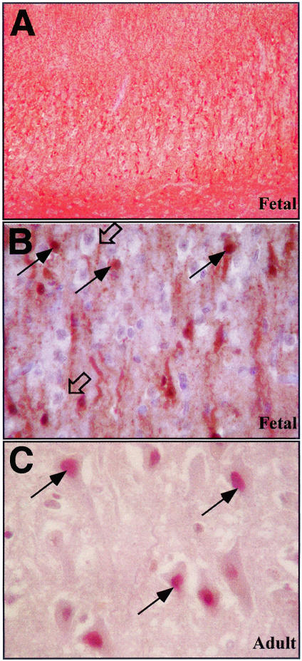
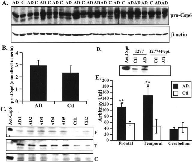
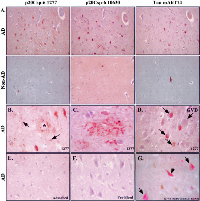
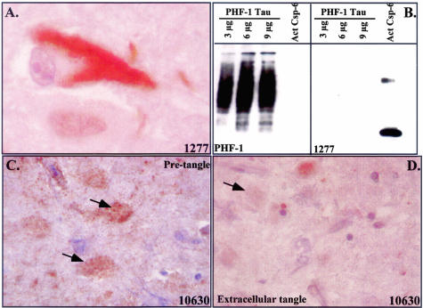
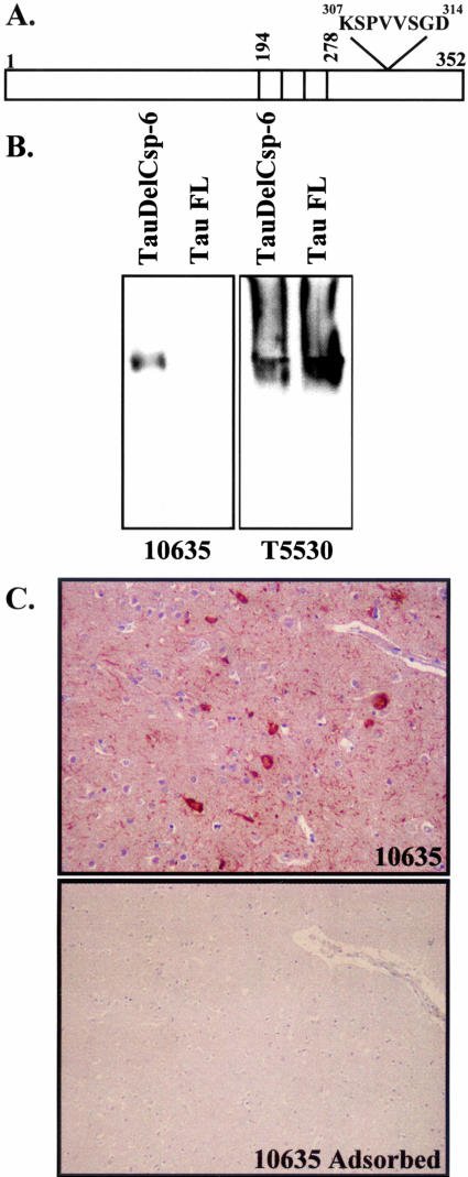
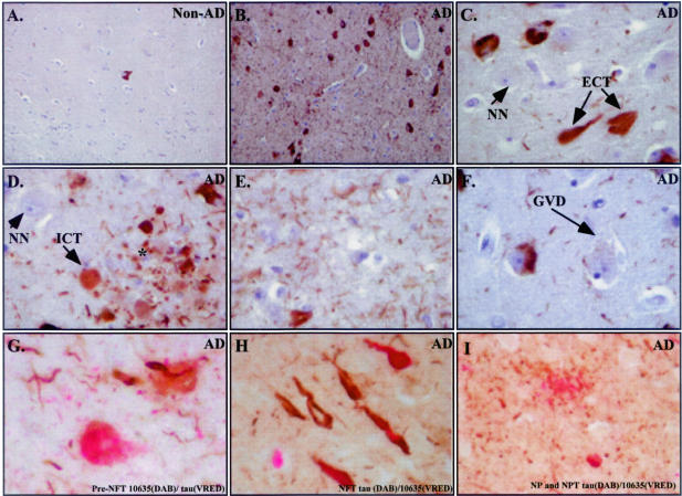
Previously, we have shown that caspase-6 but not caspase-3 is activated by serum deprivation and induces a protracted cell death in primary cultures of human neurons (LeBlanc AC, Liu H, Goodyer C, Bergeron C, Hammond J: Caspase-6 role in apoptosis of human neurons, amyloidogenesis and Alzheimer's disease. J Biol Chem 1999, 274:23426-23436 and Zhang Y, Goodyer C, LeBlanc A: Selective and protracted apoptosis in human primary neurons microinjected with active caspase-3, -6, -7, and -8. J Neurosci 2000, 20:8384-8389). Here, we show with neoepitope antibodies that the p20 subunit of active caspase-6 increases twofold to threefold in the affected temporal and frontal cortex but not in the unaffected cerebellum of Alzheimer's disease brains and is present in neurofibrillary tangles, neuropil threads, and the neuritic plaques. Furthermore, a neoepitope antibody to caspase-6-cleaved Tau strongly detects intracellular tangles, extracellular tangles, pretangles, neuropil threads, and neuritic plaques. Immunoreactivity with both antibodies in pretangles indicates that the caspase-6 is active early in the pathogenesis of Alzheimer's disease. In contrast to the nuclear and cytosolic localization of active caspase-6 in apoptotic neurons of fetal and adult ischemic brains, the active caspase-6 in Alzheimer's disease brains is sequestered into the tangles or neurites. The localization of active caspase-6 may strongly jeopardize the structural integrity of the neuronal cytoskeletal system leading to inescapable neuronal dysfunction and eventual cell death in Alzheimer's disease neurons. Our results suggest that active caspase-6 is strongly implicated in human neuronal degeneration and apoptosis.


Figures

Figure 1
Figure 1
The p20Csp-6 antiserum specifically recognizes the p20 subunit of active Csp-6 under denaturant and native conditions. A: Schematic diagram of pro-Csp-6 showing the sequence and location of the Csp-6 antigen PLDVVD. B: Western blot analysis of 60 μg of E. coli protein lysates from in vitro-translated recombinant Bax, pro-Csp-6, or active Csp-6 with anti-Bax, anti-pro-Csp-6, and anti-p20Csp-6Ab 1277 antisera. C: Western blot analysis of 10 μg of E. coli lysate proteins containing recombinant Csp-6 and 10 ng of purified recombinant Csp-3 with p20Csp-6Ab 1277 antiserum or anti-Csp-3 antiserum. D: Western blot analysis of nonimmunoprecipitated (lane 1) or p20Csp-6Ab 1277 immunoprecipitated p20 subunit of active Csp-6 in the absence or the presence of PLDVVD peptide (lanes 2 and 3) with p20Csp-6Ab 1277 antiserum. The right panel represents a control immunoprecipitation in the absence of E. coli lysate and recombinant Csp-6 probed with p20Csp-6Ab 1277 antiserum only to show the nonspecificity of the high-molecular weight bands. Similar results were obtained with the p20Csp-6Ab 10630 antiserum (not shown). E: Western blot analysis of active Csp-6 and catalytically inactive Csp-6 in nondenaturant conditions with p20Csp-6Ab 1277 antiserum.
Figure 2
Figure 2
Immunocytochemical analysis of fetal ischemic brain with the p20Csp-6Ab 1277 antiserum. Low (A) and high (B) magnification of fetal ischemia. C: Adult ischemia. Open arrows in B identify normal nonimmunoreactive neurons and filled arrows show apoptotic immunoreactive neurons. Arrows in C identify nuclear immunoreactivity in ischemic adult neurons.
Figure 3
Figure 3
The p20 subunit of Csp-6 is increased in AD frontal and temporal cortex compared to non-AD age-matched control tissues. A: Western blot analyses of proteins from AD and control (Ctl) frontal cortex with anti-full-length Csp-6 antibody (Pharmingen). B: Quantitative analysis of full-length Csp-6 shown in A. C: Western blot analysis of p20 Csp-6Ab-immunoprecipitated proteins from AD and non-AD frontal cortex (F), temporal cortex (T), and cerebellum (C) with the 1277 p20Csp-6Ab. Recombinant active Csp-6 was included as a control (Act. Csp-6). D: Western blot analysis with 1277 p20Csp-6Ab of the p20Csp-6Ab-immunoprecipitated or PLDVVD peptide-adsorbed p20Csp-6Ab-immunoprecipitated proteins from AD frontal cortex proteins. E: Quantitative analysis of the p20 Csp-6 subunit levels in the frontal, temporal, and cerebellum of 15 AD versus 8 non-AD control cases. Analysis of variance, <0.009. *, Statistically significant difference between AD and Ctl, P < 0.03. **, Statistically significant difference between cerebellum and frontal or temporal cortex, P < 0.001.
Figure 4
Figure 4
The p20Csp-6 antiserum immunoreacts with NPTs, degenerating neurites in plaques, and NFTs in AD brains. A: Adjacent AD hippocampal brain section immunostained with the p20Csp-6Ab 1277 or 10630 antisera compared with monoclonal Tau T14 antibody. The second row shows the control non-AD tissue stained at the same time as the AD sections. B–D: Sections stained with 1277 p20Csp-6Ab showing immunoreactivity to plaques (*), NFT, and NPTs (arrow) in B, neuritic staining of a plaque in C and intracellular pretangles (small arrows) but not GVD in D. The p20Csp-6 immunoreactivity is adsorbed with recombinant active Csp-6 (E) and is absent in prebleed serum (F). Co-localization of active Csp-6 with Tau is shown in pretangles (arrows) and tangles (arrowhead) in G.
Figure 5
Figure 5
p20-Csp-6 immunoreactivity in tangles. A: High magnification showing localization of Csp-6 immunostaining in a NFT. B: Western blot analysis of purified PHF tau with PHF-1 tau antibody or p20-Csp-6 1277 antiserum. C and D: Immunoreactivity of 10630 with pretangles in C (arrows) or 10630 with extracellular tangles in D (arrow).
Figure 6
Figure 6
Characterization of the tauΔCsp-6Ab. A: Schematic diagram showing the amino acid position of the peptide antigen used for the tauΔCsp-6Ab relative to the three microtubule binding repeats in fetal tau (352 aminoacids). B: Western blot analysis of Tau (T5530) and TauΔCsp-6 (10635) separated under nondenaturant conditions. C: Immunocytochemistry of AD brain section with 10635- and 10635-adsorbed antiserum.
Figure 7
Figure 7
Csp-6-cleaved tau antibody strongly detects NPTs, neurites in senile plaques, intracellular tangles, extracellular tangles, and pretangles in AD brains. A: Immunostaining of hippocampi from non-AD (A) or AD (B) with the 10635 tauΔCsp-6Ab. Higher magnification of certain areas shows immunostaining in extracellular tangles (ECT) in C, pretangles (pre-NFT) and degenerating neurites of senile plaques (*) in D, NPTs in E, normal unstained neurons (NN) in C and D, and absence of staining in GVD in F. Co-localization of Tau and TauΔCsp-6Ab in pretangle (G), tangles (H), neuropil threads (G and I) and neuritic plaque (I).

References

    1. LeBlanc AC, Liu H, Goodyer C, Bergeron C, Hammond J. Caspase-6 role in apoptosis of human neurons, amyloidogenesis and Alzheimer’s Disease. J Biol Chem. 1999;274:23426–23436. - PubMed
    1. Zhang Y, Goodyer C, LeBlanc A. Selective and protracted apoptosis in human primary neurons microinjected with active caspase-3, -6, -7, and -8. J Neurosci. 2000;20:8384–8389. - PMC - PubMed
    1. Boatright KM, Renatus M, Scott FL, Sperandio S, Shin H, Pedersen IM, Ricci JE, Edris WA, Sutherlin DP, Green DR, Salvesen GS. A unified model for apical caspase activation. Mol Cell. 2003;11:529–541. - PubMed
    1. Gervais F, Xu D, Robertson G, Vaillancourt J, Zhu Y, Huang J, LeBlanc A, Smith D, Rigby M, Shearman M, Clarke E, Zheng H, Van Der Ploeg L, Ruffolo S, Thornberry N, Xanthoudakis S, Zamboni R, Roy S, Nicholson D. Involvement of caspases in proteolytic cleavage of Alzheimer’s β-amyloid precursor protein and amyloidogenic β-peptide formation. Cell. 1999;97:395–406. - PubMed
    1. Weidemann A, Paliga K, Durrwang U, Reinhard F, Zchuckert O, Evin G, Masters CL. Proteolytic processing of the Alzheimer’s disease amyloid precursor protein within its cytoplasmic domain by caspase-like enzymes. J Biol Chem. 1999;274:5823–5829. - PubMed

Publication types

MeSH terms