. 2004 Aug;165(2):577-92.
doi: 10.1016/s0002-9440(10)63322-6.

The 14-3-3 protein epsilon isoform expressed in reactive astrocytes in demyelinating lesions of multiple sclerosis binds to vimentin and glial fibrillary acidic protein in cultured human astrocytes

Affiliations

The 14-3-3 protein epsilon isoform expressed in reactive astrocytes in demyelinating lesions of multiple sclerosis binds to vimentin and glial fibrillary acidic protein in cultured human astrocytes

Jun-Ichi Satoh et al. Am J Pathol. 2004 Aug.

Abstract

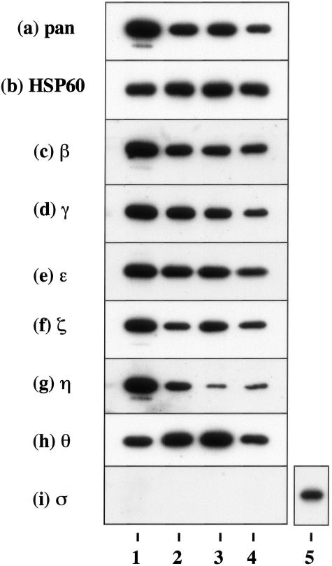
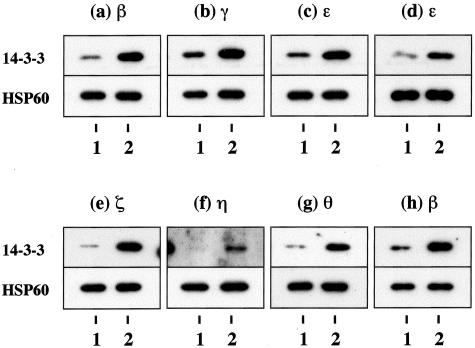
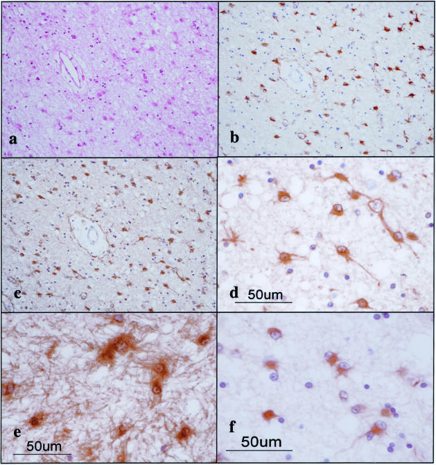
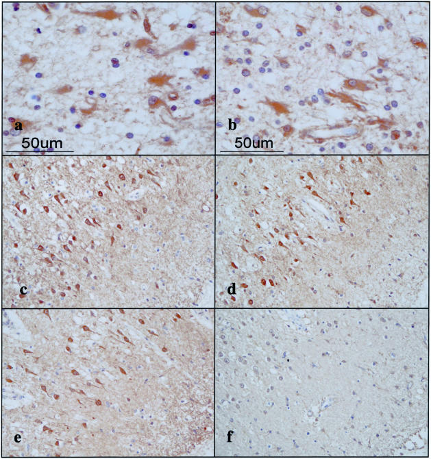
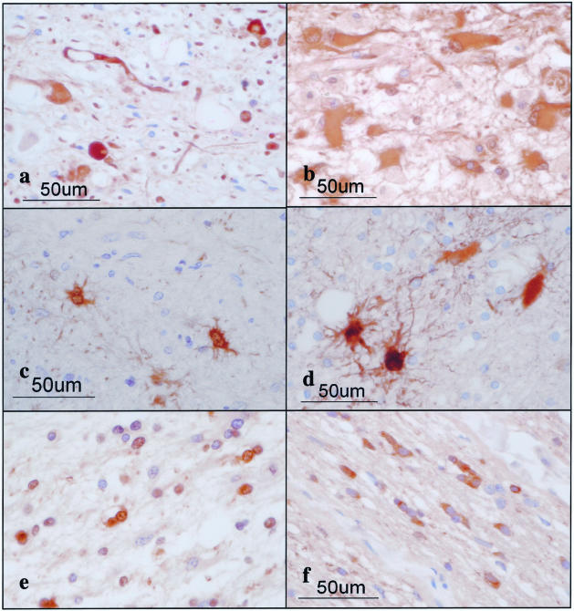
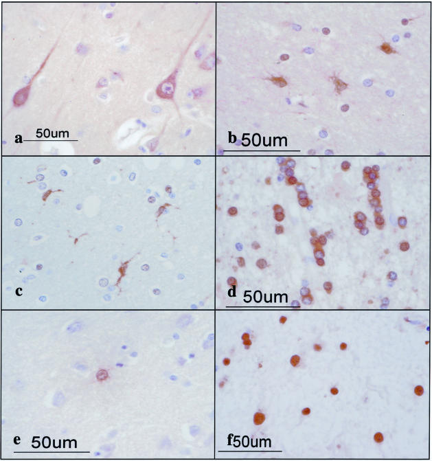
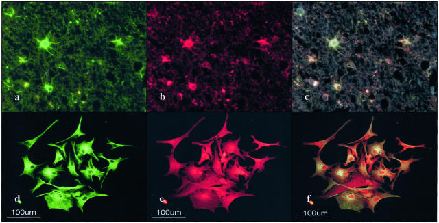
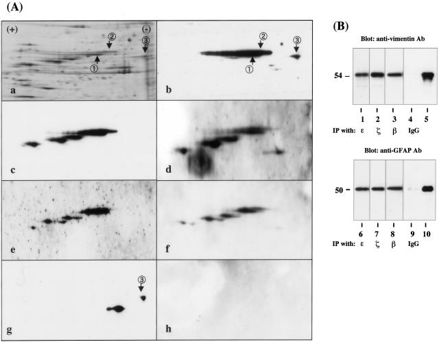
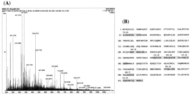
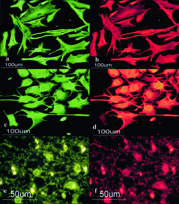
The 14-3-3 protein family consists of acidic 30-kd proteins expressed at high levels in neurons of the central nervous system. Seven isoforms form a dimeric complex that acts as a molecular chaperone that interacts with key signaling components. Recent studies indicated that the 14-3-3 protein identified in the cerebrospinal fluid of various neurological diseases including multiple sclerosis (MS) is a marker for extensive brain destruction. However, it remains unknown whether the 14-3-3 protein plays an active role in the pathological process of MS. To investigate the differential expression of seven 14-3-3 isoforms in MS lesions, brain tissues of four progressive cases were immunolabeled with a panel of isoform-specific antibodies. Reactive astrocytes in chronic demyelinating lesions intensely expressed beta, epsilon, zeta, eta, and sigma isoforms, among which the epsilon isoform is a highly specific marker for reactive astrocytes. Furthermore, protein overlay, mass spectrometry, immunoprecipitation, and double-immunolabeling analysis showed that the 14-3-3 protein interacts with both vimentin and glial fibrillary acidic protein in cultured human astrocytes. These results suggest that the 14-3-3 protein plays an organizing role in the intermediate filament network in reactive astrocytes at the site of demyelinating lesions in MS.


Figures

Figure 1
Figure 1
Constitutive expression of 14-3-3 isoforms in cultured human cells. Two μg of total protein extract isolated from brain tissues or cultured cells incubated in 10% FBS-containing medium were processed for Western blot analysis using a battery of 14-3-3 isoform-specific antibodies or the antibodies broadly reactive against all of the isoforms listed in Table 1, or the antibody against the housekeeping gene product HSP60. a to i indicate the following antibody specificity: a, all isoforms; b, HSP60; c, β; d, γ; e, ε; f, ζ; g, η; h, θ; and i, σ. Lanes 1 to 4 represent homogenate of the human cerebrum (lane 1), NTera2-derived differentiated neurons (NTera2-N) (lane 2), U-373MG astrocytoma cells (lane 3), fetal human astrocytes (AS1477) (lane 4), and HeLa cervical carcinoma cells (lane 5).
Figure 2
Figure 2
Growth-dependent expression of various 14-3-3 isoforms in cultured human astrocytes. Human and mouse astrocytes were plated at subconfluent density and incubated for 7 days in the serum-free culture medium or in 10% FBS-containing culture medium. Two μg of total protein extract was processed for Western blot analysis using a battery of 14-3-3 isoform-specific antibodies or with the antibodies broadly reactive against all isoforms (top). After stripping the antibodies, identical blots were relabeled with the antibody against HSP60 for the standardization of expression levels (bottom). a to g (top) indicate the expression of β (a), γ (b), ε (c), ζ (e), η (f), and θ (g) in human astrocytes (AS1477); ε in human astrocytes (AS-BW) (d); and β in mouse astrocytes (h). Lanes 1 and 2 represent the cells cultured under the serum-free growth-arrested condition (lane 1) or the serum-containing growth-promoting condition (lane 2). Additional data are shown in supplementary Figure 2 on the American Journal of Pathology website.
Figure 3
Figure 3
The 14-3-3 ε isoform is expressed in reactive astrocytes in chronic demyelinating lesions of MS. MS brain tissues were processed for immunohistochemical analysis using ε isoform-specific antibody or the antibody against GFAP or vimentin. a to f represent the following: a: no. 744 MS, chronic active demyelinating lesions in the subcortical white matter of the frontal lobe (H&E). b: No. 744 MS, the area corresponding to a (GFAP). Many reactive astrocytes are stained. c: No. 744 MS, the area corresponding to a (ε). Many reactive astrocytes are stained. d: No. 744 MS, a higher magnification view of c (ε). Reactive astrocytes are stained. e: No. 544 MS, chronic inactive demyelinating lesions in the optic nerve (ε). Reactive astrocytes and the glial scar are stained. f: No. 744 MS, chronic active demyelinating lesions in the subcortical white matter of the frontal lobe (vimentin). Reactive astrocytes are stained.
Figure 4
Figure 4
Expression of various 14-3-3 isoforms in reactive astrocytes and cortical neurons in MS brain. MS brain tissues were processed for immunohistochemical analysis using a battery of 14-3-3 isoform-specific antibodies. a to f represent the following: a: no. 744 MS, chronic active demyelinating lesions in the subcortical white matter of the frontal lobe (β). Reactive astrocytes are stained. b: No. 744 MS, chronic active demyelinating lesions in the subcortical white matter of the frontal lobe (ζ). Reactive astrocytes are stained. c: No. 744 MS, the cerebral cortex of the frontal lobe (γ). Cortical neurons are stained. d: No. 744 MS, the area corresponding to c (η). Cortical neurons are stained. e: No. 744 MS, the area corresponding to c (ζ). Cortical neurons are stained. f: No. 744 MS, the area corresponding to c (ε). Cortical neurons are devoid of staining.
Figure 5
Figure 5
Expression of various 14-3-3 isoforms in reactive astrocytes, surviving oligodendrocytes, and injured axons in chronic demyelinating lesions of MS and in infarcted lesions. The brains of MS and non-MS control cases were processed for immunohistochemical analysis using a battery of 14-3-3 isoform-specific antibodies. a to f represent the following: a: no. 609 MS, chronic active demyelinating lesions in the medulla oblongata (γ). Disrupted axons are stained. b: No. 719 acute cerebral infarction, infarcted lesions in the parietal cerebral cortex (ε). Reactive astrocytes are stained. c: No. 791 MS, chronic inactive lesions in the pons (σ). Reactive astrocytes are stained. d: No. 719 acute cerebral infarction, infarcted lesions in the parietal cerebral cortex (σ). Reactive astrocytes are stained. e: No. 609 MS, chronic active demyelinating lesions in the periventricular white matter of the frontal lobe (θ). Surviving oligodendrocytes are stained. f: No. 744 MS, chronic active demyelinating lesions in the optic nerve (θ). Surviving oligodendrocytes are stained.
Figure 6
Figure 6
Expression of various 14-3-3 isoforms in neurons, astrocytes, oligodendrocytes, and microglia in non-MS brains. The brains of non-MS control cases were processed for immunohistochemical analysis using a battery of 14-3-3 isoform-specific antibodies. a to f represent the following: a: no. G9 neurologically normal subject, the frontal cerebral cortex (γ). Cortical neurons are stained. b: No. 523 schizophrenia, frontal cerebral cortex (ε). Astrocytes are stained. c: No. 826 schizophrenia, the frontal cerebral cortex (η). Microglia are stained. d: No. 786 acute cerebral infarction, the subcortical white matter of the parietal lobe (θ). Surviving oligodendrocytes are stained. e: No. G7 neurologically normal subject, the frontal cerebral cortex (σ). A few astrocytes are stained. f: No. 789 old cerebral infarction, the frontal cerebral cortex (η). The nuclei of reactive astrocytes are stained.
Figure 7
Figure 7
Co-expression of the 14-3-3 ε isoform and GFAP in reactive astrocytes in chronic demyelinating lesions of MS and in cultured human astrocytes. Cultured human astrocytes and MS brain tissues were processed for double immunolabeling with anti-GFAP antibody and ε isoform-specific antibody followed by labeling with fluorescein isothiocyanate- and rhodamine-conjugated secondary antibodies. a to f represent no. 744, chronic active demyelinating lesions in the subcortical white matter of the frontal lobe (ac), cultured human astrocytes (AS-BW) (d–f), GFAP (a, d), ε (b, e), and the overlay (c, f).
Figure 8
Figure 8
Two-dimensional gel electrophoresis and immunoprecipitation analysis of 14-3-3 ε isoform-binding proteins in cultured human astrocytes. A: Two-dimensional gel analysis. Human astrocytes (AS-BW) were incubated in 10% FBS-containing culture medium. Twenty-one μg of total protein extract was separated on a two-dimensional PAGE gel, transblotted onto a polyvinylidene difluoride membrane, and processed for overlay analysis with recombinant human 14-3-3ε protein possessing the Xpress tag (rh14-3-3ε) followed by labeling with anti-Xpress antibody. After the probe and antibody were stripped, the blot was repeatedly relabeled six times with the antibodies against GFAP, vimentin, and vimentin with specific phosphorylated serine epitopes, and with recombinant human interferon-stimulated protein ISG15 having the Xpress tag (rhISG15). a to g represent silver staining (a), rh14-3-3ε labeling followed by staining with anti-Xpress antibody (b), vimentin (c), vimentin with phosphorylated Ser-39 (d), vimentin with phosphorylated Ser-72 (e), vimentin with phosphorylated Ser-83 (f), GFAP (g), and rhISG15 labeling followed by staining with anti-Xpress antibody (h). Two major spots labeled with rh14-3-3ε and anti-vimentin antibody are named spot no. 1 and no. 2, while a spot labeled with rh14-3-3ε and anti-GFAP antibody is designated spot no. 3. Spots no. 1 and no. 2 were excised from the gel and processed for mass spectrometry (MS) analysis. B: Immunoprecipitation analysis. Total protein extract of cultured human astrocytes was immunoprecipitated with ε isoform-specific antibody (lanes 1 and 6), ζ isoform-specific antibody (lanes 2 and 7), β-isoform-specific antibody (lanes 3 and 8), with the same amount of normal rabbit IgG (lanes 4 and 9), or untreated with any antibodies (lanes 5 and 10; 2 μg of total protein extract before processing for immunoprecipitation). Then, the immunoprecipitates were processed for Western blot analysis using anti-vimentin (top) or anti-GFAP antibody (bottom).
Figure 9
Figure 9
Mass spectrometry analysis of the 14-3-3 ε isoform-binding proteins in cultured human astrocytes. Spots no. 1 and no. 2 labeled with the rh14-3-3ε probe (Figure 8A, a and b) were excised from the gel, trypsinized, and processed for nanoESI-MS/MS analysis. A: The spectra of nanoESI-MS/MS analysis of spot no. 1. Each peak indicates individual peptide fragments. The position of several peaks was automatically numbered on the spectra. Peptides derived from the autolytic fragments of trypsin (eg, 412, 421, and 523) were omitted to be processed for further analysis. The peptide fragments were selected for MS analysis in order of their signal intensity. B: Amino acid sequence of human vimentin. Eight peptide fragments of spot no. 1 identified by nanoESI-MS/MS analysis (shadowed) showed a perfect match with the amino acid sequence encompassing residues 51 to 466 of vimentin. The number indicated on each fragment represents the position in the horizontal axis of the spectra (A).
Figure 10
Figure 10
Co-expression of the 14-3-3 ε isoform and vimentin in cultured human astrocytes and reactive astrocytes in chronic demyelinating lesions of MS. Cultured human astrocytes and MS brain tissues were processed for double immunolabeling with anti-vimentin antibody and ε isoform-specific antibody or anti-GFAP antibody followed by labeling with fluorescein isothiocyanate- and rhodamine-conjugated secondary antibodies. a to f represent cultured human astrocytes (AS-BW) (a–d); no. 744, chronic active demyelinating lesions in the subcortical white matter of the frontal lobe (e, f); vimentin (a, c, e); ε (b, f); and GFAP (d).
Figure 11
Figure 11
Putative role of the 14-3-3 protein in reactive gliosis in MS. Reactive gliosis is characterized by hypertrophy and proliferation of astrocytes associated with enhanced expression of GFAP (green) and vimentin (orange), which are co-polymerized in assembled filaments. Cultured human astrocytes expressed β, γ, ε, ζ, η, and θ isoforms, whose levels were markedly up-regulated under the growth-promoting culture condition, in which the 14-3-3 protein (red) interacted with vimentin (orange) and GFAP (green). These observations suggest that the 14-3-3 protein (red) might act as an adaptor that connects vimentin (orange) and GFAP (green) in reactive astrocytes at the site of demyelinating lesions in MS.

Similar articles

Cited by

References

    1. Fu H, Subramanian RR, Masters SC. 14-3-3 proteins: structure, function, and regulation. Annu Rev Pharmacol Toxicol. 2000;40:617–647. - PubMed
    1. van Hemert MJ, Steensma HY, van Heusden GPH. 14-3-3 proteins: key regulators of cell division, signaling and apoptosis. Bioessays. 2001;23:936–947. - PubMed
    1. Aitken A, Baxter H, Dubois T, Clokie S, Mackie S, Mitchell K, Peden A, Zemlickova E. 14-3-3 proteins in cell regulation. Biochem Soc Trans. 2002;30:351–360. - PubMed
    1. Berg D, Holzmann C, Riess O. 14-3-3 proteins in the nervous system. Nature Rev Neurosci. 2002;4:752–762. - PubMed
    1. Boston PF, Jackson P, Kyonoch PAM, Thompson RJ. Purification, properties, and immunohistochemical localisation of human brain 14-3-3 protein. J Neurochem. 1982;38:1466–1474. - PubMed

Publication types

MeSH terms