

Loss of LFA-1, but not Mac-1, protects MRL/MpJ-Fas(lpr) mice from autoimmune disease
- PMID: 15277234
- PMCID: PMC1618580
- DOI: 10.1016/S0002-9440(10)63325-1
Loss of LFA-1, but not Mac-1, protects MRL/MpJ-Fas(lpr) mice from autoimmune disease
Abstract
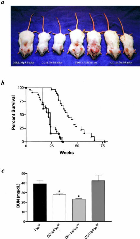
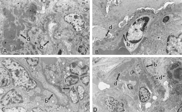
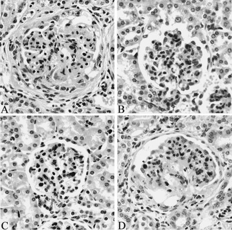
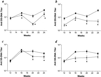
Systemic lupus erythematosus (SLE) is an autoimmune disease characterized by immune complex-mediated tissue injury. Many different adhesion molecules are thought to participate in the development of SLE; however, few studies have directly examined the contributions of these proteins. Here we demonstrate that LFA-1 plays an essential role in the development of lupus in MRL/MpJ-Fas(lpr) mice. Mice deficient in LFA-1, but not Mac-1, showed significantly increased survival, decreased anti-DNA autoantibody formation, and reduced glomerulonephritis. The phenotype of the LFA-1-deficient mice was similar to that observed in beta(2) integrin-deficient (CD18-null) MRL/MpJ-Fas(lpr) mice, suggesting a lack of redundancy among the beta(2) integrin family members and other adhesion molecules. These studies identify LFA-1 as a key contributor in the pathogenesis of autoimmune disease in this model, and further suggest that therapeutic strategies targeting this adhesion molecule may be beneficial for the treatment of SLE.
Figures

Similar articles
-
Intercellular adhesion molecule-1 deficiency protects MRL/MpJ-Fas(lpr) mice from early lethality.J Immunol. 1997 Aug 15;159(4):2058-67. J Immunol. 1997. PMID: 9257874
-
FcgammaRIIB deficiency with Fas mutation is sufficient for the development of systemic autoimmune disease.Eur J Immunol. 2003 Apr;33(4):1020-9. doi: 10.1002/eji.200323794. Eur J Immunol. 2003. PMID: 12672068
-
IFN-gamma receptor signaling is essential for the initiation, acceleration, and destruction of autoimmune kidney disease in MRL-Fas(lpr) mice.J Immunol. 1998 Jul 1;161(1):494-503. J Immunol. 1998. PMID: 9647261
-
Use of genetic knockouts to modulate disease expression in a murine model of lupus, MRL/lpr mice.Immunol Res. 2002;25(2):143-53. doi: 10.1385/ir:25:2:143. Immunol Res. 2002. PMID: 11999168 Review.
-
Alterations in leucocyte trafficking in lupus-prone mice: an examination of the MRL/faslpr mouse.Immunol Cell Biol. 2003 Oct;81(5):390-6. doi: 10.1046/j.1440-1711.2003.01186.x. Immunol Cell Biol. 2003. PMID: 12969327 Review.
Cited by
-
Multiple lupus-associated ITGAM variants alter Mac-1 functions on neutrophils.Arthritis Rheum. 2013 Nov;65(11):2907-16. doi: 10.1002/art.38117. Arthritis Rheum. 2013. PMID: 23918739 Free PMC article.
-
Knockout of the KH-Type Splicing Regulatory Protein Drives Glomerulonephritis in MRL-Faslpr Mice.Cells. 2021 Nov 14;10(11):3167. doi: 10.3390/cells10113167. Cells. 2021. PMID: 34831390 Free PMC article.
-
Neutrophils: game changers in glomerulonephritis?Trends Mol Med. 2010 Aug;16(8):368-78. doi: 10.1016/j.molmed.2010.06.002. Epub 2010 Jul 29. Trends Mol Med. 2010. PMID: 20667782 Free PMC article. Review.
-
Mice expressing the variant rs1143679 allele of ITGAM (CD11b) show impaired DC-mediated T cell proliferation.Mamm Genome. 2019 Oct;30(9-10):245-259. doi: 10.1007/s00335-019-09819-y. Epub 2019 Oct 31. Mamm Genome. 2019. PMID: 31673770 Free PMC article.
-
Differential roles for beta2 integrins in experimental autoimmune bullous pemphigoid.Blood. 2006 Feb 1;107(3):1063-9. doi: 10.1182/blood-2005-08-3123. Epub 2005 Oct 18. Blood. 2006. PMID: 16234355 Free PMC article.
References
-
- Mageed RA, Prud’homme GJ. Immunopathology and the gene therapy of lupus. Gene Ther. 2003;10:861–874. - PubMed
-
- Pollard KM, Hultman P, Kono DH. Using single-gene deletions to identify checkpoints in the progression of systemic autoimmunity. Ann NY Acad Sci. 2003;987:236–239. - PubMed
-
- Cravens PD, Lipsky PE. Dendritic cells, chemokine receptors and autoimmune inflammatory diseases. Immunol Cell Biol. 2002;80:497–505. - PubMed
-
- Sturfelt G. The complement system in systemic lupus erythematosus. Scand J Rheumatol. 2002;31:129–132. - PubMed
Publication types
MeSH terms
Substances
Grants and funding
LinkOut - more resources
Full Text Sources
Other Literature Sources
Medical
Molecular Biology Databases
Research Materials
Miscellaneous
