Comparative Study
. 2004 Aug;165(2):617-30.
doi: 10.1016/s0002-9440(10)63326-3.

Lack of integrin alpha1beta1 leads to severe glomerulosclerosis after glomerular injury

Affiliations
Comparative Study

Lack of integrin alpha1beta1 leads to severe glomerulosclerosis after glomerular injury

Xiwu Chen et al. Am J Pathol. 2004 Aug.

Abstract

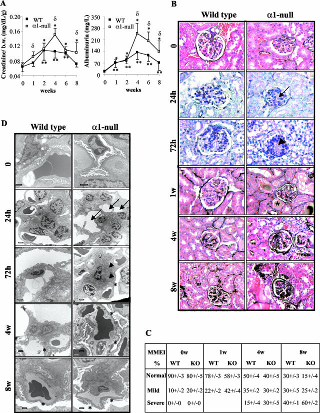
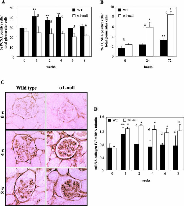
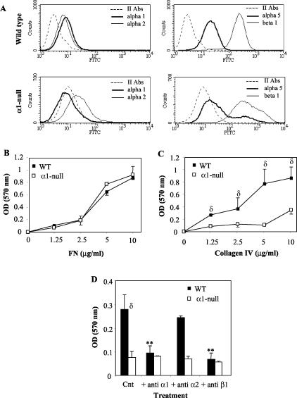
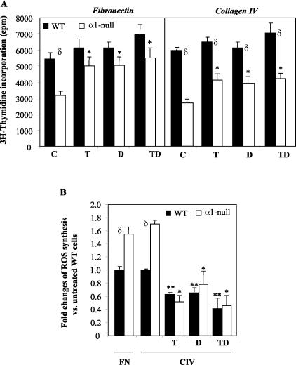
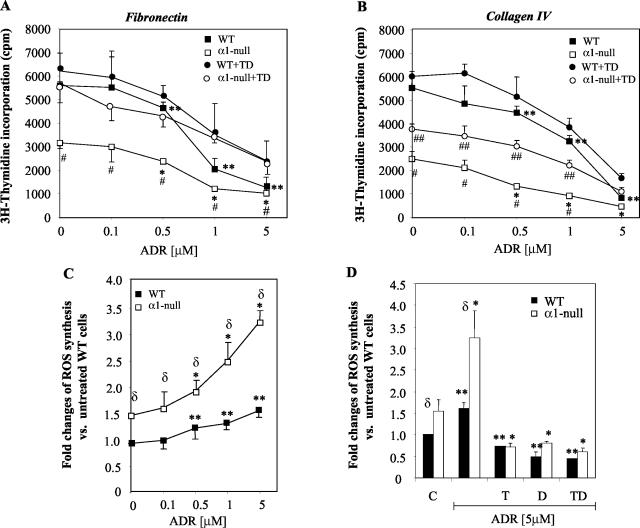
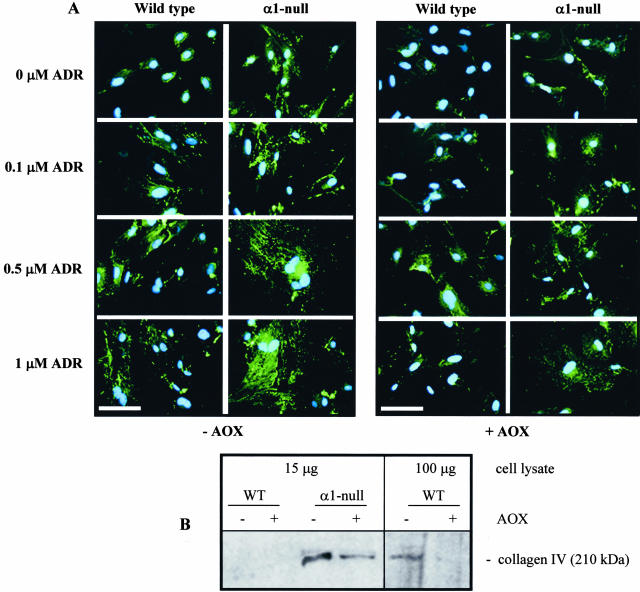
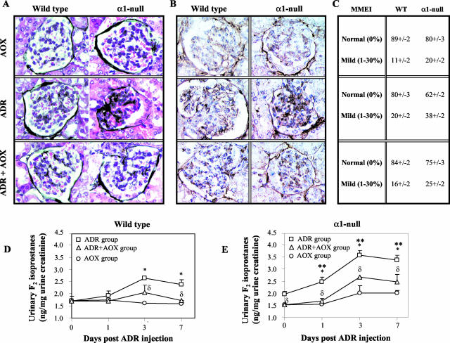
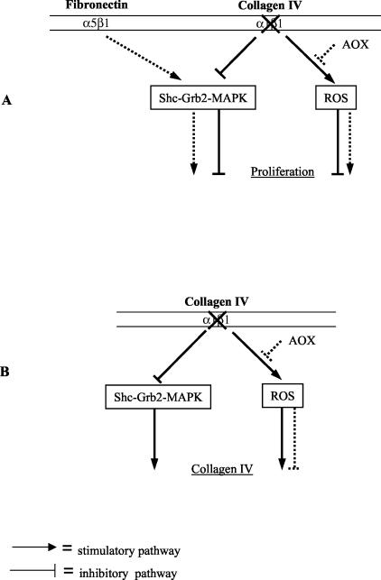
Severity of fibrosis after injury is determined by the nature of the injury and host genetic susceptibility. Metabolism of collagen, the major component of fibrotic lesions, is, in part, regulated by integrins. Using a model of glomerular injury by adriamycin, which induces reactive oxygen species (ROS) production, we demonstrated that integrin alpha1-null mice develop more severe glomerulosclerosis than wild-type mice. Moreover, primary alpha1-null mesangial cells produce more ROS both at baseline and after adriamycin treatment. Increased ROS synthesis leads to decreased cell proliferation and increased glomerular collagen IV accumulation that is reversed by antioxidants both in vivo and in vitro. Thus, we have identified integrin alpha1beta1 as a modulator of glomerulosclerosis. In addition, we showed a novel pathway where integrin alpha1beta1 modulates ROS production, which in turn controls collagen turnover and ultimately fibrosis. Because integrin alpha1beta1 is expressed in many cell types this may represent a generalized mechanism of controlling matrix accumulation, which has implications for numerous diseases characterized by fibrosis.


Figures

Figure 1
Figure 1
Integrin α1-null mice develop severe glomerular injury after ADR injection. A: Creatinine (mg/dL/g) and albuminuria (mg/L) were measured from urine or serum obtained from untreated or ADR-treated mice immediately at sacrifice. Data are presented as the mean and SD of seven wild-type (WT) and α1-null (KO) mice. Differences between untreated and treated α1-null mice (*) or untreated and treated wild-type mice (**) or wild-type and α1-null mice within the same treatment group (δ) were significant with P < 0.05. B: Five-week-old male mice received a single intravenous injection of ADR (10 mg/kg) and kidneys were evaluated at the time points indicated. Signs of early mesangiolysis (arrow), increased matrix deposition (arrowhead), and hyalinosis (*), were evident primarily in the α1-null mice 24 hours, 72 hours, and 1 week after ADR injection (periodic acid-Schiff). At later stages, increased ECM accumulation and sclerosis were more severe in the α1-null mice compared to their treated wild-type counterparts (Jones’ staining). C: One hundred glomeruli/kidney from seven untreated and ADR-treated mice were evaluated and the degree of matrix mesangial expansion (MMEI) scored and expressed as described in Materials and Methods. D: Ultrastructural examination of kidney sections revealed early mesangiolysis (arrow), matrix accumulation (arrowhead), and FPE (*) primarily in the α1-null mice 24 hours and 72 hours after ADR injection. FPE was also visible in wild-type mice 4 weeks after ADR injection, although it was much more severe in α1-null. Scale bar, 50 μm. Original magnifications, × 400 (B).
Figure 2
Figure 2
Excessive collagen IV expression is the cause of the increased mesangial expansion in the α1-null mice after ADR treatment. A and B: Kidney sections from wild-type and integrin α1-null mice were stained with anti-PCNA antibody (A) or with the Dead End colorimetric TUNEL system (B), and the percentage of proliferating or apoptotic cells expressed as (number of PCNA-positive or apoptotic cells in the glomerulus/total number of cells in the glomerulus) × 100. Ten glomeruli/kidney were evaluated. Data are presented as mean and SD of seven kidneys/genotype. C: Collagen IV staining of kidney sections from wild-type and integrin α1-null mice after ADR treatment. Note the increased collagen deposition in the glomeruli of α1-null mice at 4 weeks after ADR treatment. Maximum difference between the two genotypes was observed at 8 weeks (anti-collagen IV). D: α1(IV) collagen mRNA levels in renal cortices of wild-type and integrin α1-null mice were normalized to the β-2 tubulin mRNA levels and expressed as mRNA collagen IV/mRNA tubulin. Data are mean and SD of seven kidneys/genotype. Increased collagen IV levels were observed in kidneys of both wild-type and α1-null mice at 1 week after ADR treatment, but persisted only in the α1-null mice throughout the study period. *, **, and δ are as in Figure 1A. Original magnifications, ×400 (C).
Figure 3
Figure 3
Mesangial cell adhesion on collagen IV is integrin α1β1-dependent. A: Integrin expression by wild-type and integrin α1-null mesangial cells. Cells were incubated with antibodies to α1, α2, α5, or β1 integrin subunits. Integrin expression is displayed by a shift in mean fluorescent intensity compared with no primary antibody incubation using fluorescence-activated cell sorting analysis. B and C: Primary mesangial cells (5 × 10⁴) were plated onto 96-well plates coated with fibronectin (B) or collagen IV (C) at the concentrations indicated for 1 hour in serum-free medium. Adherent cells were then stained with crystal violet, lysed, and the OD measured. Data represent the mean and SD of quadruplicate samples of 10 pooled wild-type and integrin α1-null mice. D: Mesangial cells were plated in the presence or absence of blocking antibodies (10 μg/ml) to the integrin subunits indicated on collagen IV (5 μg/ml) for 1 hour in serum-free medium. Adhesion was then determined and expressed as described above. Note that adhesion of mesangial cells to CIV is primarily integrin α1β1-mediated. Data are expressed as in B.
Figure 4
Figure 4
α1-Null mesangial cells show reduced proliferation on both collagenous and noncollagenous substrata. A: Primary mesangial cells (5 × 10³) were plated on 10 μg/ml of fibronectin or 10 μg/ml of collagen IV in the absence or presence of 10 μmol/L TEMPOL (T), 1 μmol/L DPI (D), or a mixture of TEMPOL and DPI (TD). Incorporation of 3H-thymidine was then evaluated as described in Materials and Methods. Data represent the means and SD of quadruplicate samples derived from 10 pooled wild-type and integrin αl-null mice. B: Mesangial cells were cultured on 10 μg/ml of fibronectin (FN)- or collagen IV (CIV)-coated dishes in the absence or presence of 10 μmol/L TEMPOL (T), 1 μmol/L DPI (D), or a mixture of TEMPOL and DPI (TD) together with 2 μmol/L dihydro-rhodamine. Six hours later, ROS generation was determined by FACS as described in Materials and Methods. Data represent the mean and SD of three experiments performed in duplicates. *, **, and δ are as in Figure 1A.
Figure 5
Figure 5
ADR reduces mesangial cell proliferation that can be rescued by antioxidants. A and B: Mesangial cells were plated on 10 μg/ml fibronectin (A)- or 10 μg/ml collagen IV (B)-coated dishes in the presence of ADR at the concentration indicated in the absence of presence of mixture of 10 μmol/L TEMPOL and 1 μmol/L DPI (TD). 3H-thymidine incorporation was then evaluated as described in Materials and Methods. Data represent means and SD of quadruplicate samples of mesangial cells derived from 10 pooled wild-type and integrin αl-null mice. C and D: Wild-type and α1-null mesangial cells were plated on 10 μg/ml collagen IV in the presence of ADR at concentrations indicated (C) or with 5 μmol/L ADR and 10 μmol/L TEMPOL (T), 1 μmol/L DPI (D), or a mixture of TEMPOL and DPI (TD) (D). ROS generation was determined after incubation with 2 μmol/L dihydro-rhodamine for 6 hours as described in Materials and Methods. Data represent the mean and SD of three experiments performed in duplicates. *, **, and δ are as in Figure 1A. Differences between ADR-treated α1-null and ADR + antioxidant-treated α1-null cells (#) or wild-type and α1-null cells within the same ADR + antioxidants group (##) were significant with P < 0.05.
Figure 6
Figure 6
Collagen deposition in wild-type and integrin α1-null mesangial cells. A: Mesangial cells were plated on uncoated chamber slides and treated for 48 hours with ADR at μmol/L concentrations indicated in the absence (left) or presence of 10 μmol/L TEMPOL and 1 μmol/L DPI (right). Indirect immunofluorescence was then performed to evaluate collagen IV synthesis and deposition. α1-Null mesangial cells synthesize more collagen IV than their wild-type counterparts and this synthesis is enhanced after ADR treatment. Although treatment with antioxidants (AOX) (right) reduced collagen deposition in both untreated and ADR-treated cells, α1-null mesangial cells still produced more collagen IV than their wild-type counterparts. B: Cell lysates were prepared from untreated or antioxidant-treated mesangial cells cultured as described in A and Western blot analysis was performed using anti-mouse collagen IV-specific antibodies. Note that α1-null mesangial cells produce more collagen IV than their wild-type counterparts, and treatment with antioxidants only partially rescues collagen synthesis. In contrast, antioxidant treatment completely inhibited collagen synthesis in wild-type cells, paralleling the results obtained with immunofluorescence (A). Note that collagen IV is detectable in wild-type cells only when 100 μg of total cell lysate were used for analysis. Scale bar, 20 μm (A).
Figure 7
Figure 7
In vivo treatment with antioxidants ameliorates ADR-induced renal injury. Five-week-old male mice (n = four/genotype) received ADR, antioxidants (AOXs), or a combination of ADR and AOX as described in Materials and Methods, and kidneys were analyzed 1 week after ADR injection. The increased ECM deposition, determined by both Jones’ (A) and collagen IV (B) staining, evident in ADR-treated α1-null mice, was significantly ameliorated by administration of AOXs. C: MMEI in glomeruli from ADR-, AOX-, and ADR + AOX-treated mice was evaluated as described in Materials and Methods. D and E: F2-isoprostanes were measured in the urine as described in Materials and Methods and expressed as ng/mg urine creatinine. Values represent the mean and SD calculated for four samples/treatment. F2-isoprostanes levels in the urine of untreated α1-null mice were significantly increased compared to their untreated wild-type counterparts (P < 0.05). Pretreatment with AOXs significantly reduced the excretion of this biomarker for oxidative stress in the α1-null mice (P < 0.05). ADR injection led to increased excretion of F2-isoprostanes in both wild-type and integrin α1-null mice (*, P < 0.05 relative to untreated mice), although it was significantly higher in the latter group, starting 1 day after ADR injection (**, P < 0.05 between wild-type and α1-null mice within the same group). AOX treatment in ADR-treated mice significantly reduced F2-isoprostanes excretion in both genotypes (δ, P < 0.05 between ADR-treated and ADR + AOX-treated mice). Original magnifications, ×400 (A, B).
Figure 8
Figure 8
Regulation of cell proliferation and collagen synthesis in α1-null mesangial cells. Schematic illustration of how cell proliferation (A) or collagen synthesis (B) might be regulated in α1-null mesangial cells. Integrin α1β1 is the only collagen-binding receptor able to support cell proliferation and down-regulate endogenous collagen synthesis on collagens via activation of the Shc/Grb2/MAPK pathway. In the absence of integrin α1β1, cells plated on collagen substrata develop up-regulated synthesis of ROS and decreased activation of the Shc/Grb2/MAPK pathway that could lead to reduced cell proliferation (A) and increased collagen synthesis (B). When cells are plated on collagen, addition of antioxidants (AOXs) could only partially rescue α1-null mesangial cell growth (A) or partially decrease collagen synthesis (B) because of the persistent lack of MAPK activation. In contrast, treatment with antioxidants could completely rescue proliferation of α1-null mesangial cells on fibronectin (A) as on this matrix, other integrins (ie, α5β1) could promote cell proliferation via activation of the Shc/Grb2/MAPK pathways.

Similar articles

Cited by

References

    1. Fogo AB. Mesangial matrix modulation and glomerulosclerosis. Exp Nephrol. 1999;7:147–159. - PubMed
    1. Hynes R. Integrins: bidirectional, allosteric signaling machines. Cell. 2002;110:673–687. - PubMed
    1. Voigt S, Gossrau R, Baum O, Loster K, Hofmann W, Reutter W. Distribution and quantification of alpha 1-integrin subunit in rat organs. Histochem J. 1995;27:123–132. - PubMed
    1. Korhonen M, Ylanne J, Laitinen L, Virtanen I. Distribution of beta 1 and beta 3 integrins in human fetal and adult kidney. Lab Invest. 1990;62:616–625. - PubMed
    1. Shikata K, Makino H, Morioka S, Kashitani T, Hirata K, Ota Z, Wada J, Kanwar YS. Distribution of extracellular matrix receptors in various forms of glomerulonephritis. Am J Kidney Dis. 1995;25:680–688. - PubMed

Publication types

MeSH terms

LinkOut - more resources