. 2004 Aug 2;166(3):347-57.
doi: 10.1083/jcb.200312007. Epub 2004 Jul 26.

Muscle satellite cells adopt divergent fates: a mechanism for self-renewal?

Affiliations

Muscle satellite cells adopt divergent fates: a mechanism for self-renewal?

Peter S Zammit et al. J Cell Biol. .

Abstract

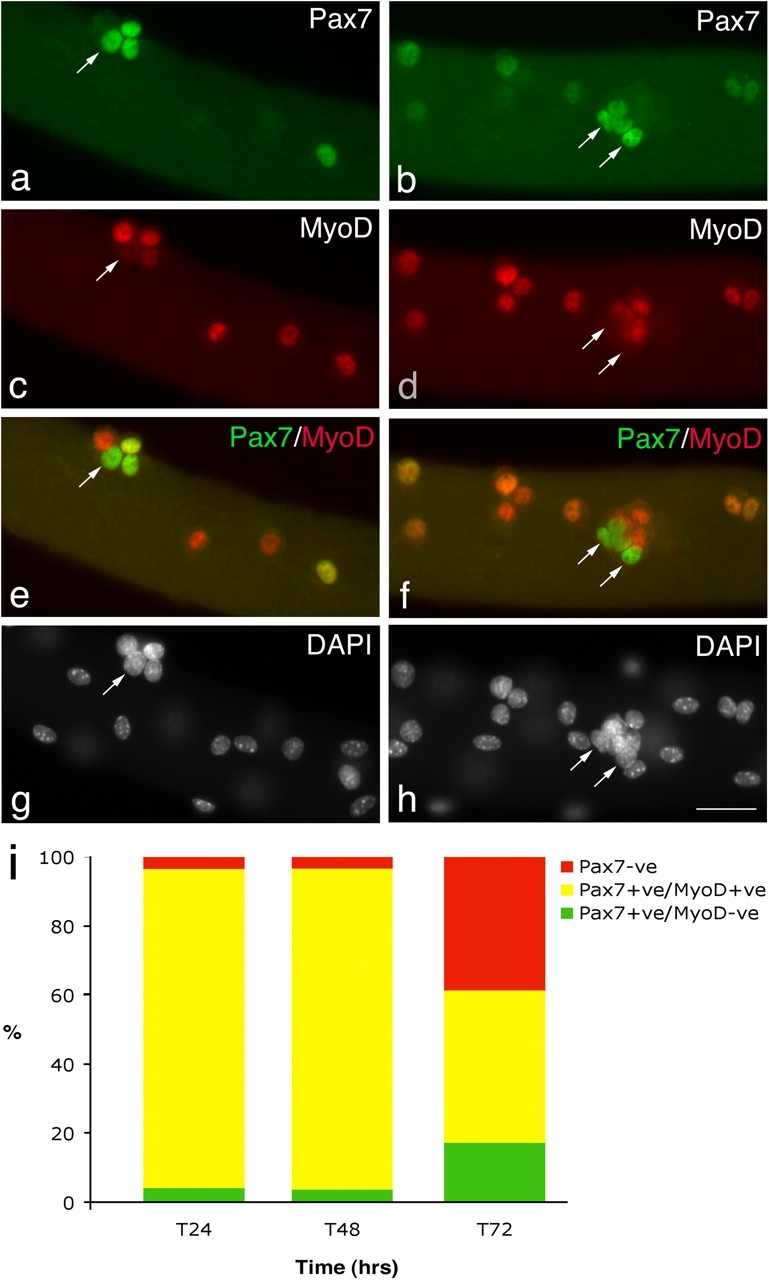
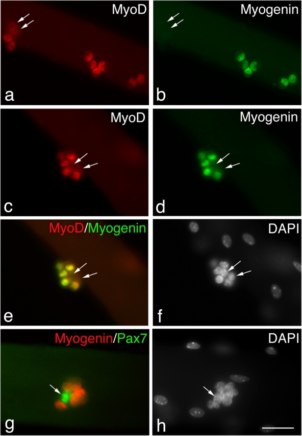
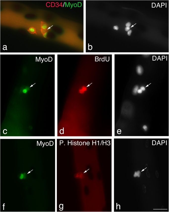
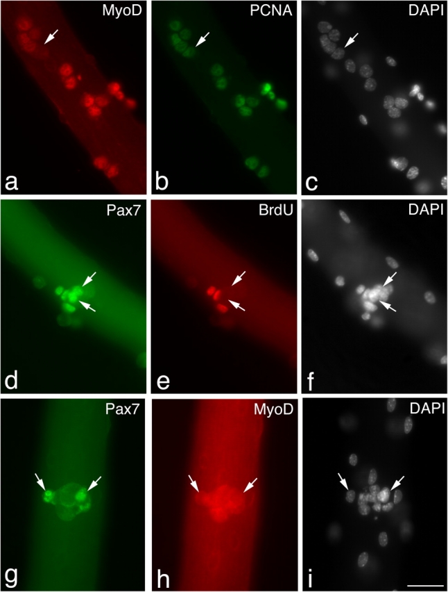
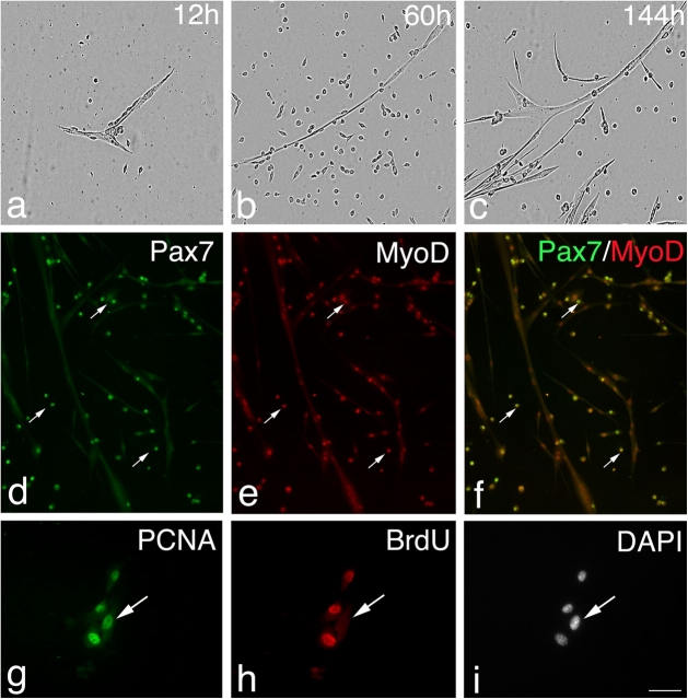
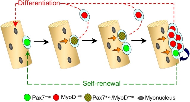
Growth, repair, and regeneration of adult skeletal muscle depends on the persistence of satellite cells: muscle stem cells resident beneath the basal lamina that surrounds each myofiber. However, how the satellite cell compartment is maintained is unclear. Here, we use cultured myofibers to model muscle regeneration and show that satellite cells adopt divergent fates. Quiescent satellite cells are synchronously activated to coexpress the transcription factors Pax7 and MyoD. Most then proliferate, down-regulate Pax7, and differentiate. In contrast, other proliferating cells maintain Pax7 but lose MyoD and withdraw from immediate differentiation. These cells are typically located in clusters, together with Pax7-ve progeny destined for differentiation. Some of the Pax7+ve/MyoD-ve cells then leave the cell cycle, thus regaining the quiescent satellite cell phenotype. Significantly, noncycling cells contained within a cluster can be stimulated to proliferate again. These observations suggest that satellite cells either differentiate or switch from terminal myogenesis to maintain the satellite cell pool.


Figures

Figure 1.
Figure 1.
Satellite cells adopt divergent fates. After 72 h in culture, coimmunostaining of EDL myofibers demonstrates that in the majority of satellite cells, Pax7 (a and b) is becoming down-regulated whereas MyoD (c and d) remains strongly expressed. However, combining the Pax7 (green) and MyoD (red) fluorescent immunosignals (e and f) clearly shows that some (∼23%) MyoD−ve daughters (arrows) maintain robust Pax7 expression. Pax7+ve/MyoD−ve satellite cell (arrows) are typically located (85%) in clusters (defined as four or more overlying/touching cells) together with both Pax7+ve/MyoD+ve and Pax7−ve/MyoD+ve cells. Counterstaining with DAPI was used to identify all nuclei present on the myofiber (g and h). Bar, 30 μm. The number of satellite cells from 3F-nlacZ-E mouse 3 and 4 (Table II) that were either Pax7+ve/MyoD−ve, Pax7+ve/MyoD+ve, or Pax7−ve at T24, T48, and T72 were counted. Expressing this data as the mean percentage in each category of the total number of satellite cells per myofiber (i) illustrates that the Pax7+ve/MyoD+ve cells present at T24 and T48 give rise to the majority of Pax7+ve/MyoD−ve and Pax7−ve populations at T72.
Figure 2.
Figure 2.
Not all satellite cells are committed to differentiation. Coimmunostaining of EDL myofibers that had been in culture for 72 h showed that most (∼73%) MyoD+ve satellite cells (a and c) were committed to differentiation, as shown by the presence of myogenin (b and d). The yellow color resulting from combining the MyoD (c) and myogenin (d) fluorescent immunosignals (e) clearly shows that most MyoD+ve daughters within this cluster also contain myogenin. However, it is significant that the MyoD−ve daughters in this cluster were also myogenin−ve (arrows), and at least some of these are presumably Pax7+ve (Fig. 1). Coimmunostaining for Pax7 (green) and myogenin (red) showed that these two antigens were essentially mutually exclusive (g) with only 6.8 ± 1.6% of Pax7+ve cells also expressing myogenin. Counterstaining with DAPI was used to identify all nuclei present (f and h). Bar, 30 μm.
Figure 3.
Figure 3.
Satellite cells can maintain Pax7, but lose MyoD. Quiescent satellite cells are generally distributed as single cells along the length of the myofiber. Between 24–48 h in culture, almost all satellite cells express both Pax7 and MyoD and begin to divide; therefore, it can be assumed that most pairs/small groups of cells present at these times are derived from a single parent satellite cell. Immunostaining followed by X-gal incubation of EDL myofibers from a 3F-nlacZ-E mouse after 48 h in culture shows that of four CD34+ve satellite cells (a, red-cell surface), three contained MyoD protein (a, green nuclear), but significantly one did not (a and b, arrows). These MyoD−ve cells (c–e, arrows) had undergone division, as shown by the incorporation of BrdU into the daughter cells (d), and were not merely quiescent satellite cells. Whether MyoD protein levels vary with cell cycle is unresolved (Kitzmann et al., 1998; Lindon et al., 1998). However, it is important that both the MyoD+ve and MyoD−ve progeny of a single satellite cell could be in the same phase of the cell cycle (f–h). An example is shown where immunostaining followed by X-gal incubation of an EDL myofiber from a 3F-nlacZ-E mouse after 48 h in culture shows that MyoD+ve (f) and MyoD−ve (f–h, arrows) daughters are both in the same phase of the cell cycle as shown by phosphorylated Histone H1/H3 immunostaining (g). Counterstaining with DAPI was used to identify all nuclei present (b, e, and h). Bar, 30 μm.
Figure 4.
Figure 4.
Pax7+ve/MyoD−ve satellite cells eventually cycle slowly, or not at all. Immunostaining of EDL myofibers after 72 h in culture showed that the majority of MyoD+ve cells (a) were also expressing PCNA (b), indicating that they were still cycling. Rare MyoD−ve cells (a–c, arrows) were also PCNA+ve (b, arrow) and would presumably be either Pax7+ve or myogenin+ve. However, a pulse of BrdU between 96–120 h in culture (d–f) followed by immunostaining showed that by 120 h, 42.5% of Pax7+ve cells (d–f, arrows) had failed to incorporate BrdU (e, arrows) over the previous 24 h. At 120 h, the Pax7+ve cells (g–i, arrow) were negative for MyoD (h, arrow), with MyoD being undetectable in almost all satellite cells at this time. Counterstaining with DAPI was used to identify all nuclei present (c, f, and i). Bar, 30 μm.
Figure 5.
Figure 5.
Isolated clusters of satellite cell progeny contain cells able to reactivate, proliferate, and differentiate. EDL myofibers were maintained in suspension for 120 h, at which time clusters were removed with a pipette, plated onto Matrigel, and photographed over the next 144 h (a–c). Clusters quickly adhered to the Matrigel substrate, and both single cells and small myotubes rapidly appeared (a). Cells proliferated over the subsequent days (b) until a second wave of myotube formation began around 96 h (c). In separate experiments, myofibers were maintained in suspension for 120 h, at which time MyoD protein was virtually absent. Clusters were then removed, plated, and fixed after a further 120 h and immunostained (d–f). The proliferating cells (d–f, arrows) expressed both Pax7 (d, green) and MyoD (e, red), seen as yellow when the red and green immunosignals are combined (f) interspersed with myocytes and myotubes. To determine if satellite cells that had stopped cycling while associated with a myofiber were able to still undergo division, a BrdU pulse was administered between 96–120 h. Clusters were then plated and 24 h later coimmunostained for PCNA and BrdU (g–i, arrows). Proliferating cells were identified by PCNA (g) expression that were BrdU−ve (h), indicating that they had been refractory to BrdU incorporation while associated with the myofiber, but were able to reenter cell cycle when dissociated/stimulated. Counterstaining with DAPI was used to identify all nuclei present (i). Bars: (d–f) 100 μm; (g–i) 30 μm.
Figure 6.
Figure 6.
Model of satellite cell self-renewal. Quiescent satellite cells (green) activate to coexpress Pax7 and MyoD (green and red tartan), and then most proliferate, down-regulate Pax7, maintain MyoD (red), and differentiate (red pathway). However, activated Pax7+ve/MyoD+ve (green and red tartan) satellite cells can also divide to give rise to cells that adopt a different fate. These give rise to clusters of cells containing both Pax7−ve/MyoD+ve (red) progeny, whereas others down-regulate MyoD expression and cycle while maintaining only Pax7 (green). These clusters may grow by the further generation of cells with divergent fates. Pax7+ve/MyoD−ve cells (green) become quiescent, thus renewing the satellite cell pool (green pathway), whereas the MyoD+ve cells (red) differentiate to produce myonuclei (red pathway). Signaling from the myofiber (orange arrows) and/or between cells within the clusters (blue arrow) may dictate which fate the satellite cell adopts.

References

    1. Andres, V., and K. Walsh. 1996. Myogenin expression, cell cycle withdrawal, and phenotypic differentiation are temporally separable events that precede cell fusion upon myogenesis. J. Cell Biol. 132:657–666. - PMC - PubMed
    1. Artavanis-Tsakonas, S., M.D. Rand, and R.J. Lake. 1999. Notch signaling: cell fate control and signal integration in development. Science. 284:770–776. - PubMed
    1. Asakura, A., P. Seale, A. Girgis-Gabardo, and M.A. Rudnicki. 2002. Myogenic specification of side population cells in skeletal muscle. J. Cell Biol. 159:123–134. - PMC - PubMed
    1. Baroffio, A., M. Hamann, L. Bernheim, M.L. Bochaton-Piallat, G. Gabbiani, and C.R. Bader. 1996. Identification of self-renewing myoblasts in the progeny of single human muscle satellite cells. Differentiation. 60:47–57. - PubMed
    1. Beauchamp, J.R., L. Heslop, D.S. Yu, S. Tajbakhsh, R.G. Kelly, A. Wernig, M.E. Buckingham, T.A. Partridge, and P.S. Zammit. 2000. Expression of CD34 and Myf5 defines the majority of quiescent adult skeletal muscle satellite cells. J. Cell Biol. 151:1221–1234. - PMC - PubMed

Publication types