. 2004 Aug 10;101(32):11668-72.
doi: 10.1073/pnas.0403499101. Epub 2004 Jul 26.

Rpe65 Leu450Met variant is associated with reduced levels of the retinal pigment epithelium lipofuscin fluorophores A2E and iso-A2E

Affiliations

Rpe65 Leu450Met variant is associated with reduced levels of the retinal pigment epithelium lipofuscin fluorophores A2E and iso-A2E

So Ra Kim et al. Proc Natl Acad Sci U S A. .

Abstract

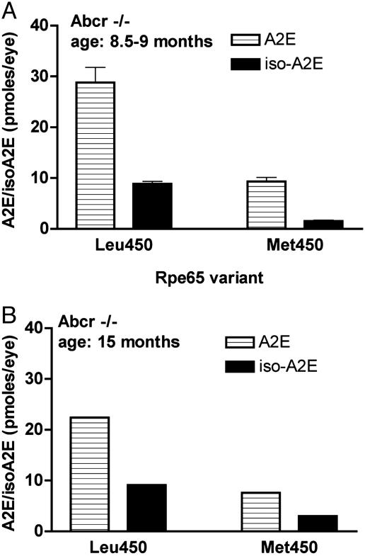
There is a growing body of evidence that the nondegradable fluorophores that accumulate as the lipofuscin of retinal pigment epithelium (RPE) are involved in mechanisms leading to the degeneration of RPE in macular degeneration. Most of the constituents of RPE lipofuscin are inadvertent products of the retinoid visual cycle, the enzymatic pathway by which the 11-cis-retinal chromophore of rhodopsin is generated. Indeed, a major constituent of RPE lipofuscin, the pyridinium bisretinoid A2E, is a diretinal conjugate that forms in photoreceptor cells and is deposited in RPE cells as a consequence of the phagocytosis of the outer segment membrane by RPE cells. Given the adverse effects of A2E, there is considerable interest in combating its deposition so as to protect against vision loss. These efforts, however, necessitate an understanding of factors that modulate its formation. Here we show that an amino acid variant in murine Rpe65, a visual-cycle protein required for the regeneration of 11-cis-retinal, is associated with reduced A2E accumulation.


Figures

Fig. 1.
Fig. 1.
Lipofuscin fluorophores as by-products of the visual cycle. (A) The retinoid cycle and formation of the lipofuscin fluorophores A2E, iso-A2E, and all-trans-retinal (ATR) dimer. Upon the photoisomerization of 11-cis-retinal, all-trans-retinal is released from rhodopsin and reduced to all-trans-retinol by all-trans-retinol dehydrogenase (atrDH). Lecithin retinol acyltransferase (LRAT) generates all-trans-retinyl esters from all-trans-retinol, and RPE65 presents retinyl esters to isomerohydrolase (IMH) for processing to 11-cis-retinol. 11-cis-retinol is then oxidized by 11-cis-retinol dehydrogenase (11-cRDH) to regenerate 11-cis-retinal. All-trans-retinal that evades reduction reacts with phosphatidylethanolamine (PE) (2:1) to generate the precursor A2-PE, from which A2E and iso-A2E form, or ATR dimer. (B) Structures of A2E and iso-A2E. These pigments interconvert under the influence of light.
Fig. 2.
Fig. 2.
Analysis of hydrophobic extracts of eye cups excised from Abcr+/+ mice (age 7 months) with the Leu-450 or Met-450 variant of Rpe65. (A and B) Typical chromatograms obtained by reverse-phase HPLC with monitoring at 430 nm illustrate the detection of A2E and iso-A2E. (C) Quantitation of A2E and iso-A2E in eye cups of Abcr+/+ mice with either the Leu-450 or Met-450 polymorphism. Levels were determined as integrated peak areas normalized to an external standard of A2E. Values are expressed as picomoles per eye and for each group are based on measures obtained after pooling eight eyes into a single sample.
Fig. 3.
Fig. 3.
Quantitation of A2E and iso-A2E in eye cups of Abcr–/– mice expressing either the Leu-450 or Met-450 variant of Rpe65. Levels were determined as integrated peak areas normalized to an external standard of A2E. (A) A2E and iso-A2E in eyes of Abcr–/– mice (age 8.5–9 months). Values (mean ± SD) are expressed as picomoles per eye and are based on two to five samples and two eye cups per sample. (B) Eye cups obtained from Abcr–/– mice (age 15 months). Values are based on single samples with two eyes per sample.
Fig. 4.
Fig. 4.
Quantitation of A2E and iso-A2E in eye cups of BALB/cByJ and C57BL/6J-c2J mice obtained as retired breeders. Levels were determined as integrated peak areas normalized to an external standard of A2E. Values (mean ± SEM) are expressed as picomoles per eye; four experiments with 8–23 eyes per group in each experiment. *, P < 0.01, unpaired Student's t test.

Similar articles

Cited by

References

    1. Rando, R. R. (2001) Chem. Rev. (Washington, D.C.) 101, 1881–1896. - PubMed
    1. Hamel, C. P., Tsilou, E., Pfeffer, B. A., Hooks, J. J., Detrick, B. & Redmond, T. M. (1993) J. Biol. Chem. 268, 15751–15757. - PubMed
    1. Gollapalli, D. R., Maiti, P. & Rando, R. R. (2003) Biochemistry 42, 11824–11830. - PubMed
    1. Jahng, W. J., David, C., Nesnas, N., Nakanishi, K. & Rando, R. R. (2003) Biochemistry 42, 6159–6168. - PMC - PubMed
    1. Mata, N. L., Weng, J. & Travis, G. H. (2000) Proc. Natl. Acad. Sci. USA 97, 7154–7159. - PMC - PubMed

Publication types

LinkOut - more resources