. 2004 Aug 3;101(31):11269-74.
doi: 10.1073/pnas.0400541101. Epub 2004 Jul 26.

Reinitiation involving upstream ORFs regulates ATF4 mRNA translation in mammalian cells

Affiliations

Reinitiation involving upstream ORFs regulates ATF4 mRNA translation in mammalian cells

Krishna M Vattem et al. Proc Natl Acad Sci U S A. .

Abstract

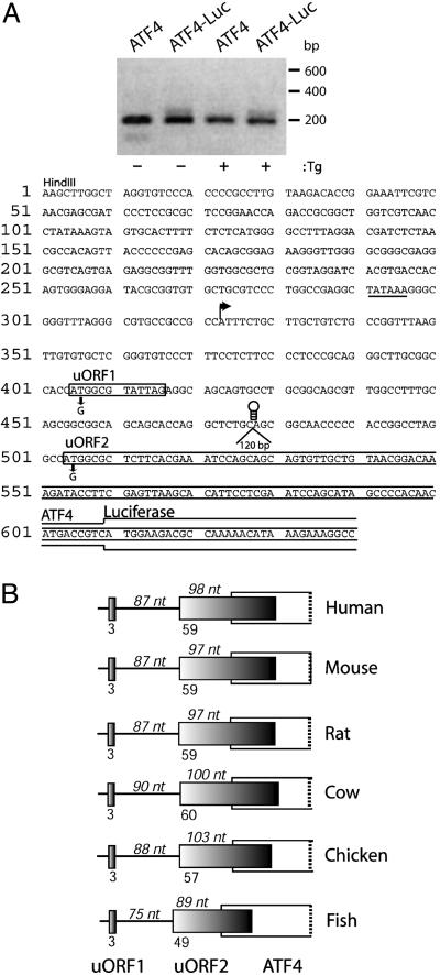
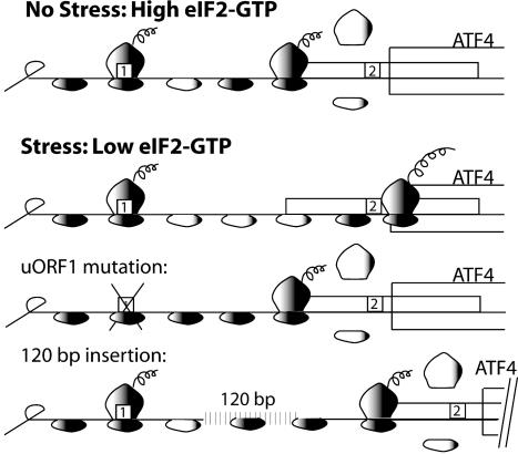
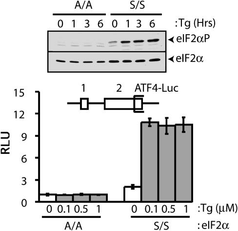
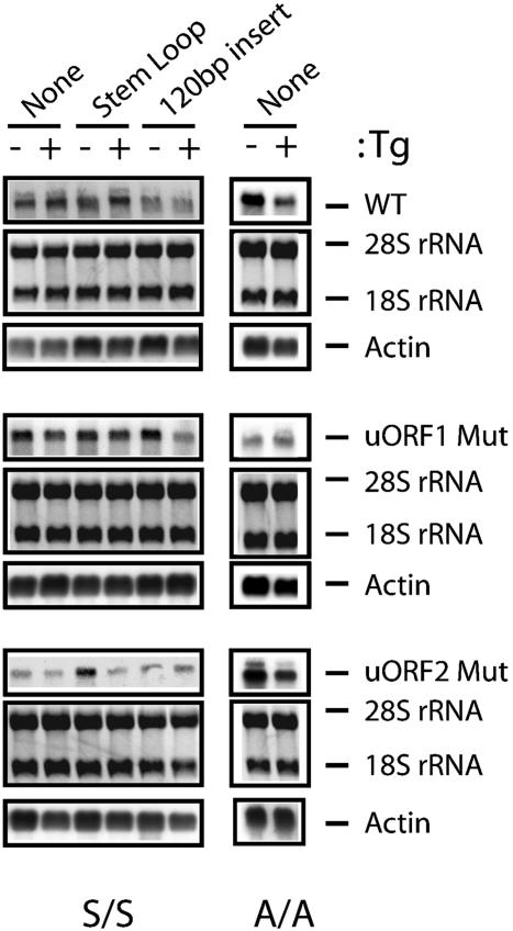
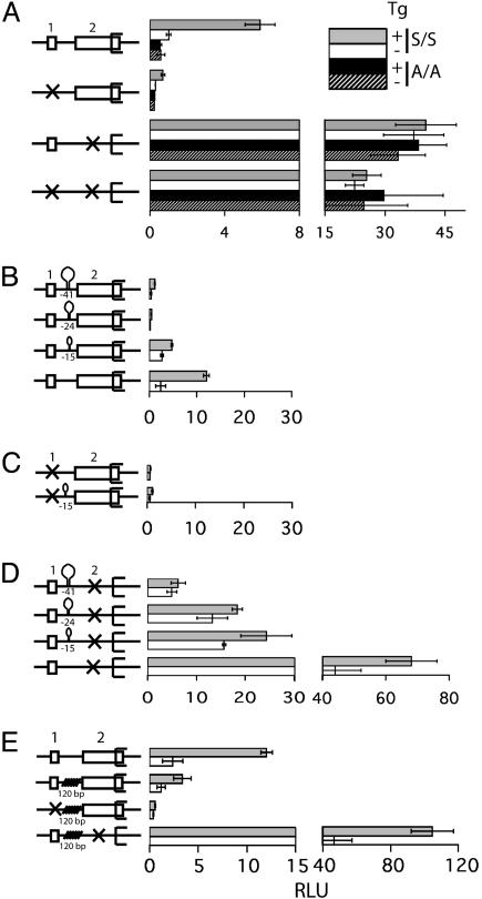
During cellular stresses, phosphorylation of eukaryotic initiation factor-2 (eIF2) elicits gene expression designed to ameliorate the underlying cellular disturbance. Central to this stress response is the transcriptional regulator activating transcription factor, ATF4. Here we describe the mechanism regulating ATF4 expression involving the differential contribution of two upstream ORFs (uORFs) in the 5' leader of the mouse ATF4 mRNA. The 5' proximal uORF1 is a positive-acting element that facilitates ribosome scanning and reinitiation at downstream coding regions in the ATF4 mRNA. When eIF2-GTP is abundant in nonstressed cells, ribosomes scanning downstream of uORF1 reinitiate at the next coding region, uORF2, an inhibitory element that blocks ATF4 expression. During stress conditions, phosphorylation of eIF2 and the accompanying reduction in the levels of eIF2-GTP increase the time required for the scanning ribosomes to become competent to reinitiate translation. This delayed reinitiation allows for ribosomes to scan through the inhibitory uORF2 and instead reinitiate at the ATF4-coding region. Increased expression of ATF4 would contribute to the expression of genes involved in remediation of cellular stress damage. These results suggest that the mechanism of translation reinitiation involving uORFs is conserved from yeast to mammals.


Figures

Fig. 1.
Fig. 1.
The two uORFs present in the noncoding portion of the ATF4 mRNA are conserved among vertebrates. (A Upper) DNA was derived by 5′ RACE by using RNA prepared from S/S MEF cells treated with Tg or no stress. DNA samples were separated by electrophoresis in a 2% agarose gel. ATF4 indicates 5′ RACE products prepared by using endogenous ATF4 mRNA, and ATF4-Luc indicates products derived from ATF4-Luc transcripts. Size markers in base pairs are indicated to the right. (A Lower) Sequence of the leader of the ATF4 mRNA. A HindIII restriction site was engineered into the ATF4 DNA. The major transcription start site of the ATF4 gene was determined by sequencing of 5′ RACE products and is indicated by an arrow. ATF4 sequences upstream of this transcription initiation site may contribute to ATF4 transcription, as illustrated by a potential TATAAA box (underlined). Boxes represent uORFs 1 and 2 sequences located upstream of the ATF4-coding region. uORF1 encodes a 3-amino acid-residue polypeptide and is 87 nt upstream of uORF2. uORF2, 180 nt in length, overlaps 83 nt of the ATF4-coding region, which matches the overlap between uORF2 and the ATF4-Luc reporter. Mutations in the uORF1 and uORF2 initiation codons rendering each nonfunctional for ATF4 translation control are indicated below the sequences. Three different stem–loop structures and a 120-bp insertion were individually introduced at PstI restriction site located between uORFs 1 and 2. It is noted that an additional small ORF is present 5′ of the major transcription start site. ATF4-luciferase activity of a reporter construct containing a mutation in the ATG of this uORF was induced in response to stress similar to that measured for WT ATF4-Luc (data not shown), supporting the idea that this region is not present in the ATF4 mRNA. (B) Representative cDNAs encoding ATF4-related sequences in GenBank (accession no.), including human (BC008090), mouse (AK003001), rat (CK601272), cow (CK960046), chicken (AB013138), and zebrafish (CA470055), reveal mRNAs with a similar two-uORF configuration as described for mouse ATF4. Each panel is drawn to scale. Dark-colored boxes represent the two uORFs. The open white-colored box overlapping the uORF2 is the ATF4-coding region. The number of nucleotides between uORF1 and 2 and between start of the uORF2 and the start of the overlapping ATF4-coding region are indicated on top of each panel. The numbers mentioned below each panel represent the number of amino acids encoded by each of the uORFs.
Fig. 5.
Fig. 5.
Model for ATF4 translational control by its leader sequences. The ATF4 mRNA is illustrated as a straight line that has uORFs 1 and 2 that are presented as boxes. The shading of the small ribosomal subunit indicates its association with eIF2-GTP bound formula image. After translation of the positive-acting uORF1, ribosomes retain the capacity to reinitiate translation at a downstream ORF. The basis for this reinitiation capacity is currently not clear. In the related GCN4 translation mechanism, the termination context of the analogous uORF1 is thought to facilitate the retention of the small ribosomal subunit with the GCN4 mRNA (1, 12, 28). After translation of the ATF4 uORF1, the 40S ribosomal subunits are proposed to resume scanning in a 5′ to 3′ direction along the ATF4 transcript. When eIF2-GTP bound formula image is plentiful during nonstressed conditions, the small ribosomal subunits quickly acquire the eIF2 ternary complex and, coupled with the 60S ribosome, reinitiate translation at uORF2. After translation of this inhibitory uORF2, ribosomes dissociate from the ATF4 mRNA, thereby reducing expression of the ATF4-coding region. When cells are subjected to ER stress or to nutrient deprivation, the levels of eIF2 phosphorylation are enhanced leading to reduced eIF2-GTP levels. After translation of uORF1, there is an increased time required for reacquisition of eIF2-GTP coupled formula image that allows a portion of the scanning 40S ribosomal subunits to scan through the negative-acting uORF2. While scanning the mRNA-leader region from beginning of uORF2 to the initiation codon of the ATF4-coding region, ribosomes reacquire the eIF2 ternary complex, facilitating translational expression of ATF4. When uORF1 is mutated, ribosomes scanning from the 5′ end of the ATF4 mRNA will initiate translation at uORF2. After translation of the inhibitory uORF2, ribosomes dissociate from the ATF4 mRNA, thus lowering translation of the ATF4-coding region even when eIF2-GTP levels are reduced in response to cellular stress. When the distance between uORF1 and uORF2 is increased compared to WT, most ribosomes are competent for reinitiation before reaching uORF2, thereby reducing ATF4 translation independent of eIF2-GTP availability.
Fig. 2.
Fig. 2.
Phosphorylation of eIF2 is required for ATF4 translational expression. (Upper)WT(S/S) or mutant MEF cells containing eIF2α with Ala substituted for Ser-51 (A/A) were treated with 1.0 μM Tg for the indicated number of hours, or no stress (0), and phosphorylation of eIF2α was measured by immunoblot by using Ab that specifically recognizes eIF2α phosphorylated at Ser-51. Total eIF2α was similarly analyzed by using Ab that recognizes both phosphorylated and nonphosphorylated forms of eIF2α. (Lower) The uORFs in the ATF4-Luc reporter construct are indicated as boxes labeled 1 or 2 (not to scale). The S/S and A/A MEF cells were cotransfected with the ATF4-Luc plasmid and a Renilla luciferase plasmid that served as an internal control for normalization. The transfected cells were subsequently treated with the indicated concentrations of Tg for 6 h or no ER stress agent (0 μM). Relative light units (RLU) is a ratio of firefly luciferse units normalized for Renilla luciferase units, and each value was derived from three independent transfections. White-colored bars represent values obtained from nonstressed MEF cells, and gray-colored bars represent values from cells subjected to ER stress.
Fig. 3.
Fig. 3.
Levels of ATF4-Luc mRNAs are similar between nonstressed and ER stressed MEF cells. Total RNA was prepared from S/S and A/A MEF cells expressing WT ATF4-Luc or mutant versions defective in uORF1 or uORF2 as indicated. The same amount of each total RNA was separated by gel electrophoresis, and 18S and 28S rRNA was visualized by staining with ethidium bromide (Middle). RNA was then transferred to membrane filters, and ATF-Luc transcripts were measured by using a radiolabeled probe complementary to the luciferase reporter gene and autoradiography (Top). Northern blot analyses were also carried out by using a radiolabeled actin probe to ensure characterization of similar amounts of RNA (Bottom). Either a stem–loop with a ΔG = –41 kcal/mol or a 120-bp insert was included in the leader of the ATF4-Luc reporter construct as indicated.
Fig. 4.
Fig. 4.
uORF1 functions as a positive regulator, and uORF2 is inhibitory in a scanning mechanism that regulates the translation of ATF4 mRNA. Schematic representation of the WT and different mutant versions of the ATF4-leader sequences fused to luciferase are shown to the left of each luciferase measurement. (A) A box represents the WT version of uORF 1 and uORF2, and an X indicates a nonfunctional uORF due to a mutation in the initiation codon. S/S and A/A MEF cells were cotransfected with the indicated ATF4-Luc plasmid and a control Renilla luciferase plasmid. The transfected cells were treated with Tg for 6 h (gray and black bars) or no ER stress agent (white and stippled bars). Relative light units (RLU) is a ratio of firefly luciferse units normalized for Renilla luciferase units, and each value was derived from three independent transfections. For clarity the histogram is represented in two different scales. (B) Three stem–loop structures with the indicated ΔG values in kcal/mol were inserted between uORF1 and uORF2 in the WT ATF4-Luc construct. Alternatively, the stem–loop structures were inserted in ATF4-leader regions containing an uORF1 mutation (C) or an uORF2 mutation (D). (E) A 120-bp sequence was inserted in the ATF4-leader region between uORF1 and uORF2. The transfected S/S MEF cells were treated with Tg for 6 h (gray bar) or no ER stress agent (white bar), and the RLU was measured as described for A.

Similar articles

Cited by

References

    1. Hinnebusch, A. G. (1997) J. Biol. Chem. 272, 21661–21664. - PubMed
    1. Hinnebusch, A. G. (2000) in Translational Control of Gene Expression, eds. Sonenberg, N., Hershey, J. W. B. & Mathews, M. (Cold Spring Harbor Lab. Press, Plainview, NY), pp. 185–244.
    1. Wek, R. C. (1994) Trends Biochem. Sci. 19, 491–496. - PubMed
    1. Dever, T. E. (2002) Cell 108, 545–556. - PubMed
    1. Williams, B. R. (1999) Oncogene 18, 6112–6120. - PubMed

Publication types

MeSH terms

LinkOut - more resources