. 2004 Apr-May;11(3):279-93.
doi: 10.1080/10739680490425985.

K+-induced dilation of hamster cremasteric arterioles involves both the Na+/K+-ATPase and inward-rectifier K+ channels

Affiliations

K+-induced dilation of hamster cremasteric arterioles involves both the Na+/K+-ATPase and inward-rectifier K+ channels

Wendy R Burns et al. Microcirculation. 2004 Apr-May.

Abstract

Objective: The mechanism by which elevated extracellular potassium ion concentration ([K+]o) causes dilation of skeletal muscle arterioles was evaluated.

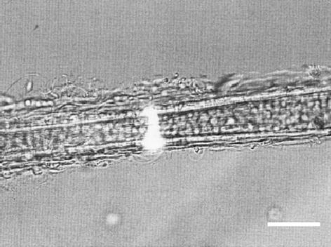
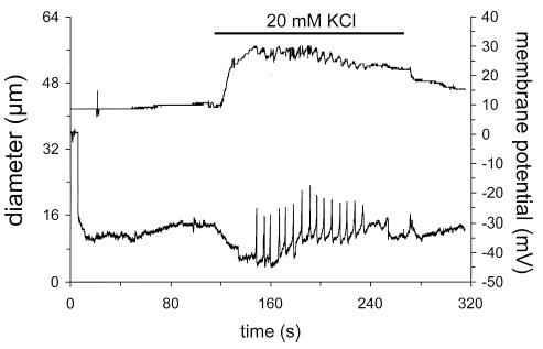
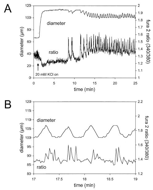
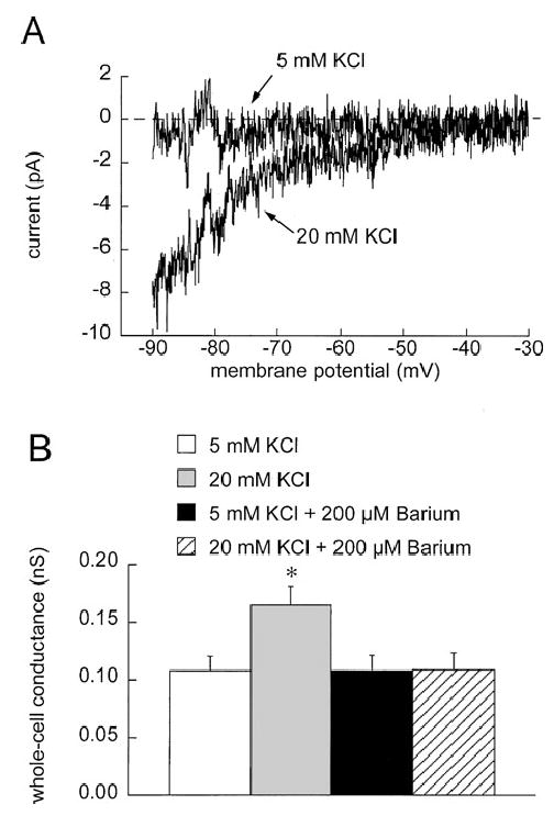
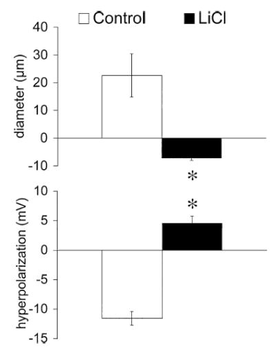
Methods: Arterioles (n = 111) were hand-dissected from hamster cremaster muscles, cannulated with glass micropipettes and pressurized to 80 cm H2O for in vitro study. The vessels were superfused with physiological salt solution containing 5 mM KCl, which could be rapidly switched to test solutions containing elevated [K+]o and/or inhibitors. The authors measured arteriolar diameter with a computer-based diameter tracking system, vascular smooth muscle cell membrane potential with sharp micropipettes filled with 200 mM KCl, and changes in intracellular Ca2+ concentration ([Ca2+]i) with Fura 2. Membrane currents and potentials also were measured in enzymatically isolated arteriolar muscle cells using patch clamp techniques. The role played by inward rectifier K+ (KIR) channels was tested using Ba2+ as an inhibitor. Ouabain and substitution of extracellular Na+ with Li+ were used to examine the function of the Na+/K+ ATPase.

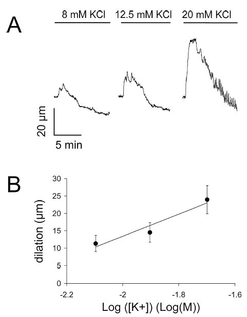
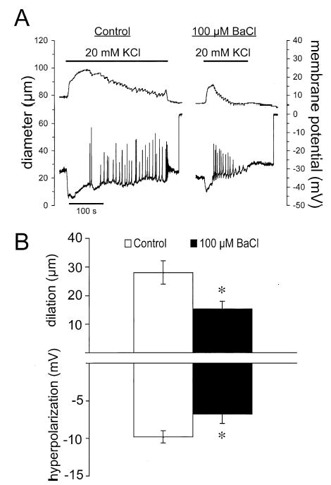
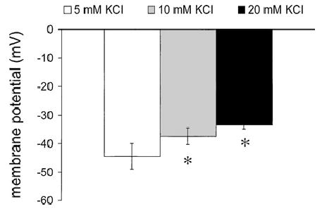
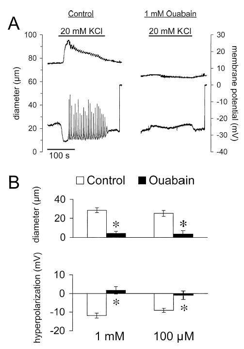
Results: Elevation of [K+]o from 5 mM up to 20 mM caused transient dilation of isolated arterioles (27 +/- 1 microm peak dilation when [K+]o was elevated from 5 to 20 mM, n = 105, p <.05). This dilation was preceded by transient membrane hyperpolarization (10 +/-1 mV, n = 23, p <.05) and by a fall in [Ca2+]i as indexed by a decrease in the Fura 2 fluorescence ratio of 22 +/- 5% (n = 4, p <.05). Ba(2+) (50 or 100 microM) attenuated the peak dilation (40 +/- 8% inhibition, n = 22) and hyperpolarization (31 +/- 12% inhibition, n = 7, p <.05) and decreased the duration of responses by 37 +/-11% (n = 20, p < 0.05). Both ouabain (1 mM or 100 microM) and replacement of Na+ with Li+ essentially abolished both the hyperpolarization and vasodilation.

Conclusions: Elevated [K+]o causes transient vasodilation of skeletal muscle arterioles that appears to be an intrinsic property of the arterioles. The results suggest that K+-induced dilation involves activation of both the Na+/K+ ATPase and KIR channels, leading to membrane hyperpolarization, a fall in [Ca2+]i, and culminating in vasodilation. The Na+/K+ ATPase appears to play the major role and is largely responsible for the transient nature of the response to elevated [K+]o, whereas KIR channels primarily affect the duration and kinetics of the response.


Figures

Figure 1
Figure 1
Elevated [K+]o causes concentration-dependent, transient dilation of skeletal muscle arterioles. (A) Digitized diameter records for a cannulated arteriole when [K+]o was elevated from 5 mM to 8, 12.5, or 20 mM as indicated. (B) Summary of the results from 11 experiments (mean resting diameter = 57 ± 5 μm, mean maximum diameter = 105 ± 5 μm) and the mean ± SE dilation induced by elevation of [K+]o from 5 mM to 8, 12.5, or 20 mM. Regression analysis showed a significant linear relationship (slope = 32 ± 11 μm/log(M), intercept = 77 ± 21, < .05, n = 33 arterioles), as indicated by the solid line.
Figure 2
Figure 2
Alexa Fluor 488-labeled arteriolar muscle cell. Shown is a combined transmitted light and epifluorescent image of an isolated arteriole that was pinned to the bottom of the chamber and from which membrane potential of arteriolar muscle cells was recorded. The image was captured with a CCD camera and a Scion image capture card controlled by a Macintosh G-4 computer running NIH Image software. Similar results were obtained in cannulated, pressurized arterioles (data not shown). Bar = 50 μm.
Figure 3
Figure 3
Elevated [K+]o causes dilation and hyperpolarization of skeletal muscle arterioles. Shown is a digitized, simultaneous recording of arteriolar diameter (top trace) and membrane potential (bottom trace) of an arteriolar muscle cell in a cannulated, pressurized cremasteric arteriole. When [K+]o was elevated from 5 mM to 20 mM (as indicated by the solid bar above the diameter trace), the arteriolar muscle cell hyperpolarized by about 10 mV and this change in membrane potential preceded arteriolar dilation. At the peak of the hyperpolarization, action potential-like spikes in membrane potential began. These membrane potential spikes preceded and correlated well with step-like decreases in arteriolar diameter. Upon return to 5 mM K+, both diameter and membrane potential recovered.
Figure 4
Figure 4
Elevated [K+]o causes dilation and decreases in intracellular Ca2+ of skeletal muscle arterioles. (A) Digitized diameter and Fura 2 fluorescence ratio traces in response to elevation of [K+]o from 5 to 20 mM as indicated by the arrow. Upon exposure to elevated K+, the Fura 2 ratio decreased, indicative of a fall in intracellular Ca2+. This change in Ca2+ occurred prior to the change in diameter (see (B)). Transient oscillations in the Fura 2 ratio were then observed that preceded but correlated well with oscillations in diameter. (B) An expanded segment of the record shown in (A) to better demonstrate the temporal relationship between the Ca2+ signal and diameter.
Figure 5
Figure 5
Barium attenuates K+-induced dilation and hyperpolarization. (A) Digitized typical records of diameter (top traces) and membrane potential (bottom traces) before (control) and during superfusion with 100 μM Ba2+ in response to elevation of [K+]o from 5 to 20 mM. Barium decreased the amplitude and the duration of both the diameter and membrane potential responses. (B) Summary data for the peak diameter and membrane potential responses. Data are means ± SE (n = 7), *significantly different from control response, p < .05.
Figure 6
Figure 6
Elevated [K+]o activates Ba2+-sensitive currents in cremasteric arteriolar muscle cells. (A) Ba2+-sensitive currents, present when cells were exposed to 5 mM K+, or 20 mM K+ were obtained by subtracting currents obtained during 500-ms ramp protocols in the absence and presence of 200 μM Ba2+. This high concentration of Ba2+ was used to insure complete inhibition of K+-activated currents. 20 mM K+ activated a Ba2+-sensitive, inwardly rectifying current. Note that no outward component of this current was observed. To summarize and quantify this K+-activated current, we performed regression analysis on the current-voltage data from −90 to −40 mV to obtain slope conductances. (B) In 6 cells, 20 mM K+ significantly (*p < .05 compared to 5 mM K+) increased the slope conductance, and this increase could be abolished by 200 μM Ba2+.
Figure 7
Figure 7
Elevated [K+]o does not hyperpolarize single cremasteric arteriolar muscle cells. Data are mean ± SE (n = 6) membrane potentials recorded in current clamp using the perforated patch technique, in 5, 10, and 20 mM K+. Elevated [K+]o was observed only to depolarize the cells. *Significantly different from potential in 5 mM K+, p < .05.
Figure 8
Figure 8
Ouabain inhibits K+-induced dilation and hyperpolarization. (A) Digitized typical records of diameter (top trace) and membrane potential (bottom trace) from a cannulated arteriole in response to elevation of [K+]o from 5 to 20 mM (solid bars over diameter trace) in the absence (control) and presence of 1 mM ouabain. In the presence of ouabain, dilation and hyperpolarization were reversed to constriction and depolarization. (B) Summary of the peak responses in the absence (control) and presence of either 1 or 100 μM ouabain. Data are means ± SE (n = 18 for diameter and n = 4 for membrane potential for 1 mM ouabain; n = 19 for diameter and n = 6 for membrane potential for 100 μM ouabain). *Significantly different from control and not significantly different from zero, p < .05.
Figure 9
Figure 9
LiCl inhibits K+-induced dilation and hyperpolarization. Data are means ± SE (n = 5), for the peak diameter and membrane potential responses when [K+]o was elevated from 5 to 20 mM, in the absence (control) or presence of PSS in which all NaCl was replaced by LiCl. *Significantly different from control and significantly greater than zero (p < .05).

Similar articles

Cited by

References

    1. Bartlett IS, Crane GJ, Neild TO, Segal SS. Electrophysiological basis of arteriolar vasomotion in vivo. J Vasc Res. 2000;37:568–575. - PubMed
    1. Blanco G, Mercer RW. Isozymes of the Na-K-ATPase: heterogeneity in structure, diversity in function. Am J Physiol. 1998;275(5 Pt 2):F633–F650. - PubMed
    1. Cohen KD, Jackson WF. Hypoxia inhibits contraction but not calcium channel currents or changes in intracellular calcium in arteriolar muscle cells. Microcirculation. 2003;10:133–141. - PMC - PubMed
    1. Duling BR. Effects of potassium ion on the microcirculation of the hamster. Circ Res. 1975;37:325–332. - PubMed
    1. Edwards FR, Hirst GDS, Silverberg GD. Inward rectification in rat cerebral arterioles: involvement of potassium ions in autoregulation. J Physiol. 1988;404:455–466. - PMC - PubMed

Publication types

Substances