

Intraepithelial lymphocytes in the villous tip: do they indicate potential coeliac disease?
- PMID: 15280404
- PMCID: PMC1770380
- DOI: 10.1136/jcp.2003.013607
Intraepithelial lymphocytes in the villous tip: do they indicate potential coeliac disease?
Abstract
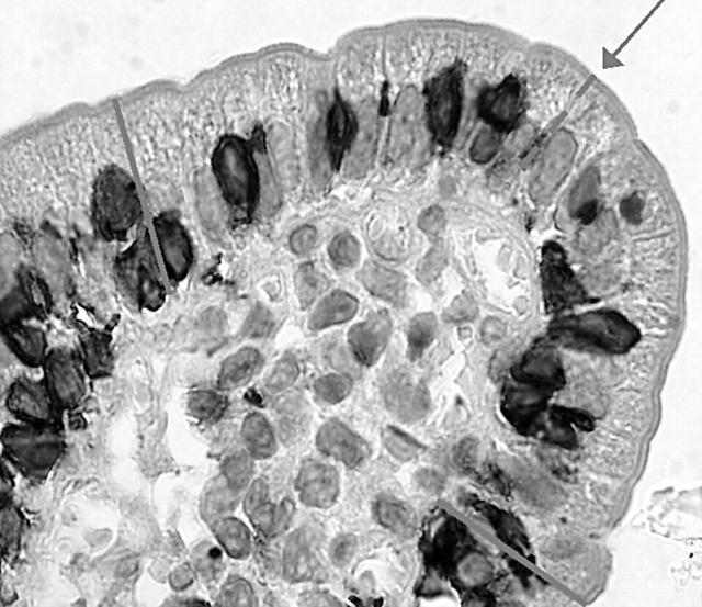
Background: The counting of intraepithelial lymphocytes (IELs) in the villous tips of architecturally normal small bowel biopsy specimens was proposed as a method to measure mucosal infiltration in gluten sensitive patients.
Aims: To apply this straightforward method in duodenal biopsy specimens from patients affected by potential coeliac disease (PCD) to verify whether it can discriminate these patients from controls.
Methods: Paraffin wax embedded duodenal sections from 11 patients affected by PCD were stained with an antihuman CD3 antibody. Sections from 19 patients affected by treated coeliac disease (TCD) and 17 patients in whom coeliac disease was excluded were stained with the same antibody to serve as controls. The slides were examined blindly. IELs/20 enterocytes in five randomly chosen villous tips were counted. Patients affected by PCD were all on a gluten containing diet. They had an architecturally normal duodenal mucosa and were positive for endomysial antibody. Both TCD and non-coeliac controls were negative for endomysial antibody.
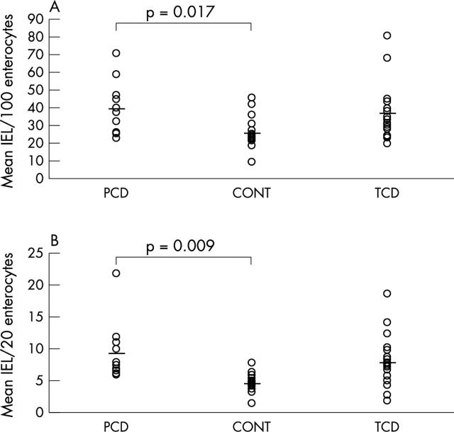
Results: The mean villous tip IEL scores were 4.6 (SD, 1.5; range, 1.4-7.8) in non-coeliac controls, 7.9 (SD, 4.0; range, 2.0-18.6) in TCD, and 9.2 (SD, 4.7; range, 5.8-21.8) in patients with PCD. The difference between PCD and non-coeliac controls was significant.
Conclusions: This is a very simple and sufficiently reliable method to count IELs. In patients with an architecturally normal duodenal mucosa, the IEL count in villous tips helps to distinguish between patients with PCD and non-coeliac controls.
Figures
References
-
- Trier JS. Celiac sprue and refractory sprue. In: Feldman M, Scharschmidt BF, Sleisenger MH, eds. Sleisenger and Fordtran’s gastrointestinal and liver disease. 6th ed. Philadelphia: WB Saunders, 1997:1557–73.
-
- Biagi F, Corazza GR. Clinical features of coeliac disease. Dig Liver Dis 2002;34:225–8. - PubMed
-
- Marsh MN, Crowe PT. Morphology of the mucosal lesion in gluten sensitivity. Baillieres Clin Gastroenterol 1995;9:273–93. - PubMed
MeSH terms
Substances
LinkOut - more resources
Full Text Sources
Medical