. 2004 Aug 2;200(3):277-85.
doi: 10.1084/jem.20040165. Epub 2004 Jul 26.

Compromised function of regulatory T cells in rheumatoid arthritis and reversal by anti-TNFalpha therapy

Affiliations

Compromised function of regulatory T cells in rheumatoid arthritis and reversal by anti-TNFalpha therapy

Michael R Ehrenstein et al. J Exp Med. .

Abstract

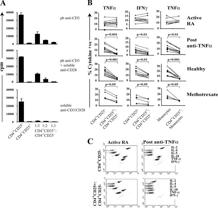
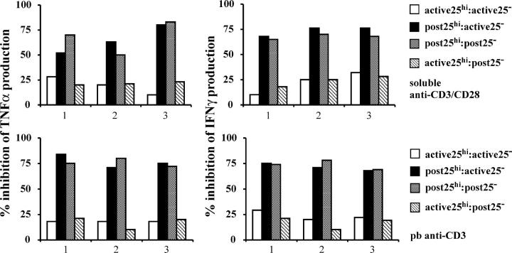
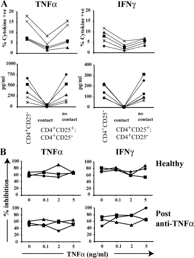
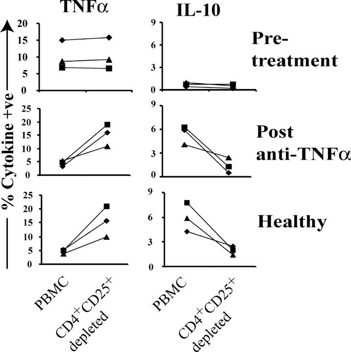
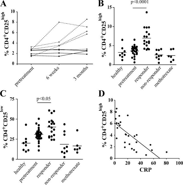
Regulatory T cells have been clearly implicated in the control of disease in murine models of autoimmunity. The paucity of data regarding the role of these lymphocytes in human autoimmune disease has prompted us to examine their function in patients with rheumatoid arthritis (RA). Regulatory (CD4(+)CD25(+)) T cells isolated from patients with active RA displayed an anergic phenotype upon stimulation with anti-CD3 and anti-CD28 antibodies, and suppressed the proliferation of effector T cells in vitro. However, they were unable to suppress proinflammatory cytokine secretion from activated T cells and monocytes, or to convey a suppressive phenotype to effector CD4(+)CD25(-) T cells. Treatment with antitumor necrosis factor alpha (TNFalpha; Infliximab) restored the capacity of regulatory T cells to inhibit cytokine production and to convey a suppressive phenotype to "conventional" T cells. Furthermore, anti-TNFalpha treatment led to a significant rise in the number of peripheral blood regulatory T cells in RA patients responding to this treatment, which correlated with a reduction in C reactive protein. These data are the first to demonstrate that regulatory T cells are functionally compromised in RA, and indicate that modulation of regulatory T cells by anti-TNFalpha therapy may be a further mechanism by which this disease is ameliorated.


Figures

Figure 1.
Figure 1.
Regulatory T cells from the PB of patients with active RA can suppress proliferation but not proinflammatory cytokine production by effector T cells and monocytes. (A) PB was collected from patients with RA; MACS®-sorted CD4+CD25+ T cells and CD4+CD25 T cells were cultured either alone or mixed at 1:3, 1:2, 1:1 ratio (105 cells/well) and stimulated with 1 μg/ml of plate-bound anti-CD3, 1 μg/ml of plate-bound anti-CD3/5 μg/ml of soluble anti-CD28, and 5 μg/ml soluble anti-CD3/anti-CD28 for 48 h. Proliferation after 5 d was determined by 3[H]Tdr incorporation. Results are expressed as mean ± SEM of triplicate cultures. One out of five independent experiments is shown. (B) MACS®-sorted CD4+CD25+ and CD4+CD25 T cells isolated from 10 patients before and after anti-TNFα therapy (3 mo) with 5 patients responding to methotrexate therapy or 10 healthy controls were cultured alone or mixed at a 1:1 ratio (105 cells/well) and stimulated with 5 μg/ml of soluble anti-CD3/anti-CD28 for 48 h. Monocytes were stimulated with 10 ng/ml LPS for 48 h. 2 μM monensin was added for the last 5 h of culture. Staining of CD4 and CD25 surface expression was performed and, subsequently, the cells were washed, permeabilized, and stained with PE-conjugated anti-TNFα or anti-IFNγ. (C) Cytokines from the supernatants collected from the experiment (B) were measured by CBA. 1 out of 10 experiments is illustrated.
Figure 2.
Figure 2.
The regulatory defect resides within the CD4+CD25high T cell population. (A) The CD4+CD25, CD4+CD25low, CD4+CD25high population were FACS®-sorted (MoFlo) from an active RA patient before treatment (white columns) and a responding RA patient (black columns) receiving anti-TNFα therapy using the indicated gates is depicted in Fig. S1. The CD4+CD25high and the CD4+CD25low T cells were cultured alone or with CD4+CD25 responder T cells (5 × 10⁴ cells/well) and stimulated with 5 μg/ml of soluble anti-CD3/anti-CD28 for 48 h. 2 μM monensin was added for the last 5 h of culture. Cells were stained to detect intracellular TNFα and IFNγ in a similar fashion to those in Fig. 1 B. Mean ± SEM of three patients before and after anti-TNFα therapy is shown. (B) Cytokines from the supernatants collected from the experiments from the three patients before and after anti-TNFα treatment (A) were measured by CBA and shown separately in each of the panels.
Figure 3.
Figure 3.
CD4+CD25high T cells from patients with active RA fail to suppress cytokines produced by CD4+CD25 T cells derived from the same patient either before or after anti-TNFα therapy. CD4+CD25high and CD4+CD25 T cells (5 × 10⁴ cells/well) isolated from patients with active RA (n = 3) or from patients responding to anti-TNFα therapy (n = 3) were mixed and stimulated with 5 μg/ml of soluble anti-CD3/anti-CD28 (top) or with 1 mg/ml of plate-bound anti-CD3 (bottom). Supernatants were analyzed by CBA. The results indicate the percentage of inhibition of the cytokine production relative to the CD4+CD25 T cells stimulated alone. One out of two experiments is shown.
Figure 4.
Figure 4.
The inhibitory effect of CD4+CD25high T cells requires cell contact and is not affected by the addition of TNFα in vitro. (A) CD4+CD25high and responder T cells derived from patients responding to anti-TNFα therapy were stimulated with 5 mg/ml of soluble anti-CD3/anti-CD28 either with or without the use of a transwell membrane to prevent cell contact. (top) 2 μM monensin was added for the last 5 h of culture, and cells were stained for detection of intracellular cytokine production. (bottom) Cytokines from the supernatants collected from the same experiment were measured by CBA. (B) CD4+CD25high and CD4+CD25 T cells isolated from healthy as well as responding patients were cultured 1:1 ratio, upon anti-CD3/anti-CD28 stimulation, in the presence of different concentrations of TNFα. The percentage of suppression of cytokine production was calculated in each individual. One out of two experiments is shown.
Figure 5.
Figure 5.
Effect of depletion of CD4+CD25+ T cells from PBMCs on TNFα and IL-10 production from the same patient before and after treatment. Total PBMCs or PBMCs depleted of CD25+ cells (by MACS® sorting) were stimulated with 5 μg/ml of soluble anti-CD3/anti-CD28 for 48 h. 2 μM monensin was added for the last 5 h of culture. Staining of CD4 and CD25 surface expression was performed. Cells were permeabilized and stained with PE-conjugated anti-TNFα or anti–IL-10. Results from three patients before and after therapy and three healthy individuals are shown.
Figure 6.
Figure 6.
Only CD4+CD25+ regulatory T cells derived from patients treated with anti-TNFα, but not from patients with active RA, convey a suppressive phenotype to effector T cells. MACS®-sorted CD4+CD25+ T cells and CD4+CD25 T cells were cultured either alone or mixed at a 1:1 ratio (105 cells/well) and stimulated with 1 μg/ml of soluble anti-CD3 and 2 μg/ml anti-CD28. After 5 d of culture, CD4+CD25+ T cells were depleted and the remaining CD4+CD25 T cells were replated 1:1 with freshly isolated autologous CD4+CD25 T cells in the presence of anti-CD3 and anti-CD28 for an additional 3 d (third column). 3[H]Tdr was added for the final 8 hof culture (expressed as mean ± SEM of triplicate cultures). Results from three patients before and after therapy and three healthy individuals are shown.
Figure 7.
Figure 7.
Increase in CD4+CD25high T cells in the PB of RA patients responding to anti-TNFα therapy. (A) The percentage of CD4+ T cells expressing CD25high was followed in four anti-TNFα responder (dashed line) and four nonresponder (solid line) patients with RA before and after treatment. (B) Analysis of larger group of patients treated with anti-TNFα (n = 27), methotrexate alone (n = 6), and healthy controls (n = 8). CD4+CD25high and (C) CD4+CD25low T cells (as defined in Fig. S1 A). (D) Scatter plot showing a significant correlation between the C reactive protein and the percentage of CD4+CD25high T cells before and after treatment (P < 0.001, r2 = 0.5278).

Comment in

  • Suppressor T cells in human diseases.
    Baecher-Allan C, Hafler DA. Baecher-Allan C, et al. J Exp Med. 2004 Aug 2;200(3):273-6. doi: 10.1084/jem.20040812. Epub 2004 Jul 26. J Exp Med. 2004. PMID: 15280423 Free PMC article.

References

    1. Sakaguchi, S., N. Sakaguchi, J. Shimizu, S. Yamazaki, T. Sakihama, M. Itoh, Y. Kuniyasu, T. Nomura, M. Toda, and T. Takahashi. 2001. Immunologic tolerance maintained by CD25+ CD4+ regulatory T cells: their common role in controlling autoimmunity, tumor immunity, and transplantation tolerance. Immunol. Rev. 182:18–32. - PubMed
    1. Bach, J.F., and J. Francois Bach. 2003. Regulatory T cells under scrutiny. Nat. Rev. Immunol. 3:189–198. - PubMed
    1. Salomon, B., D.J. Lenschow, L. Rhee, N. Ashourian, B. Singh, A. Sharpe, and J.A. Bluestone. 2000. B7/CD28 costimulation is essential for the homeostasis of the CD4+ CD25+ immunoregulatory T cells that control autoimmune diabetes. Immunity. 12:431–440. - PubMed
    1. Wu, A.J., H. Hua, S.H. Munson, and H.O. McDevitt. 2002. Tumor necrosis factor-alpha regulation of CD4+CD25+ T cell levels in NOD mice. Proc. Natl. Acad. Sci. USA. 99:12287–12292. - PMC - PubMed
    1. Kohm, A.P., P.A. Carpentier, H.A. Anger, and S.D. Miller. 2002. Cutting edge: CD4+CD25+ regulatory T cells suppress antigen-specific autoreactive immune responses and central nervous system inflammation during active experimental autoimmune encephalomyelitis. J. Immunol. 169:4712–4716. - PubMed

Publication types

MeSH terms