. 2004 Aug;78(16):8593-600.
doi: 10.1128/JVI.78.16.8593-8600.2004.

Role of JC virus agnoprotein in DNA repair

Affiliations

Role of JC virus agnoprotein in DNA repair

Armine Darbinyan et al. J Virol. 2004 Aug.

Abstract

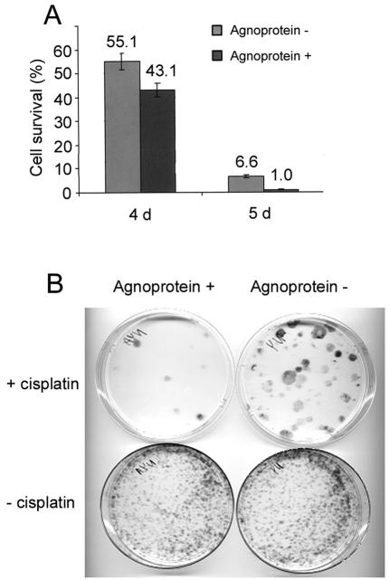
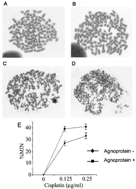
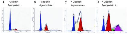
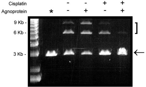
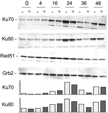
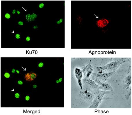
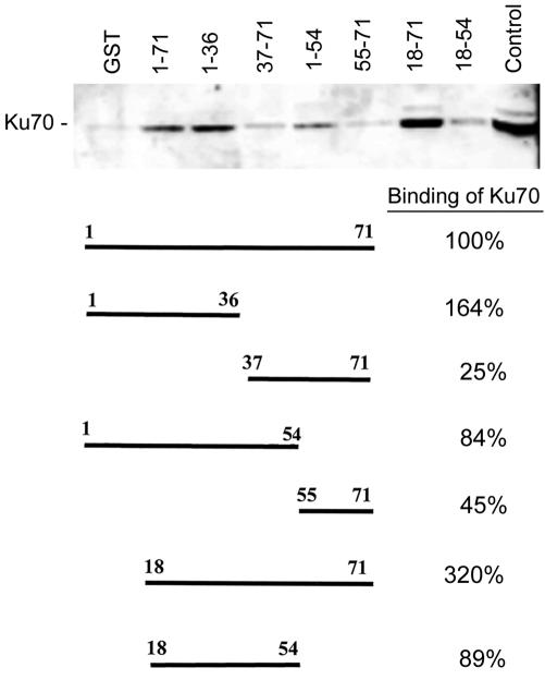
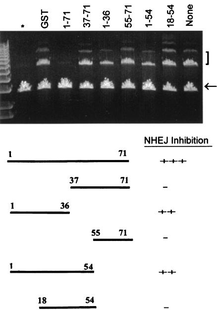
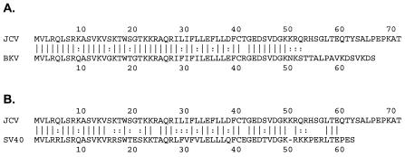
The late region of human neurotropic JC virus encodes a small 71-amino-acid agnoprotein that is also found in the polyomaviruses simian virus 40 and BK virus. Several functions of agnoprotein have been identified, including roles in regulating viral transcription and virion maturation. Earlier studies showed that agnoprotein expressed alone induced p21/WAF-1 expression and caused cells to accumulate in the G(2)/M stage of the cell cycle. Here we report that agnoprotein expression sensitized cells to the cytotoxic effects of the DNA-damaging agent cisplatin. Agnoprotein reduced the viability of cisplatin-treated cells and increased chromosome fragmentation and micronucleus formation. Whereas cisplatin-treated control cells accumulated in S phase, cells expressing agnoprotein did not, instead becoming aneuploid. Agnoprotein expression correlated with impaired double-strand-break repair activity in cellular extracts and reduced expression of the Ku70 and Ku80 DNA repair proteins. After agnoprotein expression, much of the Ku70 protein was located in the perinuclear space, where agnoprotein was also found. Results from binding studies showed an interaction of agnoprotein with Ku70 which was mediated by the N terminus. The ability of agnoprotein to inhibit double-strand break repair activity when it was added to cellular extracts was also mediated by the N terminus. We conclude that agnoprotein inhibits DNA repair after DNA damage and interferes with DNA damage-induced cell cycle regulation. Since Ku70 is a subunit of the DNA-dependent protein kinase that is responsible both for double-strand break repair and for signaling damage-induced cell cycle arrest, the modulation of Ku70 and/or Ku80 by agnoprotein may represent an important event in the polyomavirus life cycle and in cell transformation.


Figures

FIG. 1.
FIG. 1.
Effect of cisplatin treatment on survival and clonogenic ability of agnopositive and agnonegative cells. (A) Cell viability. NIH 3T3 cells expressing JCV agnoprotein and control agnonegative NIH 3T3 cells were treated with cisplatin as described in Materials and Methods. Sets of triplicates were done for each cell line under each condition (treated and untreated). Cell viability for each cell line was evaluated by counting the cells after 4 and 5 days of treatment. (B) Clonogenic ability. Cells were treated with or without cisplatin as described for panel A. After treatment, the cells were grown for 10 to 14 days, fixed, and stained with methylene blue.
FIG. 2.
FIG. 2.
Induction of chromosome breaks and micronucleus formation after cisplatin treatment. NIH 3T3 cells expressing JCV agnoprotein and control agnonegative NIH 3T3 cells were treated with cisplatin, and metaphase spreads were prepared as described in Materials and Methods. All photographs were taken at a magnification of ×700. (A) Agnonegative, untreated cells; (B) agnopositive, untreated cells; (C) agnonegative, cisplatin-treated cells; (D) agnopositive, cisplatin-treated cells. (E) Agnopositive and agnonegative cells were treated with 0, 0.125, or 0.25 μg of cisplatin/ml, and micronucleus (MIN) formation was measured as described in Materials and Methods.
FIG. 3.
FIG. 3.
Accumulation of cells with aberrant DNA content after cisplatin treatment of agnopositive and agnonegative cell lines. Agnopositive and agnonegative cells were left untreated or treated with cisplatin and subject to FACS analysis as described in Materials and Methods. (A) Agnonegative, untreated cells; (B) agnopositive, untreated cells; (C) agnonegative, cisplatin-treated cells; (D) agnopositive, cisplatin-treated cells.
FIG. 4.
FIG. 4.
NHEJ assay for DSB DNA repair. Agnopositive and agnonegative cells were left untreated or treated with cisplatin, and nuclear extracts were prepared as described in Materials and Methods. These were assayed by an NHEJ assay with a linear restriction-digested 3-kb plasmid. Lane *, control with no nuclear extract added. The arrow indicates the 3-kb monomeric linear plasmid substrate band. The 6- and 9-kb bands are the products of end joining and are indicated by a bracket.
FIG. 5.
FIG. 5.
Expression of NHEJ DNA repair proteins. Agnopositive (+) and agnonegative (−) cells were treated with cisplatin for 0, 4, 16, 24, 36, or 48 h, as indicated. Proteins were extracted and Western blotted with antibodies to Ku70, Ku80, Rad51, and Grb2 (loading control). The intensities of the Ku70 and Ku80 bands were measured by densitometry, and these data are shown in the lower panels.
FIG. 6.
FIG. 6.
Subcellular localization of agnoprotein and Ku70. U-87MG cells were transiently transfected with CMV-agnoprotein. The cells were then treated with 1 μg of cisplatin/ml for 18 h and double stained for Ku70 (green) and agnoprotein (red) as indicated. Arrows indicate an agnopositive cell; arrowheads indicate an agnonegative cell.
FIG. 7.
FIG. 7.
Analysis of agnoprotein-Ku70 interaction by using GST-agnoprotein fusion proteins. To see if Ku70 and the JCV agnoprotein could directly interact, we performed binding assays with protein extracts from U-87MG human glioblastoma cells that were negative for agnoprotein. Protein complexes were isolated by using glutathione-Sepharose 4B beads as described in Materials and Methods and were resolved by SDS-PAGE, followed by Western blotting with an anti-Ku70 antibody. The top panel shows a Western blot of the Ku70 that was pulled down by each of the GST-agnoprotein deletion mutant fusion proteins. The structure of each deletion mutant and its relative percentage of Ku70 binding are shown in the bottom panel. The lane on the far left is a negative control with GST only; the lane on the far right is a positive control with an unfractionated U-87MG cell extract. The relative intensities of the bands on the autoradiograph were measured by densitometry and the relative binding of Ku70 to each of the agnoprotein mutants was calculated as follows. The intensity of the GST-alone band (negative control) was subtracted from the intensity of each of the experimental lanes. The ratio of each of the bands relative to the full-length positive-control lane (1-71) was calculated and expressed as a percentage.
FIG. 8.
FIG. 8.
Effects of addition of full-length and C-terminal deletion mutants of agnoprotein on the end-joining activity of agnoprotein-negative cell extracts. Nuclear extracts from agnoprotein-negative NIH 3T3 cells were prepared. The extracts were incubated with GST alone or with the various GST-agnoprotein fusion proteins (full-length protein and various deletion mutants, as indicated) and assayed for NHEJ activity as described in Materials and Methods. The lane on the far left (*) is a negative control without nuclear extract. The lane on the far right (none) is a positive control that has nuclear extract but no GST-protein addition. The arrow indicates the 3-kb monomeric linear plasmid substrate band. The 6- and 9-kb bands are the products of end joining and are indicated by a bracket.
FIG. 9.
FIG. 9.
Comparison of amino acid sequence of JCV agnoprotein to BKV and SV40 agnoproteins. The amino acid sequence of the JCV agnoprotein was compared to the sequence of the agnoproteins of BKV (A) and SV40 (B). Sequence identity is indicated with parallel lines, and conservative sequence changes are indicated with colons.

References

    1. Baumann, P., and S. C. West. 1998. DNA end-joining catalyzed by human cell-free extracts. Proc. Natl. Acad. Sci. USA 95:14066-14070. - PMC - PubMed
    1. Berger, J. R. 2003. Progressive multifocal leukoencephalopathy in acquired immuno-deficiency syndrome: explaining the high incidence and disproportionate frequency of the illness relative to other immunosuppressive conditions. J. Neurovirol. 9(Suppl. 1):38-41. - PubMed
    1. Berger, J. R., and M. Concha. 1995. Progressive multifocal leukoencephalopathy: the evolution of a disease once considered rare. J. Neurovirol. 1:5-18. - PubMed
    1. Cole, C. N. 1996. Polyomavirinae: the viruses and their replication, p. 917-946. In B. N. Fields, D. M. Knipe, and P. M. Howley (ed.), Fundamental virology, 3rd ed. Lippincott Williams & Wilkins, Philadelphia, Pa.
    1. Cromie, G. A., J. C. Connelly, and D. R. Leach. 2001. Recombination at double-strand breaks and DNA ends: conserved mechanisms from phage to humans. Mol. Cell 8:1163-1174. - PubMed

Publication types

MeSH terms