. 2004 Aug;78(16):8630-40.
doi: 10.1128/JVI.78.16.8630-8640.2004.

Characterization of nucleocapsid binding by the measles virus and mumps virus phosphoproteins

Affiliations

Characterization of nucleocapsid binding by the measles virus and mumps virus phosphoproteins

Richard L Kingston et al. J Virol. 2004 Aug.

Erratum in

  • J Virol. 2005 Aug;79(15):10097

Abstract

We report an analysis of the interaction between the P protein and the RNA-associated N protein (N-RNA) for both measles and mumps viruses with proteins produced in a bacterial expression system. During this study, we verified that the C-terminal tail of the N protein is not required for nucleocapsid formation. For both measles and mumps virus N, truncated proteins encompassing amino acids 1 to 375 assemble into nucleocapsid-like particles within the bacterial cell. For measles virus N, the binding site for the P protein maps to residues 477 to 505 within the tail of the molecule, a sequence relatively conserved among the morbilliviruses. For mumps virus N, a binding site for the P protein maps to the assembly domain of N (residues 1 to 398), while no strong binding of the P protein to the tail of N was detected. These results suggest that the site of attachment for the polymerase varies among the paramyxoviruses. Pulldown experiments demonstrate that the last 50 amino acids of both measles virus and mumps virus P (measles virus P, 457 to 507; mumps virus P, 343 to 391) by themselves constitute the nucleocapsid-binding domain (NBD). Spectroscopic studies show that the NBD is predominantly alpha-helical in both viruses. However, only in measles virus P is the NBD stable and folded, having a lesser degree of tertiary organization in mumps virus P. With isothermal titration calorimetry, we demonstrate that the measles virus P NBD binds to residues 477 to 505 of measles virus N with 1:1 stoichiometry. The dissociation constant (K(d)) was determined to be 13 microM at 20 degrees C and 35 microM at 37 degrees C. Our data are consistent with a model in which an alpha-helical nucleocapsid binding domain, located at the C terminus of P, is responsible for tethering the viral polymerase to its template yet also suggest that, in detail, polymerase binding in morbilliviruses and rubulaviruses differs significantly.


Figures
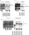
FIG. 1.
FIG. 1.
Short sequence (amino acids 477 to 505) within the tail of the measles virus N protein (MEN374-525) is responsible for measles virus P protein (MEP) binding, demonstrated by a GST pulldown assay. The results of this pulldown assay (and all others described in this paper) were analyzed by SDS-PAGE, and protein masses were verified by MALDI-TOF mass spectrometry. Constructs used in the pulldown assay are illustrated schematically below or above each lane. The leftmost lanes (labeled “Cell lysate following centrifugation”) show the crude cell lysates for each component used in the pulldown assay. (A) Measles virus P protein binds to the tail of the measles virus N protein. The last 49 amino acids of measles virus P are necessary for binding, as a construct in which this region is deleted (MEP1-458) does not bind. (B) Binding of measles virus P protein to the tail of measles virus N protein is abolished when 30 amino acids are deleted from the tail. (C) A measles virus N protein tail fragment encompassing amino acids 477 to 505 retains the ability to bind the measles virus P protein.
FIG. 2.
FIG. 2.
Last 51 amino acids of the measles virus P protein (MEP457-507) are sufficient for binding to the tail of the measles virus N protein (MEN374-525), demonstrated by a GST pulldown assay. MEP457-507 specifically binds to nucleocapsid-like particles resulting from expression of measles virus N protein, but binding is lost when the tail of N is truncated and the binding site (amino acids 477 to 505) is eliminated.
FIG. 3.
FIG. 3.
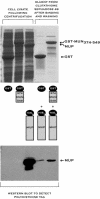
Tail of the mumps virus N protein (MUN374-549) binds weakly to the mumps virus P protein (MUP), demonstrated by a GST pulldown assay. The Western blot shown at the bottom of the figure was specific for the tag carried on the P protein.
FIG. 4.
FIG. 4.
Last 49 amino acids of the mumps virus P protein (MUP343-391) bind to the assembly domain of mumps virus N (MUN), demonstrated by a GST pulldown assay. Truncation of the tail of mumps virus N protein does not eliminate binding.
FIG. 5.
FIG. 5.
Circular dichroism spectra of MUP343-391 and MEP457-507/P458G, collected at 2°C.
FIG. 6.
FIG. 6.
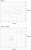
2D 1H-15N HSQC NMR spectra of MEP457-507 and MUP343-391/C356S collected at 15°C.
FIG. 7.
FIG. 7.
Binding of the measles virus P nucleocapsid-binding domain (amino acids 457 to 505) to the measles virus N tail peptide (amino acids 477 to 505) studied by isothermal titration calorimetry at 20°C. (A) Raw binding data obtained for 23 automatic injections of MEP457-507/P458G (each injection, 12 μl; protein concentration, 1.44 μM) into a cell containing MEN477-505 (initial volume, 1,800 μl; initial protein concentration, 0.24 μM). Proteins were suspended in 10 mM phosphate buffer (pH 7.0)-100 mM NaCl. (B) The integrated titration curve obtained from the raw data in panel A after baseline subtraction. The solid squares represent the experimental data, while the solid line corresponds to the standard multiple independent binding-site model that was fitted to the data.
FIG. 8.
FIG. 8.
Schematic model of polymerase attachment to the nucleocapsid in measles virus and mumps virus, incorporating the results presented in this paper. A helical nucleocapsid-binding domain (NBD) at the C terminus of the P protein (amino acids 457 to 507 in measles virus P and 343 to 391 in mumps virus P) directly mediates binding of the polymerase to the nucleocapsid. In measles virus, the NBD attaches to a short sequence (amino acids 477 to 505) within the tail of the RNA-associated N protein. In mumps virus, the NBD binds to the assembly domain (amino acids 1 to 398) of the N protein. Based on our results for measles virus, each NBD is capable of weakly associating with a single N molecule. A coiled coil within P holds the downstream NBDs in close proximity. The sequence interleaving the coiled coil and the downstream NBD is likely to be unstructured. The P protein is depicted as a tetramer, but the oligomerization state of measles virus and mumps virus P has not been determined.

References

    1. Amexis, G., S. Rubin, V. Chizhikov, F. Pelloquin, K. Carbone, and K. Chumakov. 2002. Sequence diversity of Jeryl Lynn strain of mumps virus: quantitative mutant analysis for vaccine quality control. Virology 300:171-179. - PubMed
    1. Bankamp, B., S. M. Horikami, P. D. Thompson, M. Huber, M. Billeter, and S. A. Moyer. 1996. Domains of the measles virus N protein required for binding to P protein and self-assembly. Virology 216:272-277. - PubMed
    1. Bhella, D., A. Ralph, L. B. Murphy, and R. P. Yeo. 2002. Significant differences in nucleocapsid morphology within the Paramyxoviridae. J. Gen. Virol. 83:1831-1839. - PubMed
    1. Blixenkrone-Moller, M., G. Bolt, E. Gottschalck, and M. Kenter. 1994. Comparative analysis of the gene encoding the nucleocapsid protein of dolphin morbillivirus reveals its distant evolutionary relationship to measles virus and ruminant morbilliviruses. J. Gen. Virol. 75:2829-2834. - PubMed
    1. Buchholz, C. J., C. Retzler, H. E. Homann, and W. J. Neubert. 1994. The carboxy-terminal domain of Sendai virus nucleocapsid protein is involved in complex formation between phosphoprotein and nucleocapsid-like particles. Virology 204:770-776. - PubMed

MeSH terms

LinkOut - more resources