. 2004 Aug;78(16):8878-84.
doi: 10.1128/JVI.78.16.8878-8884.2004.

Expression in a recombinant murid herpesvirus 4 reveals the in vivo transforming potential of the K1 open reading frame of Kaposi's sarcoma-associated herpesvirus

Affiliations

Expression in a recombinant murid herpesvirus 4 reveals the in vivo transforming potential of the K1 open reading frame of Kaposi's sarcoma-associated herpesvirus

Jill Douglas et al. J Virol. 2004 Aug.

Abstract

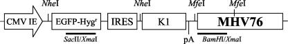
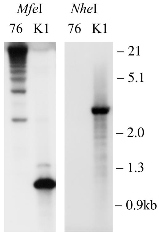
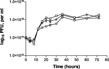
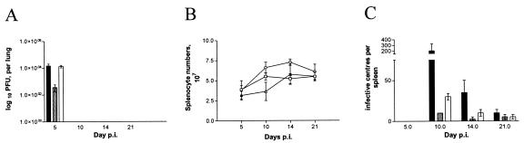
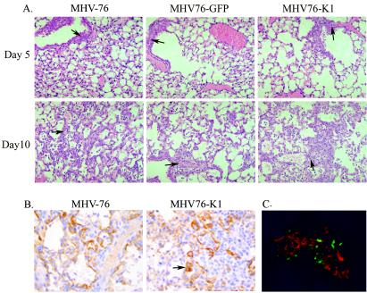
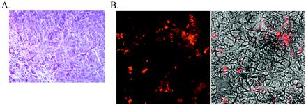
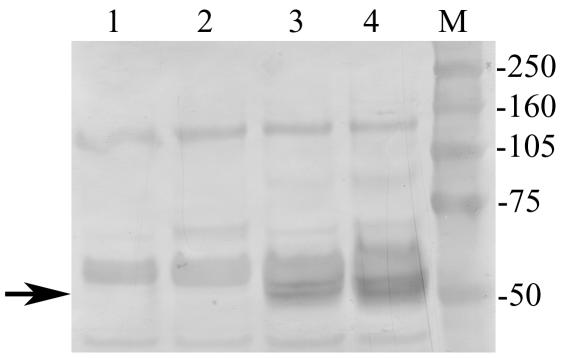
Murid herpesvirus 4 (commonly called MHV-68) is closely related to Kaposi's sarcoma-associated herpesvirus (KSHV) and provides an excellent model system for investigating gammaherpesvirus-associated pathogenesis. MHV-76 is a naturally occurring deletion mutant of MHV-68 that lacks 9,538 bp of the left end of the unique portion of the genome encoding nonessential pathogenesis-related genes. The KSHV K1 protein has been shown to transform rodent fibroblasts in vitro and common marmoset T lymphocytes in vivo. Using homologous recombination techniques, we successfully generated recombinants of MHV-76 that encode green fluorescent protein (MHV76-GFP) and KSHV K1 (MHV76-K1). The replication of MHV76-GFP and MHV76-K1 in cell culture was identical to that of MHV-76. However, infection of BALB/c mice via the intranasal route revealed that MHV76-K1 replicated to a 10-fold higher titer than MHV76-GFP in the lungs at day 5 postinfection (p.i.). We observed type 2 pneumocyte proliferation in areas of consolidation and interstitial inflammation of mice infected with MHV76-K1 at day 10 p.i. MHV76-K1 established a 2- to 3-fold higher latent viral load than MHV76-GFP in the spleens of infected mice on days 10 and 14 p.i., although this was 10-fold lower than that established by wild-type MHV-76. A salivary gland tumor was present in one of four mice infected with MHV76-K1, as well as an increased inflammatory response in the lungs at day 120 p.i. compared with that of mice infected with MHV-76 and MHV76-GFP.


Figures

FIG. 1.
FIG. 1.
Schematic diagram of plasmid used to generate recombinant MHV76-K1 virus. The expression cassette consists of the CMV-IE promoter and the EGFP-Hygr gene from pEGFPHyg (Clontech), followed by the IRES element from EMCV. Cloned immediately downstream from the IRES is the KSHV K1 ORF followed by the SV40 poly(A) signal (pA). These elements were cloned into a BamHI site next to the MHV-76 genomic DNA (nucleotides 9539 to 12569 of MHV-68) in pBluescript (Stratagene). The MHV76-GFP virus was generated by using an identical plasmid lacking the KSHV K1 gene. The positions of the probes used for Southern hybridization (BamHI-XmaI and SacII-XmaI) and the restriction sites for NheI and MfeI are indicated.
FIG. 2.
FIG. 2.
Southern blot showing purified MHV76-K1 recombinant virus compared with parental MHV-76. The left panel shows MHV-76 and MHV76-K1 digested with MfeI and probed with the BamHI-XmaI probe (Fig. 1). The right panel shows MHV-76 and MHV76-K1 digested with NheI and probed with the SacII-XmaI probe (Fig. 1).
FIG. 3.
FIG. 3.
In vitro single-step growth curves of MHV-76 (□), MHV76-GFP (▵), and MHV76-K1 (○) on BHK-21 cells at a multiplicity of infection of 5. Data are shown as mean log10 virus titers ± standard errors and are representative of two separate experiments, with each performed in duplicate.
FIG. 4.
FIG. 4.
(A) Viral replication in lungs of BALB/c mice infected intranasally with 4 × 10⁵ PFU of MHV-76 (black bars), MHV76-GFP (gray bars), or MHV76-K1 (white bars). The mean log10 virus titer ± standard error for four mice per group is shown for each time point. MHV76-GFP had a significantly lower titer than MHV-76 or MHV76-K1 on day 5 p.i. (P < 0.01). (B) Numbers of spleen cells during intranasal infection of BALB/c mice with 4 × 10⁵ PFU of MHV-76 (□), MHV76-GFP (▵), or MHV76-K1 (○). The mean total number of splenocytes ± standard error for four mice per group is shown for each time point. MHV76-K1-infected mice had a significantly higher splenocyte number than MHV76-GFP- or MHV-76-infected mice on day 14 p.i. (P < 0.05). (C) Latent virus in spleens of BALB/c mice infected intranasally with 4 × 10⁵ PFU of MHV-76 (black bars), MHV76-GFP (gray bars), or MHV76-K1 (white bars), as determined by an infective-center assay. The mean number of infective centers per spleen ± standard error for four mice per group is shown for each time point. MHV76-K1 had a significantly higher latent viral load than MHV76-GFP on day 10 p.i. (P < 0.001).
FIG. 5.
FIG. 5.
(A) Hematoxylin and eosin staining of lung sections of mice infected with MHV-76, MHV76-GFP, or MHV76-K1 at days 5 and 10 p.i., showing perivascular inflammatory cell infiltrates (arrows). (B) Cytokeratin staining of lung sections of mice infected with MHV-76 or MHV76-K1 at day 10 p.i., showing strong staining of type 2 pneumocytes in chronic proliferative inflammatory foci (arrow). (C) Mice infected with MHV76-K1 express GFP in the lungs at day 5 p.i. MHV-76 antigens were stained with polyclonal antisera and an anti-rabbit tetramethyl rhodamine isocyanate conjugate (red).
FIG. 6.
FIG. 6.
(A) Hematoxylin and eosin staining of the salivary gland adenocarcinoma present in an MHV76-K1-infected mouse at day 120 p.i. (B) Expression of MHV-76 antigens in the salivary gland adenocarcinoma detected with rabbit polyclonal antisera. The left panel shows the immunofluorescent image, and the right panel shows a phase-contrast image merged with the immunofluorescent image.
FIG. 7.
FIG. 7.
Western blot of immunoprecipitated proteins showing the expression of Myc-tagged K1 in the lungs of MHV76-K1-infected mice at day 5 p.i. (lane 3) and in a salivary gland adenocarcinoma at day 120 p.i. (lane 4). K1 expression was not detected in either the lungs (lane 1) or salivary glands (lane 2) of MHV76-GFP-infected mice. The position of the K1 protein at ∼50 kDa is indicated with an arrow.

Similar articles

Cited by

References

    1. Beral, V., and R. Newton. 1998. Overview of the epidemiology of immunodeficiency-associated cancers. J. Natl. Cancer Inst. Monogr. 1998:1-6. - PubMed
    1. Blackman, M. A., E. Flano, E. Usherwood, and D. L. Woodland. 2000. Murine gamma-herpesvirus-68: a mouse model for infectious mononucleosis? Mol. Med. Today 6:488-490. - PubMed
    1. Blaskovic, D., M. Stancekova, J. Svobodova, and J. Mistrikova. 1980. Isolation of five strains of herpesviruses from two species of free living small rodents. Acta Virol. 24:468. - PubMed
    1. Boshoff, C., and R. A. Weiss. 2001. Epidemiology and pathogenesis of Kaposi's sarcoma-associated herpesvirus. Philos. Trans. R. Soc. Lond. B Biol. Sci. 356:517-534. - PMC - PubMed
    1. Chang, Y., E. Cesarman, M. S. Pessin, F. Lee, J. Culpepper, D. M. Knowles, and P. S. Moore. 1994. Identification of herpesvirus-like DNA sequences in AIDS-associated Kaposi's sarcoma. Science 266:1865-1869. - PubMed

Publication types

MeSH terms

LinkOut - more resources