. 2004 Aug;78(16):8935-41.
doi: 10.1128/JVI.78.16.8935-8941.2004.

The gene encoding the nucleocapsid protein of Gill-associated nidovirus of Penaeus monodon prawns is located upstream of the glycoprotein gene

Affiliations

The gene encoding the nucleocapsid protein of Gill-associated nidovirus of Penaeus monodon prawns is located upstream of the glycoprotein gene

Jeff A Cowley et al. J Virol. 2004 Aug.

Abstract

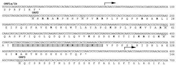
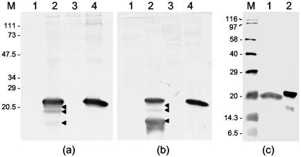
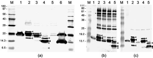
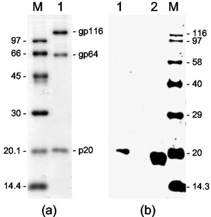
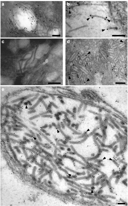
The ORF2 gene of Gill-associated virus (GAV) of Penaeus monodon prawns resides 93 nucleotides downstream of the ORF1a-ORF1b gene and encodes a 144-amino-acid hydrophilic polypeptide (15,998 Da; pI, 9.75) containing 20 basic (14%) and 13 acidic (9%) residues and 19 prolines (13%). Antiserum to a synthetic ORF2 peptide or an Escherichia coli-expressed glutathione S-transferase-ORF2 fusion protein detected a 20-kDa protein in infected lymphoid organ and gill tissues in Western blots. The GAV ORF2 fusion protein antiserum also cross-reacted with the p20 nucleoprotein in virions of the closely related Yellow head virus. By immuno-gold electron microscopy, it was observed that the ORF2 peptide antibody localized to tubular GAV nucleocapsids, often at the ends or at lateral cross sections. As GAV appears to contain only two structural protein genes (ORF2 and ORF3), these data indicate that GAV differs from vertebrate nidoviruses in that the gene encoding the nucleocapsid protein is located upstream of the gene encoding the virion glycoproteins.


Figures

FIG. 1.
FIG. 1.
Nucleotide and predicted amino acid sequences of the GAV ORF2 gene and genome regions extending into the upstream ORF1a-ORF1b and downstream ORF3 genes. The sequence is the consensus of three clones, pP18/20-1, pP18/20-9, and pP18/20-16. Indicated are the 5′-terminal positions of the 3′-coterminal sgmRNAs (arrows), the ORFa/1b and ORF2 gene termination codons (*), the in-frame AUG codons (underlined) and basic amino acids (bold) in ORFZ, and the sequence of the synthetic peptide PN1 (shaded).
FIG. 2.
FIG. 2.
Western blot of uninfected and GAV-infected P. monodon tissues carried out with antiserum to E. coli-expressed GST-ORF2 fusion protein (a) and KLH-linked peptide PN1 (b and c). (a and b) LO (lanes 1 and 2) and gill tissues (lanes 3 and 4) from an uninfected prawn (lanes 1 and 3) and a GAV-infected prawn (lanes 2 and 4). Three proteins smaller than the ORF2 protein detected in LO tissue are indicated (◂). (c) GAV-infected gill tissue (lane 1) and the E. coli-expressed His6-ORF2 fusion protein (lane 2). Proteins were separated by SDS-PAGE in 12% (a and b) or 15% (c) polyacrylamide gels. The molecular masses (in kilodaltons) of prestained protein standards (Bio-Rad) (a and b) and biotinylated protein standards (Amersham) (c) are indicated (lanes M).
FIG. 3.
FIG. 3.
Western blot of native ORF2 in GAV-infected LO and gill tissues compared to His6-ORF2 and N-terminally truncated His6-ORF2 fusion proteins expressed in E. coli. (a) GAV-infected gill tissue (lane 1) and LO tissue (lane 6) and IPTG-induced E. coli cultures transformed with pQE-ORF2 (lane 2), pQE-ORF2-M11 (lane 3), pQE-ORF2-M61 (lane 4), and pQE10 (lane 5) were resolved in a 15% polyacrylamide gel and immunodetected with KLH-PN1 peptide antiserum. Lane M contains biotinylated protein standards. (b and c) GAV-infected gill (lane 1) and IPTG-induced E. coli cultures transformed with pQE-ORF2 (lane 2), pQE10 (lane 3), pQE-ORF2-M11 (lane 4), and pQE-ORF2-M61 (lane 5) resolved in a 12% polyacrylamide gel and immunodetected using antiserum to GST-ORF2 (b) or KLH-PN1 peptide (c). Lanes M contain BenchMark prestained protein ladders (Invitrogen). The predominant His6-ORF2 fusion proteins (◂) and minor larger and smaller His6-ORF2 fusion protein forms that were also immunodetected (◃) are indicated.
FIG. 4.
FIG. 4.
(a) Structural proteins (gp116, gp64, and p20) of purified YHV (lane 1) detected by staining with Coomassie blue. (b) Western blot of purified YHV (lane 1) and GAV-infected gill tissues (lane 2) with GAV GST-ORF2 antiserum. Proteins were resolved in a 12% polyacrylamide gel. Lanes M contain protein standards (a) and biotinylated protein standards (Amersham) (b), with molecular masses (in kilodaltons) indicated.
FIG. 5.
FIG. 5.
Immunoelectron microscopy of ultrathin sections of GAV-infected LO cells using prebleed (c) or KLH-PN1 peptide (a, b, d, and e) antiserum. No gold particles (10 nm in diameter) associated with GAV nucleocapsids with prebleed sera. With the PN1 peptide antiserum, gold particles associated with free striated nucleocapsids (a and b) and nucleocapsids within mature rod-shaped virions (d) and newly formed virions (e) that were observed budding into a membranous endoplasmic vesicle. Good examples of gold particles bound to nucleocapsid ends or to areas where the nucleocapsids had been cross-sectioned laterally are highlighted (arrowheads). Bars represent 100 nm.

Similar articles

Cited by

References

    1. Altschul, S. F., T. L. Madden, A. A. Schaffer, J. Zhang, Z. Zhang, W. Miller, and D. J. Lipman. 1997. Gapped BLAST and PSI-BLAST: a new generation of protein database search programs. Nucleic Acids Res. 25:3389-3402. - PMC - PubMed
    1. Boonyaratpalin, S., K. Supamattaya, J. Kasornchandra, S. Direkbusaracom, U. Aekpanithanpong, and C. Chantanachookin. 1993. Non-occluded baculo-like virus, the causative agent of yellow-head disease in the black tiger shrimp (Penaeus monodon). Fish Pathol. 28:103-109.
    1. Chantanachookin, C., S. Boonyaratpalin, J. Kasornchandra, D. Sataporn, U. Ekpanithanpong, K. Supamataya, S. Riurairatana, and T. W. Flegel. 1993. Histology and ultrastructure reveal a new granulosis-like virus in Penaeus monodon affected by yellow-head disease. Dis. Aquat. Org. 17:145-157.
    1. Chen, Z., L. Kuo, R. R. Rowland, C. Even, K. S. Faaberg, and P. G. Plagemann. 1993. Sequences of 3′ end of genome and of 5′ end of open reading frame 1a of lactate dehydrogenase-elevating virus and common junction motifs between 5′ leader and bodies of seven subgenomic mRNAs. J. Gen. Virol. 74:643-659. - PubMed
    1. Cowley, J. A., and P. J. Walker. 2002. The complete genome sequence of gill-associated virus of Penaeus monodon prawns indicates a gene organisation unique among nidoviruses. Arch. Virol. 147:1977-1987. - PMC - PubMed