. 2004 Aug 10;101(32):11839-44.
doi: 10.1073/pnas.0404474101. Epub 2004 Jul 27.

Transplanted human fetal neural stem cells survive, migrate, and differentiate in ischemic rat cerebral cortex

Affiliations

Transplanted human fetal neural stem cells survive, migrate, and differentiate in ischemic rat cerebral cortex

S Kelly et al. Proc Natl Acad Sci U S A. .

Abstract

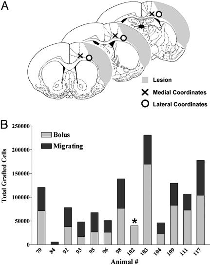
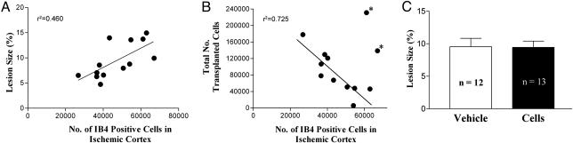
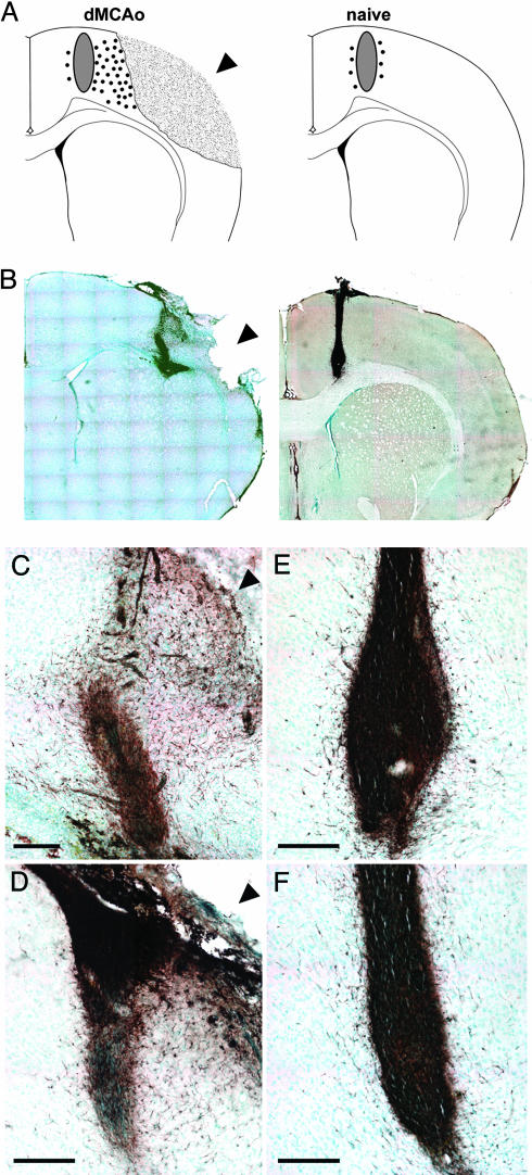
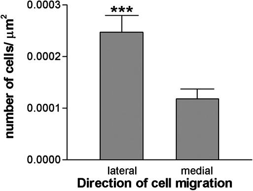
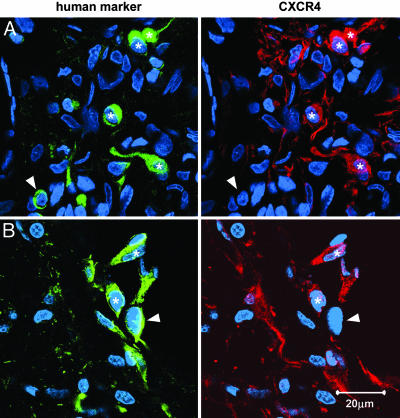
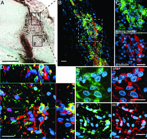
We characterize the survival, migration, and differentiation of human neurospheres derived from CNS stem cells transplanted into the ischemic cortex of rats 7 days after distal middle cerebral artery occlusion. Transplanted neurospheres survived robustly in naive and ischemic brains 4 wk posttransplant. Survival was influenced by proximity of the graft to the stroke lesion and was negatively correlated with the number of IB4-positive inflammatory cells. Targeted migration of the human cells was seen in ischemic animals, with many human cells migrating long distances ( approximately 1.2 mm) predominantly toward the lesion; in naive rats, cells migrated radially from the injection site in smaller number and over shorter distances (0.2 mm). The majority of migrating cells in ischemic rats had a neuronal phenotype. Migrating cells between the graft and the lesion expressed the neuroblast marker doublecortin, whereas human cells at the lesion border expressed the immature neuronal marker beta-tubulin, although a small percentage of cells at the lesion border also expressed glial fibrillary acid protein (GFAP). Thus, transplanted human CNS (hCNS)-derived neurospheres survived robustly in naive and ischemic brains, and the microenvironment influenced their migration and fate.


Figures

Fig. 1.
Fig. 1.
Survival of hCNS-derived neurospheres in the ischemic brain. (A) Schematic of lesion (shaded area) and transplant coordinates. The three sections represent the three levels on the A-P axis where cells were transplanted. Cells transplanted close to the lesion core (○) showed reduced survival; cells transplanted more medially (×) further from the lesion showed robust survival. The medial coordinates were used for this study. (B) Quantitation of the number of surviving cells in rats subjected to dMCAo 4 wk posttransplant. The gray bar indicates the number of cells in the graft bolus; the black bar represents the number of cells migrating from the graft (the asterisk denotes that the migrating population was not counted in this animal). On average, 100,147 ± 28,944 cells survived.
Fig. 2.
Fig. 2.
Lesion size and inflammatory response. (A) IB4-positive inflammatory cells in the cortex of cell-transplanted rats were counted in eight sections adjacent to those used to measure lesion size. Lesion size was directly correlated with the number of IB4+ cells. (B) In 10 of 12 cell-transplanted rats, the total number of surviving human cells (i.e., cells in the graft bolus plus migrating cells) was negatively correlated with the number of IB4-positive cells. An asterisk indicates animals excluded from the correlation. (C) There was no difference in lesion area in cell-treated and buffer-treated rats. Lesion area (mean ± SEM of 12 or 13 animals) was measured over eight levels per brain to get a total lesion area per animal.
Fig. 3.
Fig. 3.
Migration of transplanted human cells. (A) Schematic showing extensive migration of human cells (small circles) toward the lesion in dMCAo brains compared to very little migration away from the graft (gray oval) in naive brains. (B) 4wk posttransplant, sections were labeled with the human-specific antibody SC121, to identify human cells, and visualized with diaminobenzidine (DAB) (brown). Low magnification images depict the cells migrating long distances primarily toward the lesion in dMCAo brains. Cells transplanted into naive rats migrated little and equally in all directions. Shown are higher-magnification images of dMCAo (C and D) and naive (E and F) brains. Arrowhead indicates lesion. (Scale bar = 200 μM.)
Fig. 4.
Fig. 4.
Quantification of the human cell migration in dMCAo brains. Human cells stained with the human-specific antibody SC121 4 wk posttransplant in dMCAo brains were counted by using unbiased stereology. The number of cells migrating from the graft toward the lesion (laterally) was greater than the number migrating away from the lesion (medially) (***, P < 0.0001). Data are means ± SEM from 11 animals.
Fig. 5.
Fig. 5.
Transplanted human cells express the chemokine receptor CXCR4. Shown are confocal images from lesioned brains of human cells labeled with the human-specific antibody SC121 (green) and an anti-CXCR4 antibody (red); nuclei are stained with Hoechst (blue). (A) Representative image of migrating human cells. (B) Representative image of human cells at the lesion border. An asterisk indicates double-labeled cells; arrowhead indicates human cells that did not express CXCR4.
Fig. 6.
Fig. 6.
Marker profile of human cells depends on proximity to the lesion. (A) Immunostaining of human cells (brown) with human-specific antibody SC121. Boxes show the location of the cells in the other panels. (Scale bar = 500 μm.) (BF) Confocal images: human cells labeled with SC121 are green, nuclei stained with Hoechst are blue, and various phenotypic markers are in red. An asterisk denotes double-labeled cells. (Scale bar = 20 μM.) (B) Many human cells at the lesion border (box 1 in A) express β-tubulin III. (C) Higher magnification of demarked area in B.(D) Orthogonal view to confirm double labeling with the human marker and β-tubulin III. (E) Cells at the lesion border also express GFAP. (F) The migrating cells (box 2 in A) express doublecortin.

References

    1. American Heart Association (2002) Heart Disease and Stroke Statistics 2003 Update (American Heart Association, Dallas, TX).
    1. Nishino, H. & Borlongan, C. V. (2000) Prog. Brain Res 127, 461-476. - PubMed
    1. Mattsson, B., Sorensen, J. C., Zimmer, J. & Johansson, B. B. (1997) Stroke 28, 1225-1231; discussion 1231-1232. - PubMed
    1. Riolobos, A. S., Heredia, M., de la Fuente, J. A., Criado, J. M., Yajeya, J., Campos, J. & Santacana, M. (2001) Neurobiol. Learn. Mem. 75, 274-292. - PubMed
    1. Savitz, S. I., Rosenbaum, D. M., Dinsmore, J. H., Weschler, L. R. & Caplan, L. R. (2002) Ann. Neurol. 53, 266-275. - PubMed

Publication types