

S100A6 (Calcyclin) is a prostate basal cell marker absent in prostate cancer and its precursors
- PMID: 15280928
- PMCID: PMC2364790
- DOI: 10.1038/sj.bjc.6602034
S100A6 (Calcyclin) is a prostate basal cell marker absent in prostate cancer and its precursors
Abstract
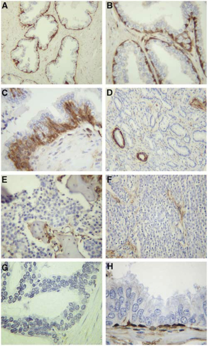
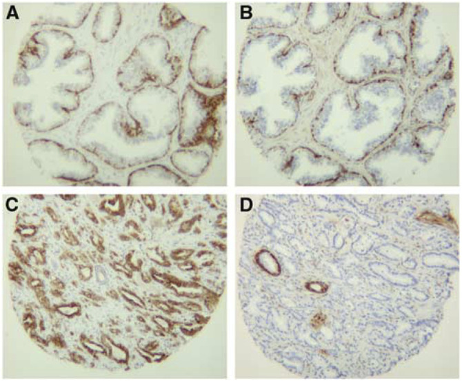
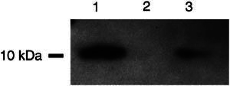
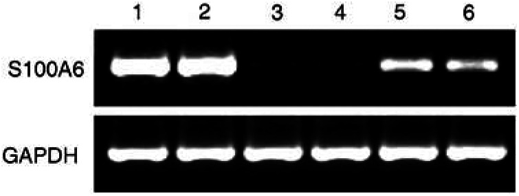
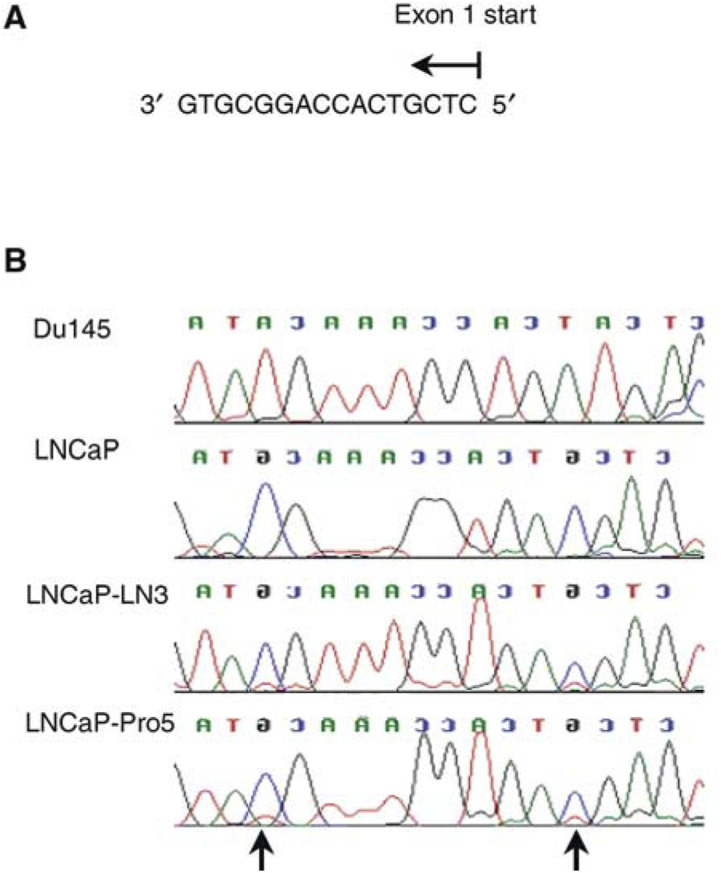
S100A6 (Calcyclin) is a calcium-binding protein that has been implicated in a variety of biological functions as well as tumorigenesis. The aim of our study was to investigate the involvement of S100A6 during prostate cancer development and progression. Using immunohistochemistry, the expression of S100A6 was examined in benign (n=66), premalignant (n=10), malignant (n=66) and metastatic prostate (n=5) tissues arranged in a tissue-microarray or whole sections as well as in prostate cancer cell lines. The S100A6 immunostaining pattern in tissues was compared with that of cytokeratin 5 (a basal cell marker) and 18 (a benign luminal cell marker). In all cases of benign epithelium, intense S100A6 expression was seen in the basal cell layer with absent staining in luminal cells. In all cases of prostatic adenocarcinoma (matched), metastatic lesions and 3/10 high-grade prostatic intraepithelial neoplasia lesions, an absence of S100A6 was seen. Western blotting and RT-PCR analysis of cell lines showed S100A6 expression to be absent in LNCaP, LNCaP-LN3 and LNCaP-Pro5 but present in Du145, PC3, PC-3M and PC-3M-LN4. LNCaP cells treated with 5-Azacytidine, caused re-expression of S100A6 mRNA. Sequencing of bisulphite modified DNA showed CpG methylation within the S100A6 promoter region and exon 1 of LNCaP, LNCaP-LN3 and LNCaP-Pro5 cell lines but not in Du145 cells. Our data suggest that loss of S100A6 protein expression is common in prostate cancer development and may occur at an early stage. The mechanism of loss of expression may involve hypermethylation of CpG sites. The finding of intense S100A6 expression in the basal cells of benign glands but loss of expression in cancer could be useful as a novel diagnostic marker for prostate cancer.
Figures
References
-
- Bender CM, Pao MM, Jones PA (1998) Inhibition of DNA methylation by 5-aza-2′-deoxycytidine suppresses the growth of human tumor cell lines. Cancer Res 58: 95–101 - PubMed
-
- Bostwick DG, Pacelli A, Lopez-Beltran A (1997) Ultrastructure of prostatic intraepithelial neoplasia. Prostate 33: 32–37 - PubMed
-
- Bovenzi V, Le NL, Cote S, Sinnett D, Momparler LF, Momparler RL (1999) DNA methylation of retinoic acid receptor beta in breast cancer and possible therapeutic role of 5-aza-2′-deoxycytidine. Anticancer Drugs 10: 471–476 - PubMed
-
- Calabretta B, Battini R, Kaczmarek L, de Riel JK, Baserga R (1986) Molecular cloning of the cDNA for a growth factor-inducible gene with strong homology to S-100, a calcium-binding protein. J Biol Chem 25: 12628–12632 - PubMed
-
- Camby I, Nagy N, Lopes MB, Schafer BW, Maurage CA, Ruchoux MM, Murmann P, Pochet R, Heizmann CW, Brotchi J, Salmon I, Kiss R, Decaestecker C (1999) Supratentorial pilocytic astrocytomas, astrocytomas, anaplastic astrocytomas and glioblastomas are characterized by a differential expression of S100 proteins. Brain Pathol 9: 1–19 - PMC - PubMed
MeSH terms
Substances
LinkOut - more resources
Full Text Sources
Medical
Research Materials
