. 2004 Sep;23(1):53-72.
doi: 10.1002/hbm.20032.

Estimation of multiscale neurophysiologic parameters by electroencephalographic means

Affiliations

Estimation of multiscale neurophysiologic parameters by electroencephalographic means

P A Robinson et al. Hum Brain Mapp. 2004 Sep.

Abstract

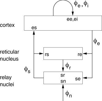
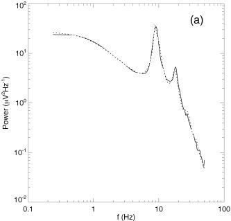
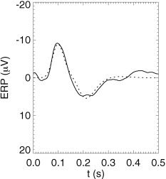
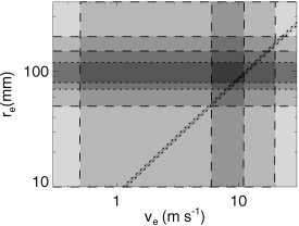
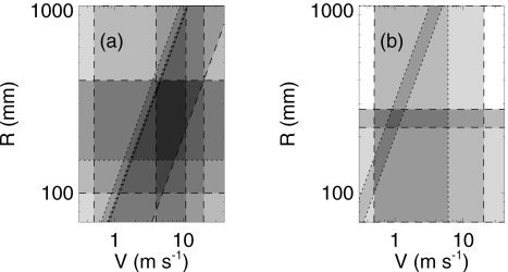
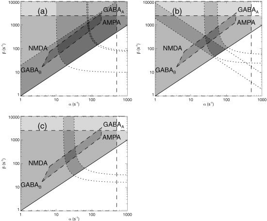
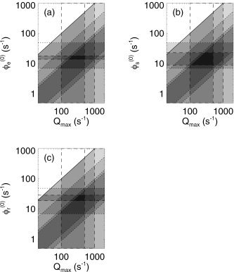
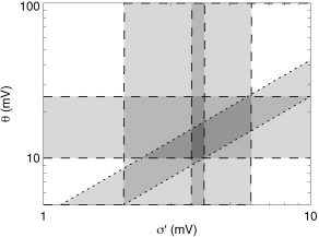
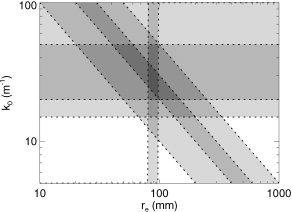
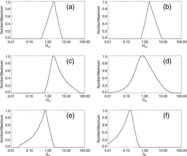
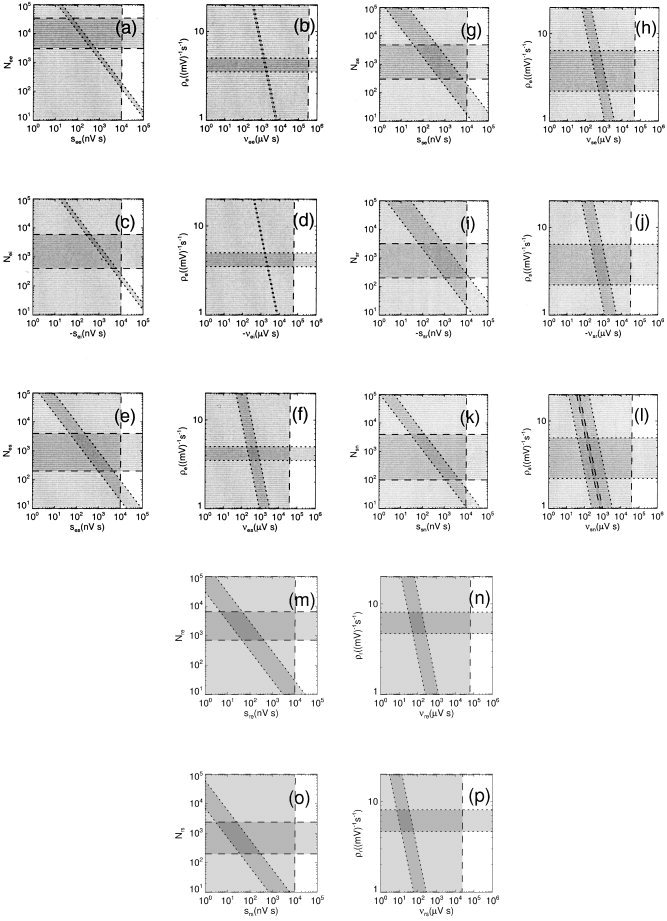
It is shown that new model-based electroencephalographic (EEG) methods can quantify neurophysiologic parameters that underlie EEG generation in ways that are complementary to and consistent with standard physiologic techniques. This is done by isolating parameter ranges that give good matches between model predictions and a variety of experimental EEG-related phenomena simultaneously. Resulting constraints range from the submicrometer synaptic level to length scales of tens of centimeters, and from timescales of around 1 ms to 1 s or more, and are found to be consistent with independent physiologic and anatomic measures. In the process, a new method of obtaining model parameters from the data is developed, including a Monte Carlo implementation for use when not all input data are available. Overall, the approaches used are complementary to other methods, constraining allowable parameter ranges in different ways and leading to much tighter constraints overall. EEG methods often provide the most restrictive individual constraints. This approach opens a new, noninvasive window on quantitative brain analysis, with the ability to monitor temporal changes, and the potential to map spatial variations. Unlike traditional phenomenologic quantitative EEG measures, the methods proposed here are based explicitly on physiology and anatomy.


Figures

Figure 1
Figure 1
Schematic of corticothalamic interactions, showing the connectivities and locations ab of the gains G ab for impulses from neurons b incident on neurons of type a. The cortex and the reticular and relay nuclei of the thalamus are shown as rectangles, along with the main neural projections between them (the latter are indicated by arrows, labeled with the type of activity that projects). External activity is indicated by ϕn.
Figure 2
Figure 2
Model predictions vs. data, showing predicted EEG spectrum for a normal adult in the relaxed eyes‐closed state (dotted curve) vs. data (solid) [Robinson et al., 2003a].
Figure 3
Figure 3
Model predictions vs. data, showing predicted background auditory ERP (dotted) vs. data (solid) [Robinson et al., 2003a].
Figure 4
Figure 4
Parameter space defined by the corticocortical axonal range r e and axonal velocity νe. Dashed lines indicate standard physiologic limits obtained from the literature, whereas dotted lines show model‐based constraints (see text for details). Dark shading indicates parameter zones that are consistent with multiple constraints.
Figure 5
Figure 5
Parameter space defined by the corticothalamic loop length R and axonal velocity V. Dashed lines indicate standard physiologic limits obtained from the literature, whereas dotted lines show model‐based constraints (see text for details). Dark shading generally indicates parameter zones that are consistent with multiple constraints.
Figure 6
Figure 6
Parameter space defined by the rates β and α of soma potential rise and fall, respectively, allowing for combined synaptic and dendritic dynamics. Dashed lines indicate physiologic limits obtained from the literature, whereas dotted lines show model‐based constraints (see text for details), and the solid line bounds the unphysical region with β < α. Dark shading indicates parameter zones that are consistent with multiple constraints.
Figure 7
Figure 7
Parameter space defined by the maximum and steady‐state firing rates. Dashed lines indicate physiologic limits obtained from the literature, whereas dotted lines show model‐based constraints (see text for details), and the solid line bounds the unphysical region with Q max < ϕa(0). The uppermost diagonal bound is a superposition of both types of constraint. Dark shading indicates parameter zones that are consistent with multiple constraints. a: Cortex. b: Thalamic relay nuclei. c: Thalamic reticular nucleus.
Figure 8
Figure 8
Parameter space defined by the spread in thresholds σ′ and the mean threshold relative to resting θ. Dashed lines indicate physiologic limits obtained from the literature, whereas dotted lines show model‐based constraints (see text for details). Dark shading indicates parameter zones that are consistent with multiple constraints.
Figure 9
Figure 9
Parameter space defined by the mean corticocortical excitatory axonal range r e and the volume conduction low‐pass wave number k 0. Dotted lines show model‐based constraints (see text for details). Dark shading indicates parameter zones that are consistent with multiple constraints.
Figure 10
Figure 10
Histograms of gains obtained by Monte Carlo inversion of equations (41) to (46), using linear fit parameters from Rowe et al. [2003] and values of ϕa(0) in physiologic ranges (see text). Each histogram is normalized by dividing by its maximum value. a: G es. b: G se. c: G sr. d: G sn. e: G re. f: G rs.
Figure 11
Figure 11
Parameter spaces defined by synaptic strengths s ab and numbers of connections N ab (left column) and by synaptic products v ab and sigmoidal slopes ρa (right column). Dashed lines indicate standard physiologic limits obtained from the literature, whereas dotted lines show model‐based constraints (see text for details). Dark shading indicates parameter zones that are consistent with multiple constraints. a, b: ab = ee. c, d: ab = ei. e, f: ab = es. g, h: ab = se. i, j: ab = sr. k, l: ab = sn. m, n: ab = re. o, p: ab = rs.

Similar articles

Cited by

References

    1. Bal T, Debay D, Destexhe A (2000): Cortical feedback controls the frequency and synchrony of oscillations in the visual thalamus. J Neurosci 20: 7478–7488. - PMC - PubMed
    1. Bal T, von Krosigk M, McCormick DA (1995): Synaptic and membrane mechanisms underlying synchronized oscillations in the ferret lateral geniculate nucleus in vitro. J Physiol 483.3: 641–663. - PMC - PubMed
    1. Barrionuevo G, Benoit O, Tempier P (1981): Evidence for two types of firing pattern during the sleep‐waking cycle in the reticular thalamic nucleus of the cat. Exp Neurol 72: 486–501. - PubMed
    1. Braitenberg V, Schüz A (1998): Cortex: statistics and geometry of neuronal connectivity (2nd ed). Berlin: Springer.
    1. Breakspear M (2002): Nonlinear phase desynchronization in human electroencephalographic data. Hum Brain Mapp 15: 175–198. - PMC - PubMed

Publication types

LinkOut - more resources