Comparative Study
. 2004 Jul 28:5:25.
doi: 10.1186/1471-2202-5-25.

Biochemical enrichment and biophysical characterization of a taste receptor for L-arginine from the catfish, Ictalurus puntatus

Affiliations
Comparative Study

Biochemical enrichment and biophysical characterization of a taste receptor for L-arginine from the catfish, Ictalurus puntatus

William Grosvenor et al. BMC Neurosci. .

Abstract

Background: The channel catfish, Ictalurus punctatus, is invested with a high density of cutaneous taste receptors, particularly on the barbel appendages. Many of these receptors are sensitive to selected amino acids, one of these being a receptor for L-arginine (L-Arg). Previous neurophysiological and biophysical studies suggested that this taste receptor is coupled directly to a cation channel and behaves as a ligand-gated ion channel receptor (LGICR). Earlier studies demonstrated that two lectins, Ricinus communis agglutinin I (RCA-I) and Phaseolus vulgaris Erythroagglutinin (PHA-E), inhibited the binding of L-Arg to its presumed receptor sites, and that PHA-E inhibited the L-Arg-stimulated ion conductance of barbel membranes reconstituted into lipid bilayers.

Results: Both PHA-E and RCA-I almost exclusively labeled an 82-84 kDa protein band of an SDS-PAGE of solubilized barbel taste epithelial membranes. Further, both rhodamine-conjugated RCA-I and polyclonal antibodies raised to the 82-84 kDa electroeluted peptides labeled the apical region of catfish taste buds. Because of the specificity shown by RCA-I, lectin affinity was chosen as the first of a three-step procedure designed to enrich the presumed LGICR for L-Arg. Purified and CHAPS-solubilized taste epithelial membrane proteins were subjected successively to (1), lectin (RCA-I) affinity; (2), gel filtration (Sephacryl S-300HR); and (3), ion exchange chromatography. All fractions from each chromatography step were evaluated for L-Arg-induced ion channel activity by reconstituting each fraction into a lipid bilayer. Active fractions demonstrated L-Arg-induced channel activity that was inhibited by D-arginine (D-Arg) with kinetics nearly identical to those reported earlier for L-Arg-stimulated ion channels of native barbel membranes reconstituted into lipid bilayers. After the final enrichment step, SDS-PAGE of the active ion channel protein fraction revealed a single band at 82-84 kDa which may be interpreted as a component of a multimeric receptor/channel complex.

Conclusions: The data are consistent with the supposition that the L-Arg receptor is a LGICR. This taste receptor remains active during biochemical enrichment procedures. This is the first report of enrichment of an active LGICR from the taste system of vertebrata.


Figures
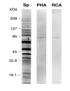
Figure 1
Figure 1
Lectin labelling of solubilized barbel epithelial proteins SDS-PAGE (4–20%) of 10 μg of solubilized barbel homogenate stained with silver (Lane "Sp") or probed with PHA-E lectin (Lane "PHA") or RCA-I lectin (Lane "RCA") using lectins at 10 μg/ml with ABC detection. Both lectins label a band at 82 – 84 kDa and lightly label at least two other bands, one near 88 kDa, the other near 120 kDa.
Figure 2
Figure 2
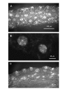
RCA-I lectin histochemistry on barbel of catfish, I. punctatus (albino). Barbels were fixed in 4% PFA, PBS, cryostat sectioned at 10 microns and histochemically probed with conjugated RCA-I at 1/200 dilution of the manufacturer's stock. (A). Surface labelling by RCA-I shows preferential recognition of binding sites primarily at the apical endings of taste buds. (B). Labelling by RCA-I of the apical region of two taste buds. (C). Labelling by RCA-I of horizontal section through the barbel showing reactive taste buds lining the epithelium.
Figure 3
Figure 3
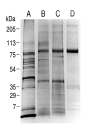
SDS-PAGE (4–20%) of solubilized barbel protein, visualized with silver stain, before (A) and after (B, C, D) each enrichment step. Each lane was loaded with 5 μg of protein. Lane A: Total protein of fraction Sp. Lane B: Galactose eluted protein from the RCA column containing L-Arg stimulated ion channel activity; Lane C: Protein of the first peak fractions of the Sephacryl gel filtration step containing L-Arg stimulated ion channel activity; and Lane D: Protein containing L-Arg stimulated ion channel activity from the ion exchange chromatography enrichment step.
Figure 4
Figure 4
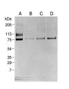
Western blots of SDS-PAGE protein samples before (A) and after (B, C, and D) each enrichment step. Western blots were performed using GP1. Although the identity of the samples applied to each lane was the same as illustrated in Figure 3, the amount of protein applied to each lane differed. (A). Fraction Sp, 5 μg, (B). Galactose eluted protein from the RCA column containing L-Arg stimulated ion channel activity, 1.6 μg, (C). Protein from the first peak fractions of the gel filtration step containing L-Arg stimulated ion channel activity, 0.2 μg, (D). Protein fraction containing L-Arg stimulated ion channel activity from the pH 9 fraction of the ion exchange procedure, 0.2 μg. Note that in spite of lowering protein amounts in lanes A – D, the intensity of the Western blot increases, indicative of an enrichment of the antigen protein(s) near 82–84 kDa.
Figure 5
Figure 5
Immunohistochemistry of catfish barbel taste buds using antibodies GP1 and GP2. Barbels were fixed in 4% PFA, PBS, and cryostat sectioned at 10 microns. Sections were probed using indicated dilutions of antibodies GP1 and GP2. (A). Section through a barbel immuno-stained with GP1 at 1/8000 dilution. The lines from "TB" point to labelled taste buds. (B). The apical aspects of three taste buds immuno-stained with 1/12000 dilution of GP2. (C). Cross section through a taste bud immuno-stained with 1/16000 dilution of GP1. (D). A single taste bud immuno-stained with 1/8000 dilution of GP2. (E). Second antibody control showing barbel without exposure to primary antibodies.
Figure 6
Figure 6
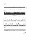
Single channel recording of the activity of the putative L-ArgR in planar lipid bilayers. Proteoliposomes containing protein from the first peak fractions off the Sephacryl S-300 elution were fused to planar lipid bilayers (DOPS:DOPE, 1:1) as described in Methods. (A). An initial control trace was obtained after addition proteoliposomes to the membrane bathing solution, before the addition of L-Arg. The three rows are from a continuous recording. (B). The addition of 10 μM L-Arg to the cis-side of the bilayer evoked regular periodic channel activity. A portion of this current record is shown at an expanded scale. (C). After several minutes of recording, the addition of 100 μM D-Arg to the cis-side resulted in the cessation of activity. Transmembrane potential was -100 mV. Traces shown in all panels are continuous records of that specific condition.
Figure 7
Figure 7
Amplitude histogram of current fluctuations of the putative L-ArgR in lipid bilayers. Current fluctuations were calculated from studies similar to those presented in Figure 6, part B, using the same protein fraction, measured at -100 mV transmembrane potential. The histogram was fit by a Gaussian distribution and mean current values were obtained from the center of the distribution. Bilayer was DOPS:DOPE, 1:1.
Figure 8
Figure 8
Cation-anion selectivity of L-ArgR channels. The figure shows current elicited by voltage ramps across a lipid bilayer (DOPS:DOPE, 1:1) containing several channels from the Sephacryl S-300 step, in the presence of a 4-fold gradient of NaCl across the membrane (cis and trans chambers contained 100 and 25 mM NaCl respectively). The reversal potential is -14 mV, corresponding to weak cation selectivity (PNa/PCl = 2.2).
Figure 9
Figure 9
The unitary current/voltage (I-V) relationship of channels formed by protein of each enrichment step. The I-V relationships were obtained using L-Arg – active protein from the RCA-I lectin column (o) from the first peak fractions off the Sephacryl S-300 HR column (■) and from the pH 9 elution of the ion-exchange column (▲). Measurements were made under symmetrical conditions of 100 mM NaCl, 1 mM CaCl2 and 5 mM MOPS (pH = 7.2). Data points indicate the Mean ± S.D. The data sets are well fit by a linear regression (r = 0.99 solid and dotted lines) with slopes of 58, 67, and 73 pS respectively. Bilayer was DOPS:DOPE, 1:1.

Similar articles

Cited by

  • Characterization of ligands for fish taste receptors.
    Oike H, Nagai T, Furuyama A, Okada S, Aihara Y, Ishimaru Y, Marui T, Matsumoto I, Misaka T, Abe K. Oike H, et al. J Neurosci. 2007 May 23;27(21):5584-92. doi: 10.1523/JNEUROSCI.0651-07.2007. J Neurosci. 2007. PMID: 17522303 Free PMC article.
  • Pseudogenization of a sweet-receptor gene accounts for cats' indifference toward sugar.
    Li X, Li W, Wang H, Cao J, Maehashi K, Huang L, Bachmanov AA, Reed DR, Legrand-Defretin V, Beauchamp GK, Brand JG. Li X, et al. PLoS Genet. 2005 Jul;1(1):27-35. doi: 10.1371/journal.pgen.0010003. Epub 2005 Jul 25. PLoS Genet. 2005. PMID: 16103917 Free PMC article.
  • Genetics of taste receptors.
    Bachmanov AA, Bosak NP, Lin C, Matsumoto I, Ohmoto M, Reed DR, Nelson TM. Bachmanov AA, et al. Curr Pharm Des. 2014;20(16):2669-83. doi: 10.2174/13816128113199990566. Curr Pharm Des. 2014. PMID: 23886383 Free PMC article. Review.

References

    1. Witt M. Reutter, K. Miller, I.J. Morphology of the peripheral taste system. In: Doty RL, editor. Handbook of Olfaction and Gustation. Second. New York, Marcel Dekker, Inc.; 2003. pp. 651–677.
    1. Lindemann B. Receptors and transduction in taste. Nature. 2001;413:219–225. doi: 10.1038/35093032. - DOI - PubMed
    1. Bachmanov AA, Li X, Li S, Neira M, Beauchamp GK, Azen EA. High-resolution genetic mapping of the sucrose octaacetate taste aversion (Soa) locus on mouse Chromosome 6. Mamm Genome. 2001;12:695–699. doi: 10.1007/s00335-001-2061-7. - DOI - PMC - PubMed
    1. Kitagawa M, Kusakabe Y, Miura H, Ninomiya Y, Hino A. Molecular genetic identification of a candidate receptor gene for sweet taste. Biochem Biophys Res Commun. 2001;283:236–242. doi: 10.1006/bbrc.2001.4760. - DOI - PubMed
    1. Max M, Shanker YG, Huang L, Rong M, Liu Z, Campagne F, Weinstein H, Damak S, Margolskee RF. Tas1r3, encoding a new candidate taste receptor, is allelic to the sweet responsiveness locus Sac. Nat Genet. 2001;28:58–63. doi: 10.1038/88270. - DOI - PubMed

Publication types

MeSH terms

LinkOut - more resources