. 2004 Jul 28:5:28.
doi: 10.1186/1471-2121-5-28.

Calcineurin activation influences muscle phenotype in a muscle-specific fashion

Affiliations

Calcineurin activation influences muscle phenotype in a muscle-specific fashion

Robert J Talmadge et al. BMC Cell Biol. .

Abstract

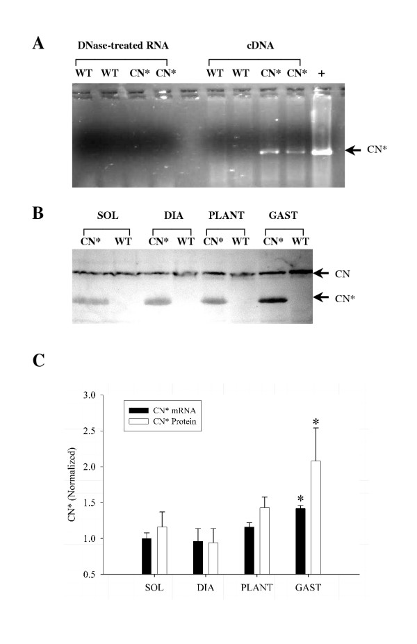
Background: The calcium activated protein phosphatase 2B, also known as calcineurin, has been implicated as a cell signaling molecule involved with transduction of physiological signals (free cytosolic Ca2+) into molecular signals that influence the expression of phenotype-specific genes in skeletal muscle. In the present study we address the role of calcineurin in mediating adaptations in myosin heavy chain (MHC) isoform expression and muscle mass using 3-month old wild-type (WT) and transgenic mice displaying high-level expression of a constitutively active form of calcineurin (MCK-CN* mice).

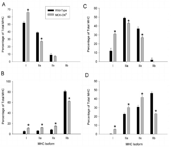
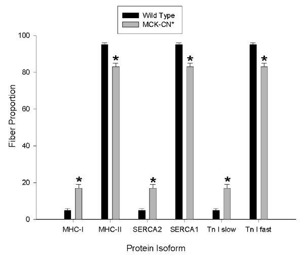
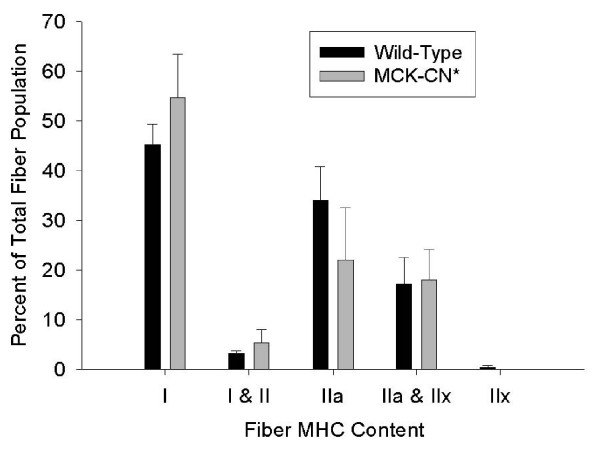
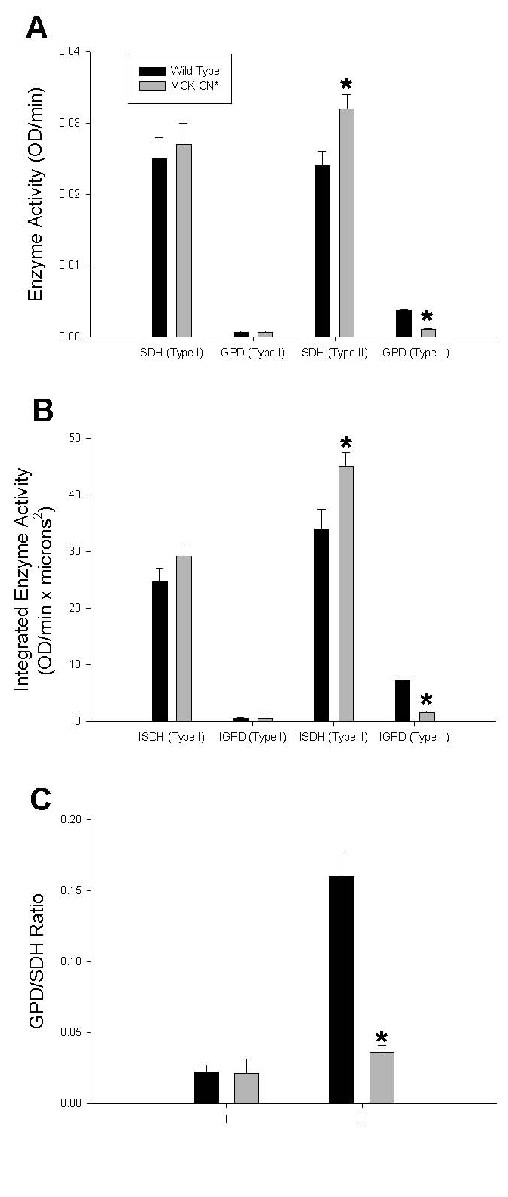
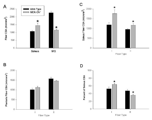
Results: Slow muscles, e.g., soleus, were significantly larger (by ~24%), whereas fast muscles, e.g., medial gastrocnemius (MG) and tibialis anterior were significantly smaller (by ~26 and ~16%, respectively) in MCK-CN* mice compared to WT. The masses of mixed phenotype muscles, such as the plantaris and the extensor digitorum longus, were not significantly changed from WT. The soleus, plantaris, MG and diaphragm displayed shifts toward slower MHC isoforms, e.g., soleus from WT mice contained ~52% MHC-I, ~39% MHC-IIa, and ~9% MHC-IIx, whereas MCK-CN* mice had ~67% MHC-I, ~26% MHC-IIa, and ~7% MHC-IIx. The specific isoforms that were either up or down-regulated were muscle-specific. For instance, the proportion of MHC-IIa was decreased in the soleus and diaphragm, but increased in the plantaris and MG of MCK-CN* mice. Also, the proportion of MHC-IIx was unchanged in the soleus, decreased in the diaphragm and increased in the plantaris and MG of MCK-CN* relative to WT mice. Fast to slow shifts in fiber type proportions were evident for the plantaris, but not the soleus. Fast, but not slow, plantaris fibers of MCK-CN* mice had higher oxidative and lower glycolytic properties than WT.

Conclusion: These data suggest that calcineurin activation can influence muscle phenotype and that the specific influence of calcineurin activation on the phenotypic and mass characteristics of a muscle is dependent upon the original phenotypic state of the muscle.


Figures

Figure 1
Figure 1
A) SYBR® Green I stained agarose gel of PCR products following RT-PCR for the MCK-CN* mRNA product. No specific product (~900 bp, arrow) is observed in the DNase-treated RNA samples (first 4 lanes) of either wild-type (WT) or MCK-CN* transgenic (CN*) mouse gastrocnemius. Therefore, there is no contaminating genomic DNA in the RNA samples used for RT-PCR. As expected, RT-PCR of the cDNA (second four lanes) reveals that expression of the transgene is only observed in the MCK-CN* mice. A positive (+) control of tail DNA from an MCK-CN* mouse is shown on the right. B) Western blot of CN* and WT mouse soleus (SOL), diaphragm (DIA), plantaris (PLANT) and gastrocnemius (GAST) muscles for calcineurin. The polyclonal MAb recognized both the endogenous (CN, ~60 kDa) and constitutively active (CN*, ~45 kDa) forms. As expected, muscle from WT muscles did not contain the CN* protein. C) Relative semi-quantitative analysis of CN* mRNA (via RT-PCR) and protein (western blotting) in muscles of MCK-CN* mice. All values were normalized to the same soleus muscle (n = 3 – 6 per group). The asterisk (*) denotes significantly different (p ≤ 0.05) from other muscles.
Figure 2
Figure 2
Adult myosin heavy chain (MHC) isoform proportions of the soleus (A), medial gastrocnemius (B), diaphragm (C), and plantaris (D) muscles of wild-type (dark bars) and MCK-CN* transgenic (gray bars) mice as determined by SDS-PAGE of whole muscle extracts. The * denotes significantly different from wild-type at p ≤ 0.05. Note the elevated proportions of slower MHC isoforms in the transgenic mice compared to wild-type.
Figure 3
Figure 3
Proportions of plantaris fibers staining positively for slow and fast isoforms of MHC, sarco(endo)plasmic reticulum Ca2+ ATPase (SERCA), and troponin I (Tn I) in wild-type and MCK-CN* transgenic mice. MHC-I, SERCA2, and TN I slow are the slow muscle isoforms and MHC-II, SERCA1 and Tn I fast are the fast muscle isoforms. The * denotes significantly different from wild-type at p ≤ 0.05. Note the elevated proportions of fibers staining positively for the slow isoforms in the transgenic mice compared to wild-type. All fibers that stained positively for the slow MHC (MHC-I) were positive for the slow isoforms of SERCA and Tn I and all fibers that stained positively for the fast MHC (MHC-II) were positive for the fast isoforms of SERCA and Tn I.
Figure 4
Figure 4
Proportions of soleus fibers staining positively for specific fast and slow MHC isoforms in wild-type and MCK* transgenic mice. No fibers stained positively for MHC-IIb with mAb BF-F3, thus antibody RT-D9 which is specific for MHC-IIx and MHC-IIb indicated the presence of MHC-IIx. No significant differences in fiber proportions of the soleus were found between wild-type and MCK-CN* mice.
Figure 5
Figure 5
Oxidative and glycolytic enzymatic profiles of individual plantaris fibers of wild-type and MCK-CN* transgenic mice. The * denotes significantly different from wild-type at p ≤ 0.05. A) The specific activity of succinate dehydrogenase (SDH, oxidative marker) was increased in fast (type II) plantaris fibers, but unchanged in slow (type I) fibers. The specific activity of α-glycerophosphate dehydrogenase (GPD, glycolytic marker) was significantly decreased in fast plantaris fibers, but not slow fibers. B) The integrated activity (see methods for explanation of integrated activity) of succinate dehydrogenase (SDH, oxidative marker) was increased in fast (type II) plantaris fibers, but unchanged in slow (type I) fibers. The integrated activity of α-glycerophosphate dehydrogenase (GPD, glycolytic marker) was significantly decreased in fast plantaris fibers, but not slow fibers. C) The GPD/SDH ratio (glycolytic/oxidative ratio) was significantly decreased in fast fibers, but unchanged in slow fibers of the plantaris of MCK-CN* mice relative to wild-type.
Figure 6
Figure 6
Individual fiber cross sectional areas (CSAs) in muscles of wild-type and MCK-CN* mice. A) Mean fiber CSAs in soleus and medial gastrocnemius (MG) muscle regardless of fiber type. B) Mean fiber CSAs of slow (type I) and fast (type II) fibers of the plantaris muscle. C) Mean fiber CSAs of slow (type I) and fast (type II) fibers of the soleus muscle. D) The percent of the entire soleus muscle cross-section occupied by slow (type I) and fast (type II) fibers. The * denotes significantly different from wild-type at p ≤ 0.05.

References

    1. Talmadge RJ. Myosin heavy chain isoform expression following reduced neuromuscular activity: potential regulatory mechanisms. Muscle Nerve. 2000;23:661–679. doi: 10.1002/(SICI)1097-4598(200005)23:5<661::AID-MUS3>3.0.CO;2-J. - DOI - PubMed
    1. Pette D, Vrbova G. Adaptation of mammalian skeletal muscle fibers to chronic electrical stimulation. Rev Physiol Biochem Pharmacol. 1992;120:115–202. - PubMed
    1. Bigard X, Sanchez H, Zoll J, Mateo P, Rousseau V, Veksler V, Ventura-Clapier R. Calcineurin Co-regulates contractile and metabolic components of slow muscle phenotype. J Biol Chem. 2000;275:19653–19660. doi: 10.1074/jbc.M000430200. - DOI - PubMed
    1. Chin ER, Olson EN, Richardson JA, Yang Q, Humphries C, Shelton JM, Wu H, Zhu W, Bassel-Duby R, Williams RS. A calcineurin-dependent transcriptional pathway controls skeletal muscle fiber type. Genes Dev. 1998;12:2499–2509. - PMC - PubMed
    1. Delling U, Tureckova J, Lim HW, De Windt LJ, Rotwein P, Molkentin JD. A calcineurin-NFATc3-dependent pathway regulates skeletal muscle differentiation and slow myosin heavy-chain expression. Mol Cell Biol. 2000;20:6600–6611. doi: 10.1128/MCB.20.17.6600-6611.2000. - DOI - PMC - PubMed

Publication types