. 2004 Jul 28;24(30):6715-23.
doi: 10.1523/JNEUROSCI.1594-04.2004.

Lipid rafts mediate the synaptic localization of alpha-synuclein

Affiliations

Lipid rafts mediate the synaptic localization of alpha-synuclein

Doris L Fortin et al. J Neurosci. .

Abstract

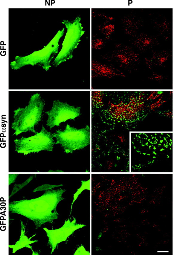
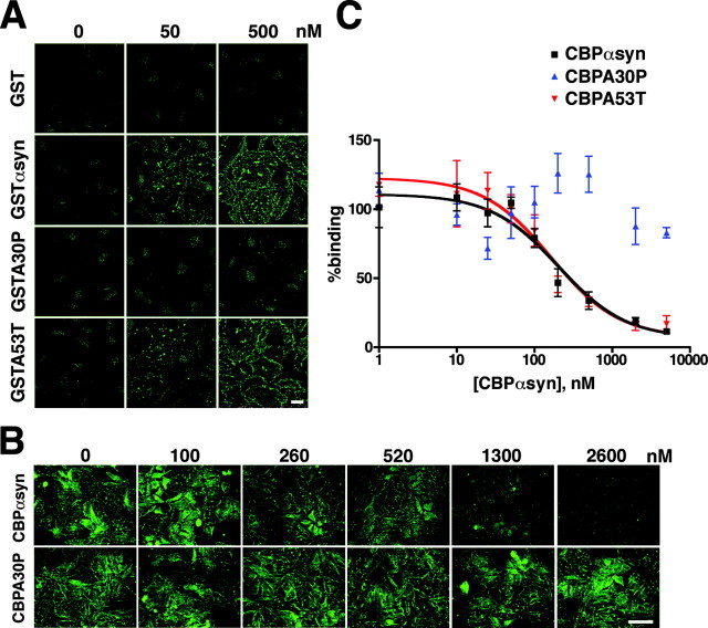
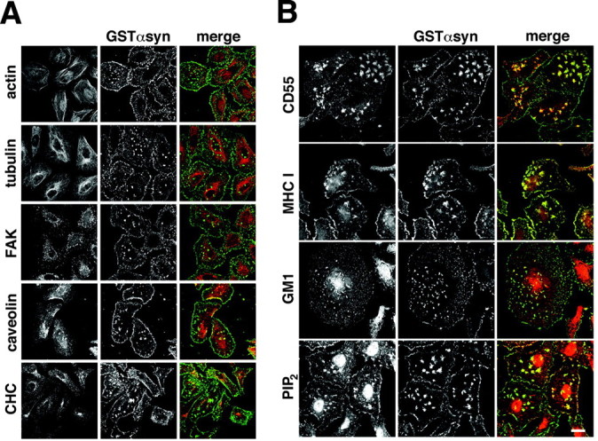
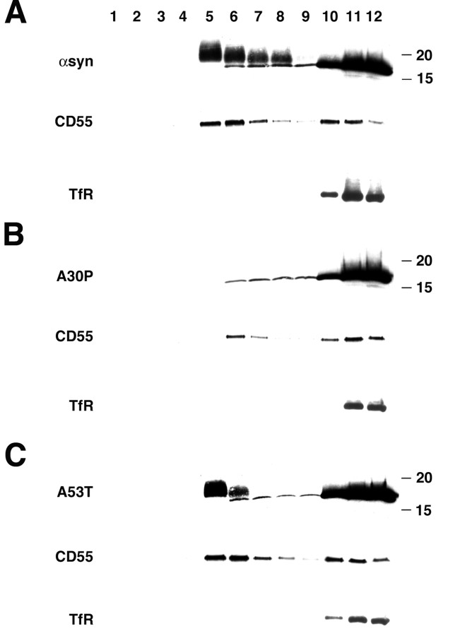
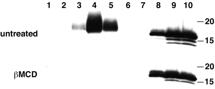
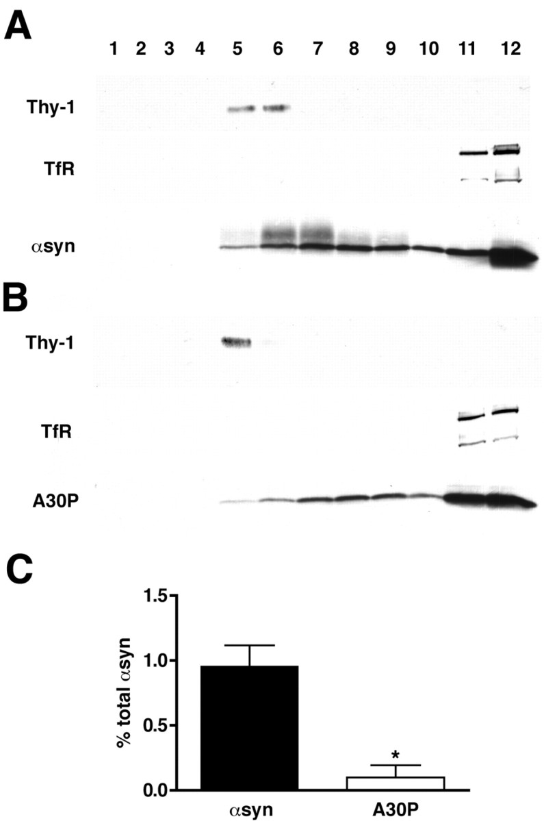
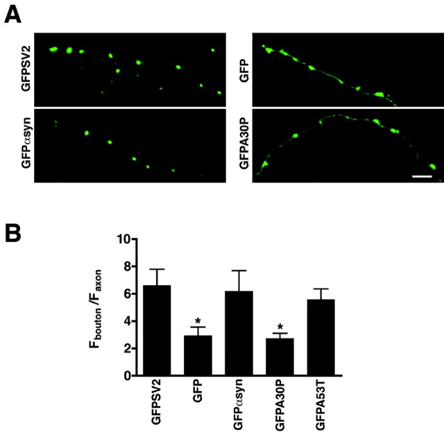
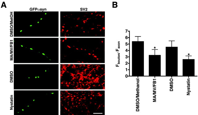
Alpha-synuclein contributes to the pathogenesis of Parkinson's disease (PD), but its precise role in the disorder and its normal function remain poorly understood. Consistent with a presumed role in neurotransmitter release and its prominent deposition in the dystrophic neurites of PD, alpha-synuclein localizes almost exclusively to the nerve terminal. In brain extracts, however, alpha-synuclein behaves as a soluble, monomeric protein. Using a binding assay to characterize the association of alpha-synuclein with cell membranes, we find that alpha-synuclein binds saturably and with high affinity to characteristic intracellular structures that double label for components of lipid rafts. Biochemical analysis demonstrates the interaction of alpha-synuclein with detergent-resistant membranes and reveals a shift in electrophoretic mobility of the raft-associated protein. In addition, the A30P mutation associated with PD disrupts the interaction of alpha-synuclein with lipid rafts. Furthermore, we find that both the A30P mutation and raft disruption redistribute alpha-synuclein away from synapses, indicating an important role for raft association in the normal function of alpha-synuclein and its role in the pathogenesis of PD.


Figures

Figure 1.
Figure 1.
Wild-type but not A30P α-synuclein binds specifically to permeabilized HeLa cells. A recombinant adenovirus was used to express GFP, α-synuclein-GFP (αsynGFP), and A30P-α-synuclein-GFP (A30PGFP) in HeLa cells. In intact (NP) cells, αsynGFP and A30PGFP have a diffuse cytosolic distribution, similar to GFP. After permeabilization with digitonin for 5 min (P), the cells specifically retain αsynGFP but not GFP. The PD-associated A30P mutation completely abolishes this retention. Permeabilized HeLa cells (P) are double stained for transferrin receptor (red). Scale bar, 20 μm.
Figure 2.
Figure 2.
Binding of α-synuclein to permeabilized HeLa cells is saturable and high affinity. A, HeLa cells were permeabilized with digitonin in the presence of 0-1000 nm recombinant GST, GST-α-synuclein (GSTαsyn), GST-A30P-α-synuclein (GSTA30P), and GST-A53T-α-synuclein (GSTA53T) and immunostained for GST. Binding is easily detectable at low concentrations of GSTαsyn (10 nm) and begins to saturate ∼0.5 μm. GST does not bind to permeabilized HeLa cells, whereas GSTA30P shows weak binding only at high concentrations (data not shown). The other PD-associated mutation, A53T, does not affect binding in this assay. Scale bar, 20 μm. B, HeLa cells were permeabilized in the presence of 200 nm GSTαsyn and 0-5 μm CBP-α-synuclein (CBPαsyn) and CBP-A30P-α-synuclein (CBPA30P). Bound GSTαsyn was detected by immunofluorescence for GST. Increasing concentrations of CBPαsyn compete with GSTαsyn, whereas CBPA30P does not compete. Scalebar, 20 μm. C, HeLa cells permeabilized in digitonin, GSTαsyn (200 nm), and 0-5 μm CBPαsyn were harvested and the extracts immunoblotted for GST. Bound GSTαsyn was quantified using a fluorescent scanner with wide linear response range and normalized to the amount of GSTαsyn bound in the absence of added competitor. Wild-type (CBPαsyn) and A53T CBP-α-synuclein (CBPA53T) compete with wild-type GSTαsyn in a dose-dependent manner. The results were fitted for one site competition to yield an IC50 ∼200 nm for both CBPαsyn and CBPA53T. However, CBPA30P does not compete with GSTαsyn. Error bars indicate SEM. n = 4-6; p < 0.05 between CBPαsyn and CBPA30P; Student's t test.
Figure 3.
Figure 3.
α-Synuclein colocalizes with components of lipid rafts in permeabilized HeLa cells. A, HeLa cells were permeabilized in the presence of 200 nm recombinant GSTαsyn, fixed, immunostained for GST, and double stained for actin, α-tubulin, FAK, caveolin-1, and clathrin heavy chain (CHC). Merged images show GST immunoreactivity in green and the different markers in red. Cells immunostained for α-tubulin were treated with 10 μm Taxol for 30 min before permeabilization to preserve microtubule structure. α-Synuclein does not colocalize with any of these markers. B, HeLa cells permeabilized in digitonin and 200 nm GSTαsyn were immunostained for GST and CD55, MHC class I, and PIP2. GM1 was detected using cholera toxin subunit B conjugated to biotin followed by streptavidin-Alexa 594. Merged images show GST immunoreactivity in green and the different markers in red. α-Synuclein colocalizes extensively with all of these known raft components. Scale bar, 20 μm.
Figure 4.
Figure 4.
α-Synuclein fractionates with detergent-resistant membranes. HeLa cells were transfected with wild-type (A), A30P (B), and A53T (C) α-syn, solubilized in TX-100 at 4°C for 30 min, and the extracts were separated by flotation through a sucrose density gradient. Wild-type and A53T α-synuclein comigrate with CD55 in light membrane fractions 5-8, whereas the transferrin receptor (TfR) remains at the bottom of the gradient. A30P-α-synuclein does not show a peak of immunoreactivity in light fractions. Fractions are numbered from the top of the gradient, and the size markers to the right indicate kilodaltons.
Figure 5.
Figure 5.
Cholesterol depletion eliminates the association of α-synuclein with light membranes. HeLa cells transfected with wild-type α-synuclein were treated with 20 mm β-methylcyclodextrin (β-MCD) for 60 min, solubilized in TX-100, and fractionated as described above. β-MCD eliminates the α-synuclein associated with light membranes observed in untreated cells. Fractions are numbered from the top of the gradient, and the size markers to the right indicate kilodaltons.
Figure 6.
Figure 6.
α-Synuclein associates with detergent-resistant membranes in mouse brain. Synaptosomes were isolated from the cortices of transgenic mice overexpressing human wild-type (A) and A30P (B) α-synuclein, solubilized in TX-100 and fractionated as described in Figure 3. A proportion of wild-type α-synuclein comigrates with Thy-1, whereas transferrin receptor (TfR) remains at the bottom of the gradient. Fractions were collected and numbered from the top of the gradient. α-Synuclein immunoreactivity in Thy-1-positive fractions was quantified by densitometry and expressed as a percentage of total immunoreactivity in all fractions. Quantification of the binding from three independent experiments (C) shows a clear reduction in binding for the A30P mutant relative to wild-type α-syn. Bar represents averages of three mice ± SD. *p = 0.0014; Student's t test.
Figure 7.
Figure 7.
The A30P mutation eliminates synaptic localization of α-synuclein. A, Hippocampal neurons transfected with GFPSV2, GFP, GFPαsyn, GFPA30P, and GFPA53T were fixed between 17-21 d in vitro, stained, and imaged by confocal microscopy. Scale bar, 5 μm. B, The synaptic enrichment of GFP-tagged protein was determined by calculating the ratio of fluorescence at synapses (identified by synaptophys in staining) to that in neighboring axons. GFPαsyn is enriched at synapses to an extent very similar to the GFP fusion with SV2, an integral membrane protein of synaptic vesicles. The PD-associated mutation A30P decreases synaptic enrichment of α-synuclein to a level similar to that of GFP alone. For each construct, nine fields from two independent cultures were examined. Bars represent averages ± SD. *p < 0.0001 (Student's t test) between GFPαsyn and GFP as well as GFPαsyn and GFPA30P. The analysis and quantification were performed blind to the plasmid transfected.
Figure 8.
Figure 8.
Cholesterol and sphingolipid depletion reduce the synaptic localization of α-synuclein. A, Hippocampal neurons transfected with GFPαsyn were treated with 250 μm mevalonic acid (MA), 4 μm mevastatin (MV), and 10 μm fumonisin B1 (FB1) (or DMS0/methanol control) at days 12 and 15 in vitro, fixed at day 18, immunostained for SV2, and imaged by confocal microscopy. In separate cultures, nystatin (10 μg/ml, or DMSO vehicle as control) was added at day 18 for 20 min before fixation and staining for SV2. Scale bar, 5 μm. SV2 was used as synaptic marker in this case because synaptophysin has been shown to bind cholesterol (Thiele et al., 2000) and hence might be sensitive to the manipulations. B, Synaptic enrichment was quantified as the ratio of fluorescence at the bouton to fluorescence in the axon (see Fig. 7). Both chronic treatment with MA/MV/FB1 and acute treatment with nystatin substantially reduce the synaptic localization of GFPαsyn. Eight fields from two independent cultures were examined. Bars represent mean ± SD. *p < 0.0003 (Student's t test) between drugs and the corresponding vehicle controls.

References

    1. Abeliovich A, Schmitz Y, Farinas I, Choi-Lundberg D, Ho WH, Castillo PE, Shinsky N, Verdugo JM, Armanini M, Ryan A, Hynes M, Phillips H, Sulzer D, Rosenthal A (2000) Mice lacking alpha-synuclein display functional deficits in the nigrostriatal dopamine system. Neuron 25: 239-252. - PubMed
    1. Breckenridge WC, Morgan IG, Zanetta JP, Vincendon G (1973) Adult rat brain synaptic vesicles. II. Lipid composition. Biochim Biophys Acta 320: 681-686. - PubMed
    1. Brown DA (2001) Lipid droplets: proteins floating on a pool of fat. Curr Biol 11: R446-449. - PubMed
    1. Brown DA, London E (1998) Functions of lipid rafts in biological membranes. Annu Rev Cell Dev Biol 14: 111-136. - PubMed
    1. Brown DA, Rose JK (1992) Sorting of GPI-anchored proteins to glycolipid-enriched membrane subdomains during transport to the apical cell surface. Cell 68: 533-544. - PubMed

Publication types

MeSH terms

LinkOut - more resources