. 2004 Jul 28;24(30):6799-809.
doi: 10.1523/JNEUROSCI.5463-03.2004.

Beta-amyloid peptide at sublethal concentrations downregulates brain-derived neurotrophic factor functions in cultured cortical neurons

Affiliations

Beta-amyloid peptide at sublethal concentrations downregulates brain-derived neurotrophic factor functions in cultured cortical neurons

Liqi Tong et al. J Neurosci. .

Abstract

The accumulation of beta-amyloid (Abeta) is one of the etiological factors in Alzheimer's disease (AD). It has been assumed that the underlying mechanism involves a critical role of Abeta-induced neurodegeneration. However, low levels of Abeta, such as will accumulate during the course of the disease, may interfere with neuronal function via mechanisms other than those involving neurodegeneration. We have been testing, therefore, the hypothesis that Abeta at levels insufficient to cause degeneration (sublethal) may interfere with critical signal transduction processes. In cultured cortical neurons Abeta at sublethal concentrations interferes with the brain-derived neurotrophic factor (BDNF)-induced activation of the Ras-mitogen-activated protein kinase/extracellular signal-regulated protein kinase (ERK) and phosphatidylinositol 3-kinase (PI3-K)/Akt pathways. The effect of sublethal Abeta(1-42) on BDNF signaling results in the suppression of the activation of critical transcription factor cAMP response element-binding protein and Elk-1 and cAMP response element-mediated and serum response element-mediated transcription. The site of interference with the Ras/ERK and PI3-K/Akt signaling is downstream of the TrkB receptor and involves docking proteins insulin receptor substrate-1 and Shc, which convey receptor activation to the downstream effectors. The functional consequences of Abeta interference with signaling are robust, causing increased vulnerability of neurons, abrogating BDNF protection against DNA damage- and trophic deprivation-induced apoptosis. These new findings suggest that Abeta engenders a dysfunctional encoding state in neurons and may initiate and/or contribute to cognitive deficit at an early stage of AD before or along with neuronal degeneration.


Figures

Figure 1.
Figure 1.
Viability of cortical neurons after Aβ1-42 treatment. Cortical neuronal cultures at 5 DIV were treated with Aβ1-42 at the indicated concentrations and length of time. Viability was determined by the trypan blue exclusion assay. Data shown are the mean ± SE (n = 4). In three separate experiments cell viability was measured with the MTT assay, which showed no significant reduction after exposure to 10 μm1-42 for 24 hr.
Figure 2.
Figure 2.
Pretreatment with sublethal concentrations of Aβ1-42 (5 or 10 μm) for 2 hr decreased the elevation of phosphorylated CREB levels induced by BDNF (50 ng/ml, 10 min). A, Western blot analysis of CREB phosphorylated at Ser133 (P-CREB) and total CREB (T-CREB). Aβ1-42 treatment resulted in a concentration-dependent decrease in the level of P-CREB but, considering all experiments (n = 3), had no significant effect on T-CREB levels. B, Quantification of the effect of pretreatment with 1, 5, or 10 μm1-42 (A1, A5, A10; A, here and in all other figures, stands for Aβ1-42). Estimates are the mean ± SEM (n = 3) expressed in terms of P-CREB levels obtained in the BDNF-exposed cultures (B, here and in all other figures, stands for BDNF). The effect of Aβ1-42 was significant (*p < 0.05, unpaired Student's t test). C, Pretreatment with 10 μm1-42 with random amino acid sequence [Aβ(R)] had no significant influence on the BDNF-induced increase of P-CREB levels. D, Immunohistochemical analysis of P-CREB. P-CREB immunoreactivity increased in BDNF-stimulated cultures (b) compared with unstimulated control (a). BDNF-induced increase in P-CREB immunoreactivity was suppressed by Aβ1-42 (5 μm) treatment (c). E, Exposure of the cultures to Aβ1-42 for 2 hr had no effect on P-CREB levels. In three experiments P-CREB levels in the presence of 5 and 10 μm1-42, respectively, were 104 ± 2 and 105 ± 6% of basal. F, Analysis of CRE-mediated transcriptional activity. Cortical neurons at 3 DIV were transfected with plasmid pCRE-Luc containing CRE sequences and a luciferase reporter gene (see Materials and Methods). Cells transfected with a CMV-luciferase control plasmid served to normalize CRE activity. After 40 hr the cultures were switched to fresh medium and incubated for 1 hr in the presence or the absence of 5 μm1-42. Transfected cortical cultures were incubated for an additional 9 hr either with or without the addition of 50 ng/ml BDNF, and transcriptional activity was measured by the luciferase assay. Aβ1-42 treatment decreased the BDNF-induced transcriptional activity of CRE. Estimates are the mean ± SEM (n = 3) expressed in terms of BDNF-induced transcriptional activity. The effect of Aβ1-42 was significant (*p < 0.05, unpaired Student's t test).
Figure 3.
Figure 3.
Pretreatment with sublethal Aβ1-42 (5 or 10 μm) decreased the BDNF-induced increase in the level of phosphorylated p42- and p44-MAPK/ERK elicited by stimulation with BDNF (50 ng/ml, 10 min). A, Western blot analysis showed that Aβ1-42 exposure resulted in a concentration-dependent decrease in the level of phosphorylated p42- and p44-MAPK (p42- and p44-P-MAPK). B, Quantification of the effects of pretreatment with 5 or 10 μm1-42. Estimates are the mean ± SEM (n = 3) expressed in terms of p42- and p44-P-MAPK levels obtained in the BDNF-exposed cultures. Effects of 5 and 10 μm1-42 (A5, A10) were significant (*p < 0.05, unpaired Student's t test).
Figure 4.
Figure 4.
A preparation comprising diffusible Aβ1-42 oligomers interferes with high potency with BDNF-induced signaling. A, Western blots showing that pretreatment (1 hr) with an Aβ1-42 oligomer preparation at the sublethal concentration of 200 nm compromised the increase in P-CREB levels induced by BDNF (50 ng/ml, 10 min). B, Quantification of the effect of 200nm1-42 oligomers. Estimates are the mean ± SEM (n = 3) expressed in terms of P-CREB levels obtained in BDNF-treated cultures. The effect of 200 nm1-42 oligomers was significant (*p < 0.05, unpaired Student's t test). C, Western blot analysis of P-MAPK. Pretreatment with 200 nm1-42 oligomers for 1 hr attenuated the increase in P-MAPK levels by exposure to 50 ng/ml BDNF for 10 min. D, Quantification of the effect of pretreatment with 200 nm1-42 oligomers for 1 hr. Estimates are expressed in terms of P-MAPK levels obtained in the BDNF-treated cultures; they are the mean ± SEM from three independent experiments. The effect of Aβ1-42 oligomers was significant (*p < 0.05, unpaired Student's t test).
Figure 5.
Figure 5.
Pretreatment with Aβ1-42 at sublethal concentrations decreased the level of BDNF-activated transcription factor Elk-1 (P-Elk-1) and the transcriptional activity of a serum response element-containing (SRE) construct. A, The phosphorylation of Elk-1 was examined by the use of an antibody against phosphorylated Elk-1 (P-Elk). Pretreatment with 5 or 10 μm1-42 for 2 hr attenuated the BDNF-induced increase in P-Elk-1 but had no significant effect on the total amount of Elk-1 (T-Elk). B, Quantification of the effects of pretreatment with 5 or 10 μm1-42 (B + A5 or B + A10). Estimates are the mean ± SEM (n = 3) expressed in terms of P-Elk-1 levels induced by BDNF (B). Effects of 5 and 10 μm1-42 (A5, A10) were significant (*p < 0.05, unpaired Student's t test). C, Analysis of SRE-mediated transcriptional activity. Cortical neurons at 3 DIV were transfected with plasmid pSRE-Luc containing SRE sequences and a luciferase reporter gene (see Materials and Methods). Cells transfected with a CMV-luciferase plasmid served to normalize SRE activity. After 40 hr the cultures were switched to fresh medium and incubated for 1 hr in the presence or the absence of 5 μm1-42. Transfected cortical cultures were incubated for an additional 9 hr either with or without the addition of 50 ng/ml BDNF, and transcriptional activity was measured by the luciferase assay. Aβ1-42 treatment decreased the BDNF-induced transcriptional activity of SRE. Estimates are the mean ± SEM (n = 3) expressed in terms of BDNF-induced transcriptional activity. The effect of Aβ1-42 was significant (*p < 0.05, unpaired Student's t test).
Figure 6.
Figure 6.
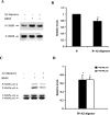
Sublethal Aβ1-42 treatment decreased the phosphorylated levels of protein kinases in the MAPK cascade (P-Raf and P-MEK). A, The phosphorylation of Raf was examined by the use of an antibody against phosphorylated Raf-1 (P-Raf). Pretreatment with 5 μm1-42 for 2 hr attenuated the BDNF-induced increase in P-Raf. B, Quantification of the effects of pretreatment with 5 μm1-42. Estimates are the mean ± SEM (n = 3) expressed in terms of P-Raf levels induced by BDNF (represented by B). Effects of 5 μm1-42 (B + A) were significant (*p < 0.05, unpaired Student's t test). C, Pretreatment with 5 μm1-42 decreased the BDNF-induced elevation in the level of phosphorylated MEK1/2 (P-MEK1/2). D, Quantification of the effects of pretreatment with 5 μm1-42 on BDNF-induced MEK1/2 phosphorylation. Estimates are the mean ± SEM (n = 3) expressed in terms of P-MEK1/2 levels induced by BDNF. Effects of 5 μm1-42 (B + A) were significant (*p < 0.05, unpaired Student's t test).
Figure 7.
Figure 7.
1-42 treatment decreased BDNF-induced Akt activation. A, Phosphorylated Akt (P-Akt) levels were determined with an antibody specific to P-Akt. Exposure to BDNF (50 ng/ml) for 10 min increased the amount of P-Akt. Pretreatment with 5 or 10 μm1-42 suppressed the effect of BDNF. Aβ1-42 had no effect on total Akt (T-Akt) levels. B, Quantification of the effect of pretreatment with 5 or 10 μm1-42. Estimates are the mean ± SEM (n = 3) expressed in terms of P-Akt levels obtained in the BDNF-exposed cultures. The effect of Aβ1-42 at 10 μm was significant (*p < 0.05, unpaired Student's t test). C, Pretreatment with 10 μm1-42 decreased BDNF-induced Akt activity, measured by the phosphorylation of glycogen synthase kinase-3α/β (GSK-3α/β), a substrate of Akt, using immunoprecipitated Akt from cell lysates as described in Materials and Methods. D, Quantification of the effects of pretreatment with 10 μm1-42. Estimates are the mean ± SEM (n = 3) expressed in terms of phosphorylated GSK-3α/β levels obtained in the BDNF-exposed cultures. The effect of Aβ1-42 was significant (*p < 0.05, unpaired Student's t test).
Figure 8.
Figure 8.
1-42 at sublethal concentrations does not interfere with the activation of TrkB and PLCγ. A, BDNF-induced phosphorylation of PLCγ at tyrosine residues. Cortical neurons were stimulated with 50 ng/ml BDNF for 10 min. Cell lysates were immunoprecipitated by agarose-linked anti-phosphotyrosine antibodies, and proteins of the washed immunoprecipitates were resolved by SDS-PAGE and subjected to Western blot analysis with a specific anti-PLCγ antibody (P-PLCγ). BDNF-induced PLCγ phosphorylation was not influenced significantly by Aβ1-42 pretreatment; in terms of phosphorylated PLCγ levels obtained in the BDNF-exposed cultures, the estimate in the Aβ1-42-treated cultures was 92 ± 7.1% (5 μm) and 97 ± 7.2% (10 μm) (n = 3). B, Phosphorylation of TrkB was examined via Western blotting with an antibody against TrkB phosphorylated at Tyr490 (P-TrkB). Pretreatment with 10 μm1-42 had no significant influence on the BDNF-induced P-TrkB content; in terms of P-TrkB levels obtained in the BDNF-exposed culture, the estimate in the Aβ1-42-pretreated cells was 94 ± 4.1% (n = 3).
Figure 9.
Figure 9.
1-42 treatment decreased the level of BDNF-activated IRS-1 and Shc. A, Pretreatment with 10 μm1-42 for 2 hr resulted in a significant reduction in the BDNF-induced increase in Tyr-phosphorylated IRS-1. IRS-1 was immunoprecipitated with an anti-IRS-1 antibody. The immunoprecipitates were analyzed by Western blotting with anti-phosphotyrosine antibody (top panel) and with anti-IRS-1 antibody (bottom panel). B, Quantification of the effects of pretreatment with 10 μm1-42. Estimates are the mean ± SEM (n = 3) expressed in terms of Tyr-phosphorylated IRS-1 levels obtained in the BDNF-exposed cultures (*p < 0.05, unpaired Student's t test). C, Pretreatment with 10 μm1-42 for 2 hr resulted in a significant reduction in BDNF-induced increase in Tyr-phosphorylated Shc isoforms as analyzed by immunoprecipitation with an Shc antibody, followed by Western blotting with anti-phosphotyrosine antibody (anti-pY; top panel) and with anti-Shc antibody (bottom panel). The isoforms are indicated as a, b, and c (approximate molecular weights, 66, 52, and 46 kDa, respectively). D, Quantification of the effects of pretreatment with 10 μm1-42. Estimates are the mean ± SEM (n = 3) expressed in terms of phosphorylated P-Shc isoform levels obtained in the BDNF-exposed cultures (*p < 0.05, unpaired Student's t test).
Figure 10.
Figure 10.
1-42 abrogated BDNF protection of cortical neurons from apoptosis induced either by exposure to camptothecin or by deprivation from trophic support. A, Cortical neurons were exposed to 5 μm camptothecin for 24 hr to induce apoptosis. Additions: Control, vehicle of BDNF solution; BDNF, 10 ng/ml; BDNF + A, 10 ng/ml BDNF + 5 μm1-42; A, 5 μm1-42. When Aβ1-42 was added, the cultures were preincubated in the presence of the peptide for 1 hr. Cell viability was assessed by trypan blue exclusion assay; data are the mean ± SE (n = 3). *p < 0.05 (ANOVA, Fisher's PLSD as the post hoc test). B, The serum-free “trophic” medium B27 was removed from cultures, and after a DMEM wash the cultures were incubated for 36 hr in DMEM in the presence or absence of 10 ng/ml BDNF ± 5 μm1-42. When Aβ was added, there was a 1 hr preincubation period in the presence of 5 μm1-42 before the removal of the B27 medium. For comparison, B27 medium was removed from sister cultures but replaced after a DMEM wash with new B27 medium containing the additives as indicated. Cell survival was assayed by MTT assay; data are the mean ± SE (n = 3). *p < 0.05 (ANOVA, Fisher's PLSD as the post hoc test).

Similar articles

Cited by

References

    1. Abe K, Saito H (2000) Amyloid beta neurotoxicity not mediated by the mitogen-activated protein kinase cascade in cultured rat hippocampal and cortical neurons. Neurosci Lett 292: 1-4. - PubMed
    1. Abel T, Kandel E (1998) Positive and negative regulatory mechanisms that mediate long-term memory storage. Brain Res Brain Res Rev 26: 360-378. - PubMed
    1. Atkins CM, Selcher JC, Petraitis JJ, Trzaskos JM, Sweatt JD (1998) The MAPK cascade is required for mammalian associative learning. Nat Neurosci 1: 602-609. - PubMed
    1. Bonni A, Brunet A, West AE, Datta SR, Takasu MA, Greenberg ME (1999) Cell survival promoted by the Ras-MAPK signaling pathway by transcription-dependent and -independent mechanisms [comments]. Science 286: 1358-1362. - PubMed
    1. Bourtchuladze R, Frenguelli B, Blendy J, Cioffi D, Schutz G, Silva AJ (1994) Deficient long-term memory in mice with a targeted mutation of the cAMP-responsive element-binding protein. Cell 79: 59-68. - PubMed

Publication types

MeSH terms

Substances

LinkOut - more resources