. 2004 Aug;24(16):7043-58.
doi: 10.1128/MCB.24.16.7043-7058.2004.

RPAP1, a novel human RNA polymerase II-associated protein affinity purified with recombinant wild-type and mutated polymerase subunits

Affiliations

RPAP1, a novel human RNA polymerase II-associated protein affinity purified with recombinant wild-type and mutated polymerase subunits

Célia Jeronimo et al. Mol Cell Biol. 2004 Aug.

Abstract

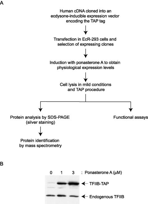
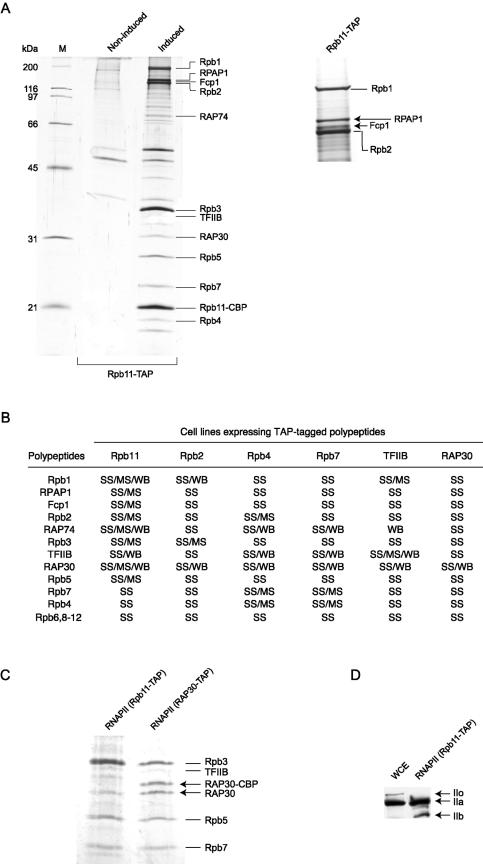
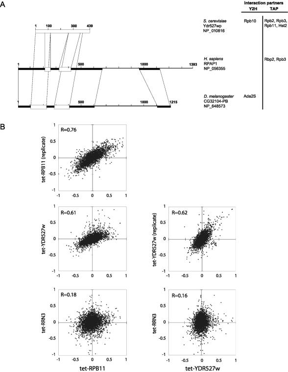
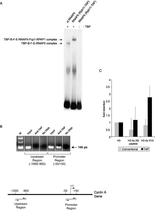
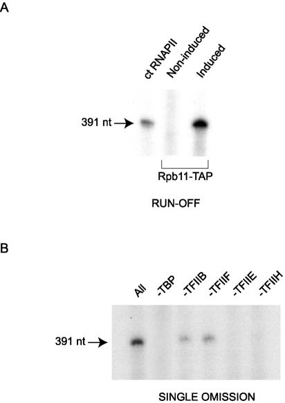
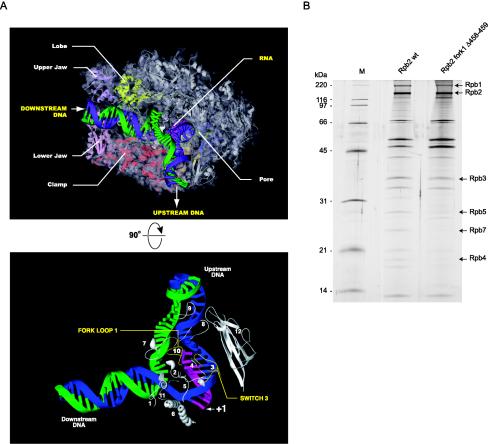
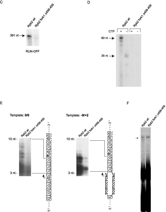
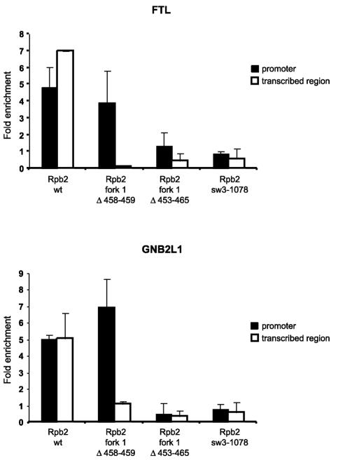
We have programmed human cells to express physiological levels of recombinant RNA polymerase II (RNAPII) subunits carrying tandem affinity purification (TAP) tags. Double-affinity chromatography allowed for the simple and efficient isolation of a complex containing all 12 RNAPII subunits, the general transcription factors TFIIB and TFIIF, the RNAPII phosphatase Fcp1, and a novel 153-kDa polypeptide of unknown function that we named RNAPII-associated protein 1 (RPAP1). The TAP-tagged RNAPII complex is functionally active both in vitro and in vivo. A role for RPAP1 in RNAPII transcription was established by shutting off the synthesis of Ydr527wp, a Saccharomyces cerevisiae protein homologous to RPAP1, and demonstrating that changes in global gene expression were similar to those caused by the loss of the yeast RNAPII subunit Rpb11. We also used TAP-tagged Rpb2 with mutations in fork loop 1 and switch 3, two structural elements located strategically within the active center, to start addressing the roles of these elements in the interaction of the enzyme with the template DNA during the transcription reaction.


Figures

FIG. 1.
FIG. 1.
TAP of human transcription factors. (A) Overview of the TAP procedure. (B) Data from a pilot experiment comparing the expression levels of a TAP-tagged polypeptide and its endogenous counterpart upon induction with ponasterone A. TFIIB-TAP expression in EcR-293 cells after induction with different concentrations of ponasterone A (0, 1, and 3 μM) for 24 h was compared to that of endogenous TFIIB by Western blotting with an antibody raised against TFIIB.
FIG.2.
FIG.2.
Purification of TAP-tagged human RNAPII complex. (A) Whole-cell extracts prepared from induced or noninduced EcR-293 cells programmed to express TAP-tagged Rpb11 (Rpb11-TAP) were purified by the TAP procedure, and the eluates were analyzed by SDS-PAGE. Gel slices containing the most abundant polypeptides were excised, digested with trypsin, and analyzed by peptide mass fingerprinting using MALDI-TOF analysis. The positions of core RNAPII subunits, including Rpb11 carrying the residual CBP domain (Rpb11-CBP), TFIIB, RAP74, RAP30, Fcp1, and RPAP1, a human gene product (DKFZP727M111 protein) of unknown function, are indicated. (B) TAP tagging of Rpb2, Rpb4, Rpb7, Rpb11, TFIIB, and RAP30 allowed for purification of the same 17-polypeptide RNAPII complex. The components of the complex were identified according to their molecular weights in SDS gels stained with silver (SS), their peptide mass fingerprints by MALDI-TOF analysis (MS), and their immunoreactivities in Western blots (WB). (C) SDS gel showing that both tagged (RAP30-CBP) and nontagged RAP30 are present in the RNAPII complex purified with TAP-tagged RAP30. Only nontagged RAP30 is found in the complex purified with TAP-tagged Rpb11. (D) The TAP-tagged RNAPII complex contains a hypophosphorylated CTD. The N-20 antibody, which specifically recognizes the N terminus of Rpb1, was used to detect both the hypophosphorylated (IIa) and hyperphosphorylated (IIo) forms of RNAPII in the whole-cell extract (WCE) and the Rpb11-TAP eluate. A truncated form of Rpb1 lacking the CTD (IIb) was also detected.
FIG. 3.
FIG. 3.
(A) Regions of homology between Homo sapiens RPAP1, D. melanogaster CG32104-PB, and S. cerevisiae Ydr527wp/RBA50, as determined by BLAST analyses. Boxes represent regions with significant alignments. Black boxes, E values ranging from 8 × 10−37 to 1 × 10⁻¹⁰; open boxes, E values ranging from 4 × 10⁻⁵ to 0.1. The M. musculus and R. norvegicus orthologues, with 80% identity with the H. sapiens RPAP1 over the full length, were not presented. Reported protein-protein interactions of RPAP1 (this article) and Ydr527wp/RBA50 (33) with transcription factors are indicated on the right. (B) Correlations among mRNA abundance profiles for Tet-promoter mutants of RPB11, YDR527w, and RRN3, measured by using oligonucleotide microarrays. Each mutant was compared to an isogenic control without the tetO7 promoter; log(ratios) are plotted. Tet-RPB11 and Tet-YDR527w were assayed twice, with the replicate cultures being grown, extracted, and assayed on different dates. The correlation of Tet-RPB11 and Tet-YDR527 to a Tet-promoter mutant of RRN3, a gene involved in polymerase I transcription, is shown for contrast.
FIG. 4.
FIG. 4.
TAP-tagged human RNAPII assembles on promoter DNA both in vitro and in vivo and binds to acetylated histones. (A) EMSA performed with 150 ng of purified calf thymus (ct) RNAPII or TAP-tagged human RNAPII in the presence of TBP, TFIIB, TFIIF, and TFIIE. A control reaction assembled in the absence of TBP was included. A radiolabeled DNA fragment containing the AdML promoter was used as a probe. (B) ChIP experiments showing that TAP-tagged RNAPII is specifically recruited to the cyclin A promoter in vivo. PCR amplification using sets of primers specific to chromosomal regions either encompassing (−50 to +50) or located upstream of (−1000 to −900) the transcription start site was used on DNA fragments enriched with IgG beads (anti-TAP). The input lanes correspond to DNA that was not subjected to immunoprecipitation. A PCR control that lacks DNA (no DNA) was included in each case. (C) Peptide binding experiment showing that TAP-tagged RNAPII is retained on acetylated histones. The results of three independent experiments were analyzed with ImageQuant software, corrected for the background, and shown relative to unmodified H3. Conventionally purified RNAPII was used as a negative control. The TAP-tagged RNAPII-bound histone tails acetylated at lysine 14 more efficiently than at lysine 9.
FIG. 5.
FIG. 5.
TAP-tagged human RNAPII complex can initiate transcription in vitro. (A) In vitro transcription reactions were reconstituted by using 165 ng of purified calf thymus (ct) RNAPII or TAP-tagged human RNAPII in the presence of TBP, TFIIB, TFIIF, TFIIE, and TFIIH. A control reaction performed with an eluate from noninduced cells was included. The linearized DNA template carries the AdML promoter and directs the synthesis of a 391-nt transcript. (B) Single omission assay in which each general transcription factor was omitted from the reconstituted system (all) described for panel A.
FIG. 6.
FIG. 6.
Purification and functional analysis of TAP-tagged human RNAPII with a mutation in fork loop 1. (A) (Top) Model showing elongating yeast RNAPII (30). The template DNA strand (blue) from −3 to +10 and the 9-nt RNA (pink) were placed according to the crystal structure. The remainder of the template strand and the coding strand (green) were modeled in Cinema 4D, and their exact positions are speculative. (Bottom) Simplified view of yeast RNAPII catalytic center. Different domains of Rpb1 and Rpb2 located near the DNA-RNA hybrid are shown. Numbers: 1 to 5, switches; 6, bridge helix; 7, rudder; 8, lid; 9, zipper; 10, fork loop 1; 11, fork loop 2; 12, wall. The parts of the lid (amino acids 250 to 258) and the rudder (amino acids 315 to 320) that are absent in the PDB files corresponding to elongating RNAPII were reconstructed based on the crystal structure of free yeast RNAPII (18). The missing parts of fork loop 1 (amino acids 468 to 476) and fork loop 2 (amino acids 503 to 508) were modeled in Cinema 4D, and their exact positions are speculative. (B) Silver-stained SDS gel of wild type TAP-tagged RNAPII (Rpb2 wt) and a mutant of human RNAPII carrying a two-amino-acid deletion in the Rpb2 fork loop 1 (Rpb2 fork1 Δ458-459) domain. The positions of some RNAPII subunits and molecular size markers are indicated. (C) In vitro transcription reactions (runoff) contained either 24 ng of wild-type RNAPII (Rpb2 wt) or 120 ng of Rpb2 fork1 Δ458-459 in the presence of TBP, TFIIB, TFIIF, TFIIE, and TFIIH. (D) Elongation assays were performed with a C-tailed template carrying a 15-C extension in the absence of general transcription factors. The coding strand lacks CMP except in the +25-to-+27 region. The positions of transcripts produced by RNAPII in the presence (+CTP; 60 nt) or absence (−CTP; 35 nt) of CTP are indicated. The reactions contained 88 ng of Rpb2 wt and 440 ng of Rpb2 fork1 Δ458-459. (E) Abortive initiation assays were performed with 100 ng of Rpb2 wt and 500 ng of Rpb2 fork1 Δ458-459 in the presence of TBP, TFIIB, TFIIF, and TFIIE on closed (0/0) or premelted (−9/+2) templates in the absence of GTP. The templates are schematized and the 3- to 10-nt abortive transcripts are indicated. (F) EMSAs were performed with the AdML promoter in the presence of TBP, TFIIB, TFIIF, and TFIIE. Two hundred nanograms of Rpb2 wt and 500 ng of Rpb2 fork 1 Δ58-459 were used in the reactions.
FIG. 6.
FIG. 6.
Purification and functional analysis of TAP-tagged human RNAPII with a mutation in fork loop 1. (A) (Top) Model showing elongating yeast RNAPII (30). The template DNA strand (blue) from −3 to +10 and the 9-nt RNA (pink) were placed according to the crystal structure. The remainder of the template strand and the coding strand (green) were modeled in Cinema 4D, and their exact positions are speculative. (Bottom) Simplified view of yeast RNAPII catalytic center. Different domains of Rpb1 and Rpb2 located near the DNA-RNA hybrid are shown. Numbers: 1 to 5, switches; 6, bridge helix; 7, rudder; 8, lid; 9, zipper; 10, fork loop 1; 11, fork loop 2; 12, wall. The parts of the lid (amino acids 250 to 258) and the rudder (amino acids 315 to 320) that are absent in the PDB files corresponding to elongating RNAPII were reconstructed based on the crystal structure of free yeast RNAPII (18). The missing parts of fork loop 1 (amino acids 468 to 476) and fork loop 2 (amino acids 503 to 508) were modeled in Cinema 4D, and their exact positions are speculative. (B) Silver-stained SDS gel of wild type TAP-tagged RNAPII (Rpb2 wt) and a mutant of human RNAPII carrying a two-amino-acid deletion in the Rpb2 fork loop 1 (Rpb2 fork1 Δ458-459) domain. The positions of some RNAPII subunits and molecular size markers are indicated. (C) In vitro transcription reactions (runoff) contained either 24 ng of wild-type RNAPII (Rpb2 wt) or 120 ng of Rpb2 fork1 Δ458-459 in the presence of TBP, TFIIB, TFIIF, TFIIE, and TFIIH. (D) Elongation assays were performed with a C-tailed template carrying a 15-C extension in the absence of general transcription factors. The coding strand lacks CMP except in the +25-to-+27 region. The positions of transcripts produced by RNAPII in the presence (+CTP; 60 nt) or absence (−CTP; 35 nt) of CTP are indicated. The reactions contained 88 ng of Rpb2 wt and 440 ng of Rpb2 fork1 Δ458-459. (E) Abortive initiation assays were performed with 100 ng of Rpb2 wt and 500 ng of Rpb2 fork1 Δ458-459 in the presence of TBP, TFIIB, TFIIF, and TFIIE on closed (0/0) or premelted (−9/+2) templates in the absence of GTP. The templates are schematized and the 3- to 10-nt abortive transcripts are indicated. (F) EMSAs were performed with the AdML promoter in the presence of TBP, TFIIB, TFIIF, and TFIIE. Two hundred nanograms of Rpb2 wt and 500 ng of Rpb2 fork 1 Δ58-459 were used in the reactions.
FIG. 7.
FIG. 7.
Purification and functional analysis of TAP-tagged human RNAPII with a mutation in switch 3. (A) Silver-stained SDS gel of wild-type TAP-tagged RNAPII (Rpb2 wt) and a mutant of human RNAPII carrying a triple alanine substitution in the Rpb2 switch 3 (Rpb2 sw3-1078) domain. (B) EMSAs were performed with the AdML promoter with TFIIB, TFIIF, and TFIIE in either the presence or the absence of TBP. The amounts used for the wild type and the mutant are indicated. (C) In vitro transcription reactions (runoff) contained different amounts of Rpb2 wt and Rpb2 sw3-1078 in the presence of TBP, TFIIB, TFIIF, TFIIE, and TFIIH.
FIG. 8.
FIG. 8.
Association of mutant forms of the RNAPII complex with chromatin in vivo. The fold enrichment over the control region of both the promoter and the transcribed region of the FTL and GNB2L1 genes after the immunoprecipitation of chromatin fragments was quantified by Q-PCR. Enrichment was normalized to an intergenic region of chromosome 17 (see Materials and Methods). Data points represent the means of four experiments, including two independent ChIP assays.

Similar articles

Cited by

References

    1. Acker, J., M. Wintzerith, M. Vigneron, and C. Kedinger. 1992. Primary structure of the second largest subunit of human RNA polymerase II (or B). J. Mol. Biol. 226:1295-1299. - PubMed
    1. Armache, K. J., H. Kettenberger, and P. Cramer. 2003. Architecture of initiation-competent 12-subunit RNA polymerase II. Proc. Natl. Acad. Sci. USA 100:6964-6968. - PMC - PubMed
    1. Awrey, D. E., R. G. Weilbaecher, S. A. Hemming, S. M. Orlicky, C. M. Kane, and A. M. Edwards. 1997. Transcription elongation through DNA arrest sites. A multistep process involving both RNA polymerase II subunit RPB9 and TFIIS. J. Biol. Chem. 272:14747-14754. - PubMed
    1. Bagby, S., S. Kim, E. Maldonado, K. I. Tong, D. Reinberg, and M. Ikura. 1995. Solution structure of the C-terminal core domain of human TFIIB: similarity to cyclin A and interaction with TATA-binding protein. Cell 82:857-867. - PubMed
    1. Bryant, G. O., and M. Ptashne. 2003. Independent recruitment in vivo by Gal4 of two complexes required for transcription. Mol. Cell 11:1301-1309. - PubMed

Publication types

MeSH terms

Grants and funding