. 2004 Aug;24(16):7091-101.
doi: 10.1128/MCB.24.16.7091-7101.2004.

Inhibition of nuclear import by the proapoptotic protein CC3

Affiliations

Inhibition of nuclear import by the proapoptotic protein CC3

Frank W King et al. Mol Cell Biol. 2004 Aug.

Abstract

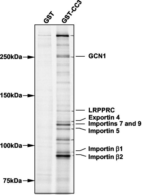
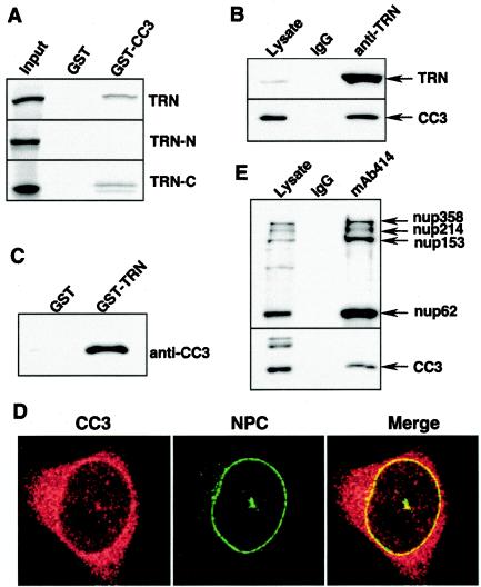
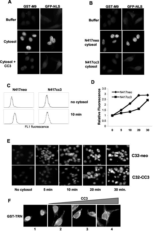
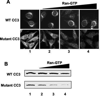
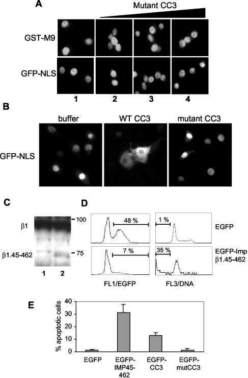
We report here that the normal cellular protein CC3/TIP30, when in excess, inhibits nuclear import in vitro and in vivo. CC3 binds directly to the karyopherins of the importin beta family in a RanGTP-insensitive manner and associates with nucleoporins in vivo. CC3 inhibits the nuclear import of proteins possessing either the classical nuclear localization signal or the M9 signal recognized by transportin. CC3 also inhibits nuclear translocation of transportin itself. Cells modified to express higher levels of CC3 have a slower rate of nuclear import and, as described earlier, show an increased sensitivity to death signals. A mutant CC3 protein lacking proapoptotic activity has a lower affinity for transportin, is displaced from it by RanGTP, and fails to inhibit nuclear import in vitro and in vivo. Together, our results support a correlation between the ability of CC3 to form a RanGTP-resistant complex with importins, inhibit nuclear import, and induce apoptosis. Significantly, a dominant-negative form of importin beta1 shown previously to inhibit multiple transport pathways induces rapid cell death, strongly indicating that inhibition of nuclear transport serves as a potent apoptotic signal.


Figures

FIG. 1.
FIG. 1.
Identification of proteins associated with CC3. HeLa cells were metabolically labeled with [35S]methionine and lysed. Cytosolic protein extracts were incubated with GST or GST-CC3. Bound proteins were recovered on glutathione-Sepharose, resolved by SDS-PAGE, and detected by autoradiography.
FIG. 2.
FIG. 2.
CC3 associates with transportin and the NPC in vitro and in vivo. (A) In vitro-translated transportin polypeptides: full-length (TRN), N-terminal amino acids 1 to 581 (TRN-N), and C-terminal amino acids 581 to 890 (TRN-C) were incubated with GST or GST-CC3. Complexes were recovered with glutathione-Sepharose, washed, resolved by SDS-PAGE, and detected by autoradiography. (B) HeLa cell extracts were subjected to immunoprecipitation with a control immunoglobulin G (IgG) or anti-transportin antibody. The immune complexes were resolved by electrophoresis and analyzed by Western blotting with antibodies to transportin (TRN) and CC3. (C) Purified CC3 at 1 μM was mixed with GST or GST-transportin for 1 h at 4°C. Complexes were recovered with glutathione-Sepharose, resolved by SDS-PAGE, and detected by Western blotting with anti-CC3 antibody. (D) HeLa cells were processed for immunofluorescence with the anti-CC3 rabbit polyclonal antibody and anti-NPC mouse monoclonal MAb414 (Covance), followed by detection with anti-rabbit IgG-CY3 (red) and anti-mouse IgG-FITC (green), and analyzed by confocal microscopy. (E) MAb414 was used to immunoprecipitate components of the NPC from HeLa cells extracts as described in Materials and Methods. The immune complexes were analyzed by Western blotting with MAb414 and antibodies to CC3.
FIG. 3.
FIG. 3.
Interactions of CC3 with NPC, transportin, and exportin 4 are not sensitive to RanGTP. (A) FITC-labeled GST-M9 or GST-CC3 at 0.5 μM were added to permeabilized HeLa cells in transport buffer with or without 50 μg of HeLa cytosolic extract and 8 μM RanQ69L. After incubation for 20 min at room temperature, the cells were fixed and examined by fluorescence microscopy. (B) In vitro-translated transportin (lane 1) was incubated with GST-CC3 in the absence (lane 2) or in the presence of 1, 4, or 8 μM RanQ69L (lanes 3 to 5). Complexes were recovered with glutathione-Sepharose, resolved by SDS-PAGE, and detected by autoradiography. (C) His-exportin 4 at 5 μM was incubated with GST (lane 1) or GST-CC3 without RanQ69L (lane 2) or in the presence of 2.5, 5, or 10 μM RanQ69L (lanes 3 to 5) for 1 h at 4°C. Complexes were recovered with glutathione-Sepharose, resolved by SDS-PAGE, and detected by staining with Coomassie blue. (D) In vitro-translated transportin was incubated with His-RanQ69L in the absence (lane 3) or presence of 1, 4, and 8 μM GST-CC3 (lanes 4 to 6). Complexes were recovered with Ni-NTA agarose and analyzed by SDS-PAGE, followed by autoradiography. Lane 2 shows the amount of transportin that was incubated without RanQ69L and bound to the Ni-NTA agarose in a nonspecific manner. (E) In vitro-translated transportin was incubated with GST-M9 in absence (lane 2) or in presence of 1, 4, or 8 μM CC3 (lanes 3 to 5). Complexes were analyzed as described for panel B.
FIG. 4.
FIG. 4.
CC3 inhibits nuclear import of substrates containing NLS or M9 import signals and nuclear translocation of transportin. (A) FITC-labeled GST-M9 at 0.5 μM or GST-GFP-NLS (1 μM) was added to permeabilized HeLa cells in transport buffer with or without 50 μg of HeLa cytosolic extract and 8 μM CC3. After incubation for 20 min at room temperature, cells were fixed, and the localization of import cargoes was examined by fluorescence microscopy. (B) FITC-GST-M9 or GST-GFP-NLS was added to permeabilized HeLa cells in transport buffer or with 150 μg of cytosolic extract prepared from either CC3-negative cells (N417neo) or from the same cells stably transfected to express high levels of CC3 (N417cc3). Cells were processed as for panel A. (C) In vitro import of FITC-GST-M9 (0.5 μM) was performed in suspension with N417neo and N417cc3 cells, and the nuclear fluorescence was analyzed by FACS. FL1 fluorescence histograms of live cells are shown for cells incubated for 30 min with GST-M9 but without cytosol (no cytosol) and for cells incubated with both GST-M9 and cytosol for 10 min. (D) Graphic representation of the results of the in vitro import assays with N417 clones performed as described for panel C. The data are shown as the relative fluorescence, where the fluorescence of cells incubated with substrate in the absence of cytosol is assigned an arbitrary value of 1. The experiment was performed twice, with nearly identical results each time. (E) FITC-labeled GST-M9 (0.5 μM) was added to permeabilized C32neo and C32cc3 cells in transport buffer with 50 μg of cytosolic extract prepared from N417neo cells. Cells were processed as for panel A after different incubation times. (F) GST-transportin at 0.5 μM was added to permeabilized HeLa cells in transport buffer alone (panel 1) or in the presence of 2, 4, or 8 μM CC3 (panels 2 to 4). After incubation for 20 min at room temperature cells were fixed and permeabilized with Triton X-100. The cellular localization of transportin was examined by immunofluorescence microscopy with polyclonal anti-GST antibodies and anti-rabbit IgG-FITC.
FIG. 5.
FIG. 5.
Interactions of mutant CC3 with transportin and the NPC are vulnerable to dissociation by RanGTP. (A) GST-CC3 or GST-mutant CC3 at 0.5 μM was added to permeabilized HeLa cells in transport buffer without (panels 1) or with 1, 4, or 8 μM RanQ69L (panels 2 to 4). After incubation for 20 min at room temperature, cells were examined by immunofluorescence microscopy with anti-GST polyclonal antibodies. (B) In vitro-translated transportin was incubated with GST-CC3 or GST-mutant CC3 alone or in the presence of 1, 4, or 8 μM RanQ69L. The presence of transportin in complexes was detected by autoradiography.
FIG. 6.
FIG. 6.
Inhibition of nuclear import by CC3 and dominant-negative importin leads to apoptosis. (A) GST-M9-FITC (0.5 μM) or GST-GFP-NLS (1 μM) was added to permeabilized HeLa cells in transport buffer with 50 μg of HeLa cytosolic extracts without (panels 1) or with 2, 4, or 8 μM of mutant CC3 protein (panels 2 to 4). After incubation for 20 min at room temperature, cells were fixed, and the cellular localization of proteins was examined by fluorescence microscopy. (B) GST-GFP-NLS (0.5 mg/ml) was injected into cytoplasm of NIH 3T3 cells with GST-CC3 (5 mg/ml) or GST-mutant CC3 (5 mg/ml), followed by incubation at 37°C. Protein localization was examined by fluorescence microscopy. (C) Analysis of expression of transiently transfected EGFP-Impβ1.45-462. HEK293 cells were transfected with empty vector (lane 1) or EGFP-Impβ1.45-462 (lane 2), harvested after 20 h, and analyzed by Western blot with antibodies to importin β1 provided by K. Weis. The migration of molecular size standards is shown. The blot was exposed for a longer period to detect the protein band corresponding to EGFP-Impβ1.45-462 (β1.45-462), which is expressed at very low levels compared to endogenous importin β1 (β1). (D) Rat1 cells were transfected with indicated constructs and analyzed for EGFP expression (FL1/EGFP, right panels) and DNA content (FL3/DNA, right panels) by FACS. The numbers show the percentage of EGFP-positive transfected cells and apoptotic cells with subdiploid DNA content. (E) Analysis of apoptotic DNA fragmentation in EGFP-positive transfected cells. The results are averages of at least three independent experiments.

Similar articles

Cited by

References

    1. Adam, S. A., and L. Gerace. 1991. Cytosolic proteins that specifically bind nuclear location signals are receptors for nuclear import. Cell 66:837-847. - PubMed
    1. Adam, S. A., R. S. Marr, and L. Gerace. 1990. Nuclear protein import in permeabilized mammalian cells requires soluble cytoplasmic factors. J. Cell Biol. 111:807-816. - PMC - PubMed
    1. Baker, M. E. 1999. TIP30, a cofactor for HIV-1 Tat-activated transcription, is homologous to short-chain dehydrogenases/reductases. Curr. Biol. 9:R471. - PubMed
    1. Baker, M. E., L. Yan, and M. R. Pear. 2000. Three-dimensional model of human TIP30, a coactivator for HIV-1 Tat-activated transcription, and CC3, a protein associated with metastasis suppression. Cell Mol. Life Sci. 57:851-858. - PMC - PubMed
    1. Ben-Efraim, I., and L. Gerace. 2001. Gradient of increasing affinity of importin beta for nucleoporins along the pathway of nuclear import. J. Cell Biol. 152:411-417. - PMC - PubMed

Publication types

MeSH terms