. 2004 Aug;24(16):7260-74.
doi: 10.1128/MCB.24.16.7260-7274.2004.

Estrogens and progesterone promote persistent CCND1 gene activation during G1 by inducing transcriptional derepression via c-Jun/c-Fos/estrogen receptor (progesterone receptor) complex assembly to a distal regulatory element and recruitment of cyclin D1 to its own gene promoter

Affiliations

Estrogens and progesterone promote persistent CCND1 gene activation during G1 by inducing transcriptional derepression via c-Jun/c-Fos/estrogen receptor (progesterone receptor) complex assembly to a distal regulatory element and recruitment of cyclin D1 to its own gene promoter

Luigi Cicatiello et al. Mol Cell Biol. 2004 Aug.

Abstract

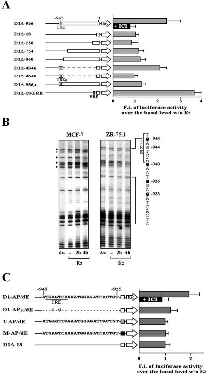
Transcriptional activation of the cyclin D1 gene (CCND1) plays a pivotal role in G(1)-phase progression, which is thereby controlled by multiple regulatory factors, including nuclear receptors (NRs). Appropriate CCND1 gene activity is essential for normal development and physiology of the mammary gland, where it is regulated by ovarian steroids through a mechanism(s) that is not fully elucidated. We report here that CCND1 promoter activation by estrogens in human breast cancer cells is mediated by recruitment of a c-Jun/c-Fos/estrogen receptor alpha complex to the tetradecanoyl phorbol acetate-responsive element of the gene, together with Oct-1 to a site immediately adjacent. This process coincides with the release from the same DNA region of a transcriptional repressor complex including Yin-Yang 1 (YY1) and histone deacetylase 1 and is sufficient to induce the assembly of the basal transcription machinery on the promoter and to lead to initial cyclin D1 accumulation in the cell. Later on in estrogen stimulation, the cyclin D1/Cdk4 holoenzyme associates with the CCND1 promoter, where E2F and pRb can also be found, contributing to the long-lasting gene enhancement required to drive G(1)-phase completion. Interestingly, progesterone triggers similar regulatory events through its own NRs, suggesting that the gene regulation cascade described here represents a crossroad for the transcriptional control of G(1)-phase progression by different classes of NRs.


Figures

FIG. 1.
FIG. 1.
(A) Analysis of CCND1 promoter responsiveness to estrogen upon stable transfection of the indicated luciferase reporter genes in hormone-responsive hBC cells, represented as fold induction by 50 nM E2 for 18 to 24 h (grey bars) versus the activity assayed in hormone-starved cells, arbitrarily set to 1. The black bar represents the fold induction measured in cells incubated simultaneously with 50 nM E2 and 5 μM ICI 182,780. On the left are schematically represented constructs used in transfections. TRE, TPA-responsive element; TREμ, same site mutated as previously described (22). Numbers at the top of the box mark the positions of the 5′-most nucleotides with respect to the transcription start site. Constructs D1Δ-138, D1Δ-754, D1Δ-860, D1Δ-956, and D1Δ-956μ were previously described (22). Constructs D1Δ-18, D1Δ-4S46, D1Δ-4S48, and D1Δ-18/ERE are described in Materials and Methods. F.i., fold induction. Bars indicate standard deviations. (B) In vivo footprinting of the CCND1 gene promoter region encompassing the TRE. The G residues marked by black arrowheads are specifically protected in E2-stimulated cells, whereas the G identified by a grey arrowhead is protected in the same cells depleted by E2. i.v., naked DNA methylated in vitro. (C) Estrogen responsiveness of luciferase reporter genes measured in hBC cells stably transfected with the plasmids schematically represented on the left. Synthetic wild-type or mutant 24-mer oligonucleotides reproducing the CCND1 gene sequence between residues −948 and −925 were cloned immediately upstream of the minimal promoter from the same gene (spanning residues −18 and + 14; white square), the thymidine kinase promoter (grey square; T-AP/dE), or the mouse mammary tumor virus promoter (black square; M-AP/dE). Mutant nucleotides in D1-APμ/dE are indicated. MCF-7 or ZR-75.1 cells were hormone deprived and restimulated as described above, including, where indicated, treatment with ICI 182,780. As for panel A, the results represent the means of three to five independent experiments. Luciferase activity assayed in starved cells transfected with each reporter was arbitrarily set to 1. Unless otherwise indicated, data displayed are from representative experiments carried out with MCF-7 cells; identical results were obtained with ZR-75.1 cells.
FIG. 2.
FIG. 2.
ChIP analysis of in vivo interactions of trans-acting factors with the TRE-containing region of the CCND1 promoter. (A) Sequence of the estrogen-responsive region between residues −948 and −925. G nucleotides with black arrowheads on top represent residues protected in hormone-stimulated cells, as detected by in vivo footprinting; the G marked with a grey arrowhead is protected instead only in quiescent cells. Consensus sequences for AP-1, Oct-1, and YY1 binding sites are aligned with the homologous sequences found here. (B and C) Soluble chromatin was prepared from hBC cells before or after 30 min of treatment with 50 nM E2 and immunoprecipitated with antibodies against the indicated proteins before DNA amplification with the indicated primers. In order to obtain optimal separation of regions immediately adjacent, only DNA samples which were not amplified by PCR with primers separated by more than 500 bp, e.g., those indicated at the top left (spanning the region between −1039 and −53), were used for the ChIP assays. Input, total chromatin before immunoprecipitation; α-Mock, control, unrelated antibodies used for immunoprecipitation; pS2, estrogen-responsive region of the pS2 gene promoter. The primers used for PCR amplifications are described in Materials and Methods. Data displayed are from representative experiments carried out with ZR-75.1 cells; identical results were obtained with MCF-7 cells.
FIG. 3.
FIG. 3.
In vitro binding of transcription factors to the estrogen-responsive region of the CCND1 gene. (A) EMSAs with nuclear extracts from hormone-responsive hBC cells treated or not treated with 50 nM E2 for 2 h and challenged with 32P-labeled AP/dE double-stranded oligonucleotide; in one case (+ICI), a 100-fold molar excess of ICI 182,780 was also added to the culture media as described in Materials and Methods. In some experiments, a 100-fold molar excess of unlabeled oligonucleotide carrying consensus binding sites for the indicated factors was used as a competitor (comp.), or the nuclear extracts were pretreated with the indicated antibodies (Ab). NIS, pool of preimmune sera; ss, supershifted complexes observed with anti-ER, anti-c-Fos, and anti-Oct-1 antibodies (antibodies against c-Jun and YY1 hamper the interactions of these factors with DNA). The protein-DNA complexes indicated as c1, c2, and c3 were consistently observed with different nuclear extracts from either MCF-7 or ZR-75.1 cells and are specific, while the smaller ones represent nonspecific complexes, which were erratic or resulted from sample degradation during preparation or handling. Data displayed are representative of results obtained in multiple tests carried out with different nuclear extracts. (B) Results of EMSAs performed with equal amounts of nuclear proteins extracted from hormone-deprived (−E2) or hormone-stimulated (+E2) hBC cells and increasing amounts of AP/dE-labeled probe. Following electrophoresis and autoradiography, the intensity of the signal corresponding to complexes c1 (Oct-1), c2 (AP-1), or c3 (YY1) in each lane was measured by densitometry. Bars represent standard deviations. (C) Representative immunoblot of nuclear extracts from hBC cells treated with E2 and analyzed by Western blotting with antibodies against the represented proteins. Only the area of the filters where each protein of interest was detected is displayed. (D) Mutant analysis of in vitro transcription factor binding to the AP/dE composite element by EMSAs with nuclear extracts from hormone-treated hBC cells. In the top panel are shown the sequences tested by direct binding, with mutant residues in evidence; numbers to the right mark the corresponding lane in the autoradiogram below. Data displayed are from representative experiments carried out with MCF-7 cells; identical results were obtained with ZR-75.1 cells.
FIG. 4.
FIG. 4.
Kinetics of in vivo transcription factor interactions with the estrogen-responsive region of the CCND1 gene in hormone-treated hBC cells and assessment of the transcriptional output. (A) ChIP analysis of CCND1 gene upstream regulatory site occupancy by different transcription factors in quiescent and estrogen-stimulated cells. (B) Summary of chemical interference assays carried out with nuclear extracts from MCF-7 cells following estrogen stimulation. The TRE sequence is boxed, and nucleotides contacting Oct-1 or YY1 proteins are indicated by black (Gs) and white (Ts) dots. (C) (Left panel) Transient transfection analysis of the effects of transcription factor overexpression on the indicated reporter gene activity in hBC cells stimulated with estrogen. A total of 50 to 200 ng of the indicated expression vectors was transfected, together with the reporter gene and an internal control; luciferase activity measured in the same cells transfected with an empty expression vector was arbitrarily set to 1. (Right panel) Effects of the AP/dE sequence on basal reporter gene activity in the absence of E2; luciferase activity of the cyclin D1 promoter-based D1Δ-18 vector was arbitrarily set to 1. Transfections were performed as described in Materials and Methods. F.i., fold induction. Bars indicate standard deviations of two to four independent assays performed in duplicate. (D) Northern blot analysis of cyclin D1 mRNA expression in ZR-75.1 cells before and at the indicated times after stimulation with E2; 36B4 ribosomal protein gene mRNA was also quantitated in the same blot as a control. (E) Sequential ChIP (ReIP) analyses of transcription factor interaction with the estrogen-responsive region of the CCND1 gene between positions −1039 and −770. II°IP, results obtained following a second immunoprecipitation with the indicated antibodies. (F) Occupancy of CCND1 gene initiator-proximal region by polymerase II, as assessed by ChIP. E2, treatment of cells with E2. For details, see Results. Unless otherwise indicated, data displayed are from representative experiments carried out with ZR-75.1 cells; identical results were obtained with MCF-7 cells.
FIG. 5.
FIG. 5.
(A) PKA inhibition does not prevent ERα recruitment to the estrogen-responsive region of the CCND1 gene promoter. Sequential ChIP (ReIP) analyses of the TRE-centered region from the CCND1 promoter. Chromatin prepared from cells treated or not treated with 50 nM E2 in the absence or presence of the PKA inhibitor H89 was subjected to the ChIP procedure with an antibody against ERα (α-ER: I°IP) and then again with anti-RNA polymerase II antibodies (α-Pol II: II°IP). In some experiments, 5 μM H89 was added to the cells 30 min before E2. Input, total chromatin before the first immunoprecipitation. As a control, antibodies against total (α-CREB) or phosphorylated (αP-CREB) CREB were used to immunoprecipitate the CRE-containing proximal region of the CCND1 promoter from hormone-deprived (−E2) and hormone-stimulated (+E2) cells. (B) In vitro binding of cyclin D1 and pRb to the upstream regulatory region of the CCND1 gene. Soluble chromatin was prepared from hormone-responsive hBC cells before and at the indicated times after treatment with 50 nM E2 and immunoprecipitated with antibodies against the indicated proteins before PCR amplification. Input, total chromatin before immunoprecipitation; α-Mock, control, unrelated antibodies used for immunoprecipitation. The presence of the E2F-containing regulatory region of the c-Myc proto-oncogene was investigated with the same immunoprecipitated samples as described in Materials and Methods. (C) Sequential ChIP (ReIP) analyses of the AP/dE region from the CCND1 promoter. Chromatin prepared from cells treated or not treated with 50 nM E2 for the indicated times was subjected to the ChIP procedure with an antibody against cyclin D1 (α-D1: I°IP) and then again with anti-pRb antibodies (α-pRb: II°IP). Input, total chromatin before the first immunoprecipitation. (D) Assays like those in panel A were carried out with chromatin extracted from hBC cells stimulated at time zero with 50 nM E2 followed, where indicated (ICI), by the addition after 60 min of 5 μM ICI 182,780 to the cell culture media. The times indicated when cells were collected for analysis. Results shown are representative of multiple independent experiments. Unless otherwise indicated, data displayed are from experiments carried out with ZR-75.1 cells; identical results were obtained with MCF-7 cells.
FIG. 6.
FIG. 6.
(A) Analysis of CCND1 promoter responsiveness to progesterone upon stable transfection of the indicated luciferase reporter genes in MCF-7 cells, reported as fold induction by 50 nM R5020 for 18 to 24 h versus the activity assayed in hormone-starved cells transfected with the same construct, arbitrarily set to 1. Plasmids used for transfections are named as shown in Fig. 1. MMTV-luc, progesterone-responsive reporter gene including the mouse mammary tumor virus long terminal repeat. Results reported represent the means of three to five independent experiments carried out several times. F.i., fold induction. Bars indicate standard deviations. (B) EMSAs with nuclear extracts from hormone-responsive hBC cells treated, where indicated (+), with 50 nM E2 or R5020 for 2 h and challenged with 32P-labeled AP-1/dE oligonucleotide. Data are representative of multiple experiments carried out with at least two different nuclear extracts from either MCF-7 or ZR 75.1 cells. (C) ChIP analysis of in vivo binding of progesterone receptors and other transcription regulatory factors to the upstream element of the CCND1 gene in ZR-75.1 cells. Input, total chromatin before immunoprecipitation. Data are representative of three experiments, one of which was carried out with MCF-7 cells.
FIG. 7.
FIG. 7.
Schematic representation of estrogen-dependent transcription factor assembly on the CCND1 gene promoter in hormone-responsive hBC cells and their interactions with each other, the distal regulatory sites, and the basal promoter. In hormone-deprived, quiescent cells, HDAC1 is present in a complex with YY1 on the ERGE. Upon estrogen (or progesterone) stimulation, these trans-repressors are displaced from DNA by a composite AP-1/ER (or PR) complex and Oct-1, inducing physical interactions of ERGE-bound complexes with the basal transcriptional machinery. At a later time (>1 h), the cyclin D1 (Cyc D1)/Cdk4 holoenzyme associates with the complex, presumably relieving the promoter from trans-repression and thereby inducing a second, longer-lasting enhancement of transcription. This event may be accompanied by Oct-1 release from ERGE. Ac, acetylated histone tails.

Similar articles

Cited by

References

    1. Abbondanza, C., N. Medici, V. Nigro, V. Rossi, L. Gallo, G. Piluso, A. Belsito, A. Roscigno, P. Bontempo, A. A. Puca, A. M. Molinari, B. Moncharmont, and G. A. Puca. 2000. The retinoblastoma-interacting zinc-finger protein RIZ is a downstream effector of estrogen action. Proc. Natl. Acad. Sci. USA 97:3130-3135. - PMC - PubMed
    1. Adams, P. D. 2001. Regulation of the retinoblastoma tumor suppressor protein by cyclin cdks. Biochim. Biophys. Acta 1471:123-133. - PubMed
    1. Albanese, C., M. D'Amico, A. T. Reutens, M. Fu, G. Watanabe, R. J. Lee, R. N. Kitsis, B. Henglein, M. Avantaggiati, K. Somasundaram, B. Thimmapaya, and R. G. Pestell. 1999. Activation of the cyclin D1 gene by the E1A-associated protein p300 through AP-1 inhibits cellular apoptosis. J. Biol. Chem. 274:34186-34195. - PubMed
    1. Altucci, L., R. Addeo, L. Cicatiello, S. Dauvois, M. G. Parker, M. Truss, M. Beato, V. Sica, F. Bresciani, and A. Weisz. 1996. 17β-Estradiol induces cyclin D1 gene transcription, p36D1-p34cdk4 complex activation and p105Rb phosphorylation during mitogenic stimulation of G1-arrested human breast cancer cells. Oncogene 12:2315-2324. - PubMed
    1. Bamberger, A. M., C. M. Bamberger, B. Gellersen, and H. M. Schulte. 1996. Modulation of AP-1 activity by the human progesterone receptor in endometrial carcinoma cells. Proc. Natl. Acad. Sci. USA 93:6169-6174. - PMC - PubMed

Publication types

MeSH terms