. 2004 Aug;24(16):7275-83.
doi: 10.1128/MCB.24.16.7275-7283.2004.

EWS/FLI-1 silencing and gene profiling of Ewing cells reveal downstream oncogenic pathways and a crucial role for repression of insulin-like growth factor binding protein 3

Affiliations

EWS/FLI-1 silencing and gene profiling of Ewing cells reveal downstream oncogenic pathways and a crucial role for repression of insulin-like growth factor binding protein 3

Alexandre Prieur et al. Mol Cell Biol. 2004 Aug.

Abstract

Ewing tumors are characterized by abnormal transcription factors resulting from the oncogenic fusion of EWS with members of the ETS family, most commonly FLI-1. RNA interference targeted to the junction between EWS and FLI-1 sequences was used to inactivate the EWS/FLI-1 fusion gene in Ewing cells and to explore the resulting phenotype and alteration of the gene expression profile. Loss of expression of EWS/FLI-1 resulted in the complete arrest of growth and was associated with a dramatic increase in the number of apoptotic cells. Gene profiling of Ewing cells in which the EWS/FLI-1 fusion gene had been inactivated identified downstream targets which could be grouped in two major functional clusters related to extracellular matrix structure or remodeling and regulation of signal transduction pathways. Among these targets, the insulin-like growth factor binding protein 3 gene (IGFBP-3), a major regulator of insulin-like growth factor 1 (IGF-1) proliferation and survival signaling, was strongly induced upon treating Ewing cells with EWS/FLI-1-specific small interfering RNAs. We show that EWS/FLI-1 can bind the IGFBP-3 promoter in vitro and in vivo and can repress its activity. Moreover, IGFBP-3 silencing can partially rescue the apoptotic phenotype caused by EWS/FLI-1 inactivation. Finally, IGFBP-3-induced Ewing cell apoptosis relies on both IGF-1-dependent and -independent pathways. These findings therefore identify the repression of IGFBP-3 as a key event in the development of Ewing's sarcoma.


Figures
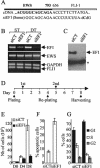
FIG. 1.
FIG. 1.
Induction of apoptosis and cell cycle arrest by inhibition of expression of the EWS/FLI-1 fusion gene using specific siRNA. (A) Scheme of type 1 EWS/FLI-1 junction and sequence of the cDNA and siEF1. (B) Inhibition of the expression of EWS/FLI-1 transcript by siEF1. RNA from Ewing cells transfected with either siEF1 or siCT were subjected to competitive RT-PCR experiments. GAPDH was used as an internal control. ST and DT, single and double transfections, respectively. (C) Immunoblot with the 7.3 monoclonal antibody. The position of a cross-reactive band used for standardization (16) is indicated by an asterisk. (D) Temporal scheme of the double-transfection strategy. The first (1st) and second (2nd) siRNA transfections are indicated. (E to G) Growth inhibition, apoptosis, and cell cycle arrest induced by the silencing of EWS/FLI-1 in the A673 Ewing cell line. Seventy-two hours after the second transfection, the cells were counted (E), analyzed by FACS for apoptosis using an annexin V detection kit assay (F), and analyzed for cell cycle progression using propidium iodide staining (G). The means ± standard deviations (error bars) of two independent experiments are shown. D0, D4, and D8, days 0, 4, and 8 after the initial plating, respectively.
FIG. 2.
FIG. 2.
Analysis of IGFBP-3 mRNA expression levels in Ewing cell lines by quantitative RT-PCR. (A) Inhibition of EWS/FLI-1 expression by siEF1 in the EW24 cell line. (B) Levels of expression of IGFBP-3 in Ewing cell lines and A673 or EW24 cells treated with siEF1 or siCT are indicated relative to the level of expression of IGFBP-3 in HeLa cells.
FIG. 3.
FIG. 3.
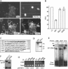
Down-regulation of IGFBP-3 expression by EWS/FLI-1 and direct binding of EWS/FLI-1 to the promoter region. (A) Inhibition of endogenous IGFBP-3 expression by ectopic EWS/FLI-1. HeLa cells transfected by EWS/FLI-1 were stained by the monoclonal 7.3 (FITC) and polyclonal anti-IGFBP-3 (cyanin-3) antibodies. Dapi, 4′,6′-diamidino-2-phenylindole. Merge, combined EWS/FLI-1 and IGFBP-3 stainings. Arrows, EWS/FLI-1-transfected cells. (B) Inhibition of the IGFBP-3 promoter by EWS/FLI-1. The IGFBP-3 promoter from positions −2584 to +63 was cloned upstream of the luciferase gene of pRep4 plasmid. Light intensity (in relative light units [RLU]) is shown on the y axis. The means ± standard deviations (error bars) from two independent duplicate experiments are shown. R2L2 and I393E are DNA binding-deficient mutants of EWS/FLI-1. Empty expression vector (−) was used as a control. (C) Potential EWS/FLI-1 binding sites within the IGFBP-3 promoter. EWS/FLI-1 consensus binding sites are indicated by bold type, and oligonucleotides used for EMSA experiments are underlined. (D) In vitro-translated EWS/FLI-1 and FLI-1. The fastest migrating FLI-1 band results from internal initiation at a downstream methionine residue (17). The positions of molecular size markers (in kilodaltons) are shown to the right of the gel. (E) EWS/FLI-1 binding to two potential sites located around positions −1686 and −1829 upstream of the transcription start site (7) in the presence (7.3) and absence (−) of the 7.3 monoclonal antibody (Ab). The arrow indicates the supershift of the EF1-DNA complex by the 7.3 antibody. E74 is the control oligonucleotide that contains a specific EWS/FLI-1 binding site (2). (F) Expression of a triple-tagged version of EWS/FLI-1 (EF1t) from the EW24-derived SFT12.1 cells. *, cross-reactive band. (G) Binding of EWS/FLI-1 to the IGFBP-3 promoter in vivo. ChIP experiments from SFT12.1 cell extracts using three different specific antibodies (anti-EWS/FLI-1 [αEWS/FLI-1] [7.3], anti-HA [αHA], or anti-Flag [αFlag]) or a control (anti-His [αHis]) antibody. The −1841/−1613 IGFBP-3 promoter fragment, according to the position of the transcription start site, is immunoprecipitated with the specific antibodies, whereas the −2704/−2545 fragment is not. The control PCR on extracts before immunoprecipitation (input) is shown.
FIG. 4.
FIG. 4.
Role of the balance of FLI-1 and EWS/FLI-1 expression in IGFBP-3 regulation. (A) Inhibition of the moderate FLI-1-induced activation of the IGFBP-3 promoter by EWS/FLI-1 but not by DNA binding-deficient mutants. Light intensity (in relative light units [RLU]) is shown on the y axis. Luciferase construct and expression vectors are identical to those used in Fig. 3B. Empty expression vector (−) was used as a control. The means ± standard deviations (error bars) of duplicate experiments are shown. (B) FLI-1 and EWS/FLI-1 binding on the −1829 probe. The reaction mixtures were incubated with no (−) or 2, 4, and 6 microliters of siEF1 or FLI-1. EMSA experiments show that FLI-1 can compete EWS/FLI-1 binding in a dose-dependent manner. Both proteins were produced by in vitro translation as shown in Fig. 3D. The two complexes observed with FLI-1 correspond to the two bands of FLI-1. (C) Increased endogenous IGFBP-3 expression induced by FLI-1 transfection in Ewing cells. IGFBP-3 levels were analyzed by quantitative RT-PCR after transfection of A673 cells with increasing amounts of the FLI-1 expression vector. Relative expression of IGFBP-3 compared to that in untransfected A673 cells is shown. The means ± standard deviations (error bars) of duplicate experiments are shown. The expression level of FLI-1 evaluated by immunoblotting is shown beneath the diagram. Wb αHA, Western blotting with anti-HA antibody.
FIG. 5.
FIG. 5.
IGFBP-3-dependent apoptosis induced by siEF1 in Ewing cells. (A) Inhibition of siEF1-induced apoptosis by silencing of IGFBP-3. The high apoptotic rate induced by siEF1 can be reduced dramatically by siRNA-based blockade of IGFBP-3 induction. The levels of expression of EWS/FLI-1 and IGFBP-3, measured by competitive RT-PCR, are shown. (B) IGF-1-dependent and -independent pathways of IGFBP-3-induced apoptosis in Ewing cells. Recombinant IGFBP-3 (rIBP3) or a version of rIBP3 with a mutated NLS (rIBP3m) were incubated in the presence (+) or absence (−) of 10% serum. The means ± standard deviations (error bars) of duplicate experiments are shown in panels A and B.(C) Regulation of the IGF-1 downstream pathways by 25 nM IGFBP-3 treatment of Ewing cells. Untreated cells (−) were used as controls. AKT-P, phosphorylated AKT; ERK-P, phosphorylated ERK; mitRAF-1, mitochondrial RAF-1.

References

    1. Arvand, A., and C. T. Denny. 2001. Biology of EWS/ETS fusions in Ewing's family tumors. Oncogene 20:5747-5754. - PubMed
    1. Bailly, R. A., R. Bosselut, J. Zucman, F. Cormier, O. Delattre, M. Roussel, G. Thomas, and J. Ghysdael. 1994. DNA-binding and transcriptional activation properties of the EWS-FLI-1 fusion protein resulting from the t(11;22) translocation in Ewing sarcoma. Mol. Cell. Biol. 14:3230-3241. - PMC - PubMed
    1. Baserga, R. 1995. The insulin-like growth factor I receptor: a key to tumor growth? Cancer Res. 55:249-252. - PubMed
    1. Bertolotti, A., Y. Lutz, D. J. Heard, P. Chambon, and L. Tora. 1996. hTAFII68, a novel RNA/ssDNA-binding protein with homology to the pro-oncoproteins TLS/FUS and EWS is associated with both TFIID and RNA polymerase II. EMBO J. 15:5022-5031. - PMC - PubMed
    1. Cohen, P., D. R. Clemmons, and R. G. Rosenfeld. 2000. Does the GH-IGF axis play a role in cancer pathogenesis? Growth Horm. IGF Res. 10:297-305. - PubMed

Publication types

MeSH terms

Substances

LinkOut - more resources