. 2004 Aug;24(16):7298-311.
doi: 10.1128/MCB.24.16.7298-7311.2004.

Protein kinase C delta blocks immediate-early gene expression in senescent cells by inactivating serum response factor

Affiliations

Protein kinase C delta blocks immediate-early gene expression in senescent cells by inactivating serum response factor

Keith Wheaton et al. Mol Cell Biol. 2004 Aug.

Abstract

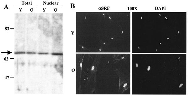
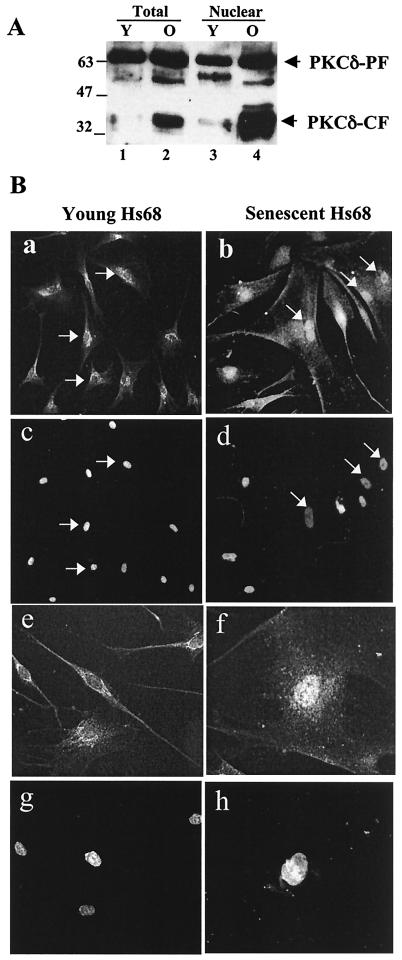
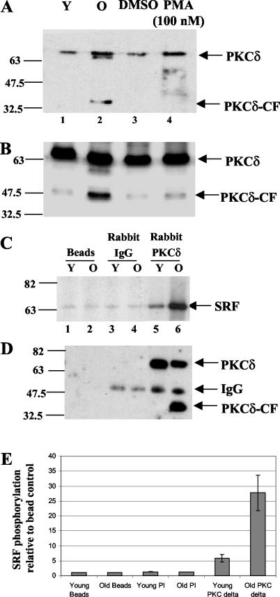
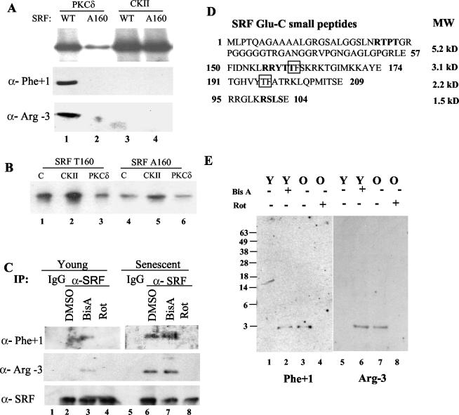
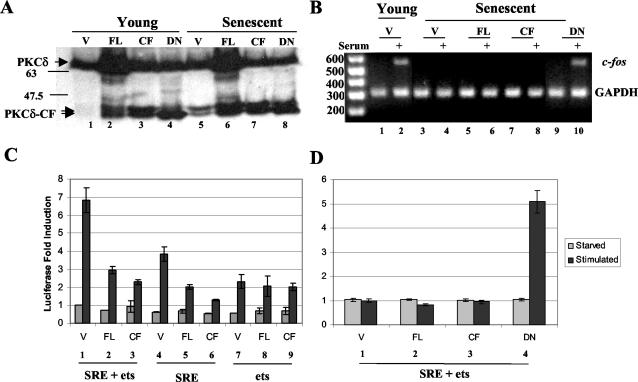
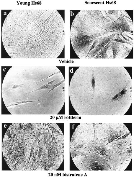
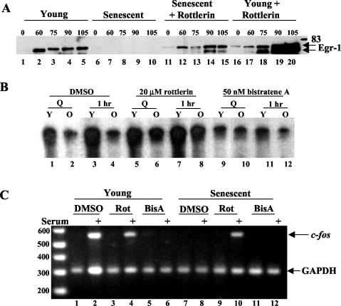
Fibroblasts lose the ability to replicate in response to growth factors and become unable to express growth-associated immediate-early genes, including c-fos and egr-1, as they become senescent. The serum response factor (SRF), a major transcriptional activator of immediate-early gene promoters, loses the ability to bind to the serum response element (SRE) and becomes hyperphosphorylated in senescent cells. We identify protein kinase C delta (PKC delta) as the kinase responsible for inactivation of SRF both in vitro and endogenously in senescent cells. This is due to a higher level of PKC delta activity as cells age, production of the PKC delta catalytic fragment, and its nuclear localization in senescent but not in low-passage-number cells. The phosphorylation of T160 of SRF by PKC delta in vitro and in vivo led to loss of SRF DNA binding activity. Both the PKC delta inhibitor rottlerin and ectopic expression of a dominant negative form of PKC delta independently restored SRE-dependent transcription and immediate-early gene expression in senescent cells. Modulation of PKC delta activity in vivo with rottlerin or bistratene A altered senescent- and young-cell morphology, respectively. These observations support the idea that the coordinate transcriptional inhibition of several growth-associated genes by PKC delta contributes to the senescent phenotype.


Figures

FIG. 1.
FIG. 1.
SRF protein levels do not change with cellular age. (A) Western blots used equal amounts of protein from young (Y, 36 mean population doublings) and old (O, 82 mean population doublings) Hs68 fibroblasts. Total (Tot) and nuclear (Nuc) extracts and a polyclonal antibody generated against full-length SRF were used. The arrow identifies a 67-kDa band characteristic of native SRF. (B) Young and senescent primary human diploid fibroblasts were fixed and stained with rabbit anti-SRF followed by goat anti-rabbit antibody-Texas Red and counterstained with DAPI to visualize DNA.
FIG. 2.
FIG. 2.
Kinase activity in senescent-cell nuclear extracts inhibits SRF DNA binding activity. (A) EMSAs with 32P-labeled SRE oligonucleotide and SRF(His)6 previously incubated with nuclear extracts from young (Y) or old (O) nuclear extracts are shown in lanes 1 and 2. Parallel kinase reactions were incubated with mutant (mut) SRE (lanes 3 and 4), competed with a 100-fold excess of unlabeled wild-type (WT) SRE (lanes 5 and 6), or incubated with a 100-fold excess of unlabeled mutant SRE (lanes 7 and 8). Preincubation with polyclonal SRF antibody (αSRF, lanes 9 and 10) supershifted or eliminated the SRF complex. Parallel reactions without SRF(His)6 did not form complexes. (B) SRF(His)6 was used in kinase reactions with equal amounts of young (Y), senescent (O), or a combination of young- and old-cell nuclear extracts (Y/O) and used in SRE EMSAs (lanes 2 to 4). A control (C) kinase reaction with SRF but without nuclear extract is shown in lane 1. Parallel reactions were also done in the presence of the phosphatase inhibitors sodium fluoride (NaF) and sodium vanadate (Na-Van) (lanes 5 to 7). The proportional addition of senescent (Old%) nuclear extracts to young nuclear extracts (Young%) were also used in SRE EMSAs (lanes 8 to 12). (C) Reactions with SRF(His)6 incubated with kinases supplied from equal amounts of young (Y), senescent (O), or a combinations of young and old nuclear extracts (Y/O) with 10 μM ATP were used in SRE EMSAs (lanes 2 to 4). A control (C) reaction with SRF but without nuclear extract is shown in lane 1. Parallel reactions were also carried out in the absence of ATP used in EMSAs (lanes 5 to 8). (D) Data were obtained from SRE EMSAs utilizing SRF kinase reactions in the presence of SRF(His)6 as the substrate and kinases supplied from equal amounts of young (Y) and senescent (O) nuclear extracts. Various amounts of the PKC inhibitors bisinodolylmaleimide II (Bis II; 25, 50, and 100 nM), chelerythrine chloride (CH-Cl; 1.25, 2.5 and 5 μM), rottlerin (Rot; 5, 10, and 20 μM), or dimethyl sulfoxide (DMSO, 1%) vehicle were used in each reaction. Control reactions with SRF but in the absence of nuclear extracts were also followed by EMSA and used to normalize experiments. Histograms show data from scanning densitometry of three independent EMSAs with the average ratio of young and old intensities relative to the control reaction under each drug concentration, with standard deviations shown by error bars.
FIG. 3.
FIG. 3.
Specific PKCδ kinase inhibitors and activators modulate SRF DNA binding activity. (A) In vitro kinase reactions with SRF(His)6 and kinases supplied from equal amounts of young (Y), senescent (O), or a combination of young- and old-cell nuclear extracts (Y/O) were performed in the presence of [γ-32P]ATP. Control (C) reactions with SRF but without nuclear extract were also incubated in the presence of [γ-32P]ATP (lanes 1, 5, and 9). Parallel sets of reactions were carried out in the presence of either 20 μM rottlerin (lanes 5 to 8) or 50 nM bistratene A (lanes 9 to 12), which inhibit and activate PKCδ, respectively (B). In vitro kinase reactions performed in parallel without radiolabel were used with labeled SRE EMSAs (lanes 13 to 24).
FIG. 4.
FIG. 4.
A 40-kDa band corresponding to the catalytic fragment of PKCδ is present in senescent cells. Western blots and indirect immunofluorescence used a polyclonal antibody (sc-937) which detects both full-length PKCδ and catalytic fragment (PKCδ-CF) of PKCδ. (A) Western blot of whole-cell (Total) and nuclear extracts collected from both young (Y) and old (O) quiescent cells. (B) PKCδ immunofluorescence with the sc-937 PKCδ antibody on young (a) and senescent (b) cells (c and d) DAPI staining of panels a and b, respectively. White arrows indicate nuclei in a typical field. A 400× magnification is also shown for young (e) and senescent (f) cells, with DAPI counterstaining (g and h, respectively).
FIG. 5.
FIG. 5.
PKCδ activity is elevated during senescence (A) Phospho-Thr-505 Western blot of total lysates of young (Y) and old (O) cells harvested after serum starvation for 48 h or when stimulated for 0.5 h with 100 nM phorbol myristate acetate. (B) The samples used in panel A were used in a PKCδ Western blot (sc-937) as a loading control for the phosphorylation-specific Western blot. (C) Lysates from young and old fibroblasts were precipitated with anti-PKCδ antibody (sc-937), nonspecific rabbit immunoglobulin G, or beads. Kinase reactions were performed with aliquots of the immunoprecipitations described in panel B, [γ-32P]ATP, and SRF(His)6. (D) Precipitated PKCδ was visualized by a Western blot with polyclonal goat anti-PKCδ (lanes 1 to 6). (E) Histogram of data obtained from scanning densitometry of three independent immunoprecipitation kinase reactions as described for panel C. Error bars indicate standard deviations.
FIG. 6.
FIG. 6.
Recombinant PKCδ inhibits SRF DNA binding activity. (A) A preparative digest of PKCδ was performed by incubation for 3 h at 37°C with recombinant caspase 3. A PKCδ Western blot shows the liberation of the PKCδ catalytic fragment (PKCδ-CF). These preparative fractions were used in subsequent phosphorylation analyses with PKCδ. (C) Phosphorylation-specific Western blot and SRE EMSA of in vitro kinase assays with activated PKCδ (lanes 2 to 4), caspase-cleaved PKCδ (lanes 5 to 7), and recombinant casein kinase II (lanes 8 to 10). The kinases were used to phosphorylate SRF(His)6 in a time course of 45 to 180 min at 37°C. The control (lane 1) used SRF(His)6 alone under the same conditions without any kinase. Reaction products were used in SRE EMSAs (B) or in a Western blot with a phosphoserine/threonine-phenylalanine (Phe +1) antibody (C).
FIG. 7.
FIG. 7.
PKCδ phosphorylates both native and recombinant SRF on T160, and mutation of this site blocks SRF inactivation. (A) The mutant form of SRF, A160, and wild-type SRF were subjected to PKCδ and casein kinase II treatment for 90 min. Kinase reactions with [γ-32P]ATP, SRF (wild type or A160), and PKCδ or casein kinase II were performed, and the results are shown in the top panel. Parallel reactions without radiolabel were analyzed by Western blotting with the Phe +1 or Arg −3 phosphorylation-specific antibodies and are shown in the bottom panels. (B) Parallel unlabeled reactions were also used in SRF-SRE EMSAs. Control (C) reactions in the EMSA used SRF T160 or A160 but were not treated with kinase. (C) Native SRF phospho-analysis was performed with young- and senescent-cell extracts treated with dimethyl sulfoxide (DMSO), rottlerin (Rot), or bistratene A (BisA) before harvesting. An immunoprecipitation with the SRF polyclonal was followed by resolution by SDS-10% PAGE and transfer. Western blots of native SRF used anti-SRF (αSRF), phospho-S/T Phe +1 (anti-Phe +1), or phospho-S/T Arg −3 (anti-Arg-3) antibodies. (D) Small peptides of SRF which are generated by Glu-C digestion and contain the consensus sequence for the anti-phospho-Phe +1 antibody (S/T-F, boxed) or anti-phospho-Arg −3 antibody (RxxS/T, bold). (E) Peptide analysis was carried out by immunoprecipitating SRF from young (Y) and senescent (O) cell extracts after vehicle, bistratene A (BisA), or rottlerin (Rot) treatment, silver staining, isolation from gels, and digestion with Glu-C. The resulting peptides were resolved on a 15% Tricine gel and Western blotted with phospho-S/T Phe +1 (Phe +1) or phospho-S/T Arg −3 (Arg-3) antibodies.
FIG. 8.
FIG. 8.
Ectopic expression of PKCδ affects SRE-dependent transcription and endogenous immediate-early gene expression. (A) A PKCδ Western blot (sc-937) with extracts of young and senescent cells previously transfected with the pkV vector (V), PKCδ-FL (FL), PKCδ-CF (CF), or dominant negative PKCδ-CF(K-R) (DN). (B) Ectopic expression of PKCδ constructs and RT-PCR analysis of endogenous c-fos transcript levels in young and senescent fibroblast RNA. Glyceraldehhyde-3-phosphate dehydrogenase (GAPDH) served as an internal control for loading, amplification, efficiency, and RNA integrity. (C and D) Ectopic expression of PKCδ constructs and SRE-driven reporter expression in young (C) and senescent (D) fibroblasts. Young and senescent fibroblasts were transfected with equal amounts of a c-fos luciferase reporter and PKCδ expression constructs. Luciferase activity was measured before and after serum induction for each cotransfection to assess c-fos SRE- plus ets-, SRE-, or ets-dependent transcription in vivo. The histograms show the effect of induction relative to each serum-starved transfected construct for three independent trials. Error bars indicate standard deviations.
FIG. 9.
FIG. 9.
PKCδ activity alters senescent-cell morphology. (a to f). Newly plated young (a, c, and e) or senescent (b, d, and f) fibroblasts were treated with dimethyl sulfoxide, 20 μM rottlerin, or 20 nM bistratene A for 10 days in culture, fixed, and assayed for acidic β-galactosidase. Images show typical fields of cells at 100× magnification.
FIG. 10.
FIG. 10.
Rottlerin restores immediate-early gene expression in senescent fibroblasts. (A) Young and senescent Hs68 cells were serum starved for 48 h prior to stimulation by serum for 60 to 105 min. All cells were treated for 4 h before harvest with 20 μM rottlerin (lanes 11 to 20) or dimethyl sulfoxide vehicle (lanes 1 to 10). Total cellular extract was harvested and used in an Egr-1 Western blot. (B) Young (Y) and senescent (O) Hs68 fibroblasts were serum starved for 48 h prior to stimulation by serum for 60 min. Cells were treated 4 h before harvest with 20 μM rottlerin, 50 nM bistratene A, or dimethyl sulfoxide vehicle. Nuclear extracts from these cells were harvested and used in an SRF-SRE EMSA (C). RNA isolated from parallel plates of cells (described for B) and used as the substrate in RT-PCRS to detect c-fos transcript levels. Glyceraldehhyde-3-phosphate dehydrogenase (GAPDH) served as an internal control for loading, amplification, efficiency, and RNA integrity.

Similar articles

Cited by

References

    1. Abate, C., D. R. Marshak, and T. Curran. 1991. Fos is phosphorylated by p34cdc2, cAMP-dependent protein kinase and protein kinase C at multiple sites clustered within regulatory regions. Oncogene 6:2179-2185. - PubMed
    1. Atadja, P. W., K. F. Stringer, and K. T. Riabowol. 1994. Loss of serum response element-binding activity and hyperphosphorylation of serum response factor during cellular aging. Mol. Cell. Biol. 14:4991-4999. - PMC - PubMed
    1. Bharti, A., S. K. Kraeft, M. Gounder, P. Pandey, S. Jin, Z. M. Yuan, S. P. Lees-Miller, R. Weichselbaum, D. Weaver, L. B. Chen, D. Kufe, and S. Kharbanda. 1998. Inactivation of DNA-dependent protein kinase by protein kinase Cδ: implications for apoptosis. Mol. Cell. Biol. 18:6719-6728. - PMC - PubMed
    1. Blake, R. A., P. Garcia-Paramio, P. J. Parker, and S. A. Courtneidge. 1999. Src promotes PKCδ degradation. Cell Growth Differ. 10:231-241. - PubMed
    1. Carlin, C., P. D. Phillips, K. Brooks-Frederich, B. B. Knowles, and V. J. Cristofalo. 1994. Cleavage of the epidermal growth factor receptor by a membrane-bound leupeptin-sensitive protease active in nonionic detergent lysates of senescent but not young human diploid fibroblasts. J. Cell Physiol. 160:427-434. - PubMed

MeSH terms