

A Ras-like GTPase is involved in hyphal growth guidance in the filamentous fungus Ashbya gossypii
- PMID: 15282338
- PMCID: PMC519154
- DOI: 10.1091/mbc.e04-02-0104
A Ras-like GTPase is involved in hyphal growth guidance in the filamentous fungus Ashbya gossypii
Abstract
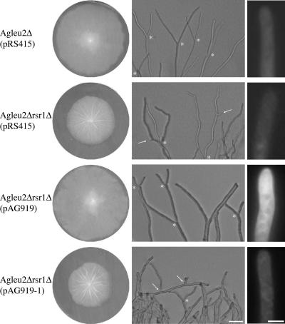
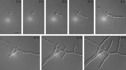
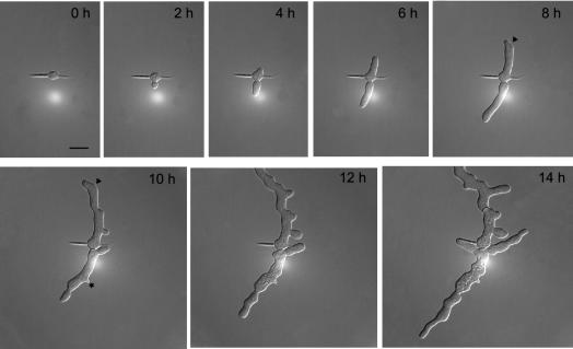
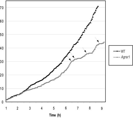
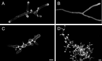
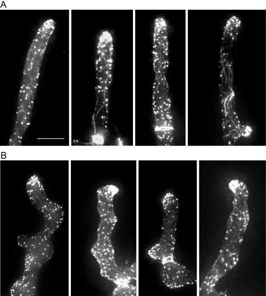
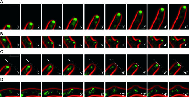
Characteristic features of morphogenesis in filamentous fungi are sustained polar growth at tips of hyphae and frequent initiation of novel growth sites (branches) along the extending hyphae. We have begun to study regulation of this process on the molecular level by using the model fungus Ashbya gossypii. We found that the A. gossypii Ras-like GTPase Rsr1p/Bud1p localizes to the tip region and that it is involved in apical polarization of the actin cytoskeleton, a determinant of growth direction. In the absence of RSR1/BUD1, hyphal growth was severely slowed down due to frequent phases of pausing of growth at the hyphal tip. During pausing events a hyphal tip marker, encoded by the polarisome component AgSPA2, disappeared from the tip as was shown by in vivo time-lapse fluorescence microscopy of green fluorescent protein-labeled AgSpa2p. Reoccurrence of AgSpa2p was required for the resumption of hyphal growth. In the Agrsr1/bud1Delta deletion mutant, resumption of growth occurred at the hyphal tip in a frequently uncoordinated manner to the previous axis of polarity. Additionally, hyphal filaments in the mutant developed aberrant branching sites by mislocalizing AgSpa2p thus distorting hyphal morphology. These results define AgRsr1p/Bud1p as a key regulator of hyphal growth guidance.
Figures
Similar articles
-
From function to shape: a novel role of a formin in morphogenesis of the fungus Ashbya gossypii.Mol Biol Cell. 2006 Jan;17(1):130-45. doi: 10.1091/mbc.e05-06-0479. Epub 2005 Oct 19. Mol Biol Cell. 2006. PMID: 16236798 Free PMC article.
-
The SH3/PH domain protein AgBoi1/2 collaborates with the Rho-type GTPase AgRho3 to prevent nonpolar growth at hyphal tips of Ashbya gossypii.Eukaryot Cell. 2006 Oct;5(10):1635-47. doi: 10.1128/EC.00210-06. Epub 2006 Sep 1. Eukaryot Cell. 2006. PMID: 16950929 Free PMC article.
-
Maximal polar growth potential depends on the polarisome component AgSpa2 in the filamentous fungus Ashbya gossypii.Mol Biol Cell. 2003 Oct;14(10):4140-54. doi: 10.1091/mbc.e03-03-0167. Epub 2003 Aug 22. Mol Biol Cell. 2003. PMID: 12937275 Free PMC article.
-
Evolution of multinucleated Ashbya gossypii hyphae from a budding yeast-like ancestor.Fungal Biol. 2011 Jun;115(6):557-68. doi: 10.1016/j.funbio.2011.02.015. Epub 2011 Feb 26. Fungal Biol. 2011. PMID: 21640319 Review.
-
Homologues of yeast polarity genes control the development of multinucleated hyphae in Ashbya gossypii.Curr Opin Microbiol. 2005 Aug;8(4):370-7. doi: 10.1016/j.mib.2005.06.021. Curr Opin Microbiol. 2005. PMID: 16023404 Review.
Cited by
-
Arf3p GTPase is a key regulator of Bud2p activation for invasive growth in Saccharomyces cerevisiae.Mol Biol Cell. 2013 Aug;24(15):2328-39. doi: 10.1091/mbc.E13-03-0136. Epub 2013 Jun 19. Mol Biol Cell. 2013. PMID: 23783029 Free PMC article.
-
From function to shape: a novel role of a formin in morphogenesis of the fungus Ashbya gossypii.Mol Biol Cell. 2006 Jan;17(1):130-45. doi: 10.1091/mbc.e05-06-0479. Epub 2005 Oct 19. Mol Biol Cell. 2006. PMID: 16236798 Free PMC article.
-
The Ashbya Genome Database (AGD)--a tool for the yeast community and genome biologists.Nucleic Acids Res. 2005 Jan 1;33(Database issue):D348-52. doi: 10.1093/nar/gki009. Nucleic Acids Res. 2005. PMID: 15608214 Free PMC article.
-
The SH3/PH domain protein AgBoi1/2 collaborates with the Rho-type GTPase AgRho3 to prevent nonpolar growth at hyphal tips of Ashbya gossypii.Eukaryot Cell. 2006 Oct;5(10):1635-47. doi: 10.1128/EC.00210-06. Epub 2006 Sep 1. Eukaryot Cell. 2006. PMID: 16950929 Free PMC article.
-
Hyphal guidance and invasive growth in Candida albicans require the Ras-like GTPase Rsr1p and its GTPase-activating protein Bud2p.Eukaryot Cell. 2005 Jul;4(7):1273-86. doi: 10.1128/EC.4.7.1273-1286.2005. Eukaryot Cell. 2005. PMID: 16002653 Free PMC article.
References
-
- Ayad-Durieux, Y., Knechtle, P., Goff, S., Dietrich, F., and Philippsen, P. (2000). A PAK-like protein kinase is required for maturation of young hyphae and septation in the filamentous ascomycete Ashbya gossypii. J. Cell Sci. 113, 4563-4575. - PubMed
Publication types
MeSH terms
Substances
LinkOut - more resources
Full Text Sources
