. 2004 Oct;15(10):4597-608.
doi: 10.1091/mbc.e04-04-0324. Epub 2004 Jul 28.

Defective epidermal barrier in neonatal mice lacking the C-terminal region of connexin43

Affiliations

Defective epidermal barrier in neonatal mice lacking the C-terminal region of connexin43

Karen Maass et al. Mol Biol Cell. 2004 Oct.

Abstract

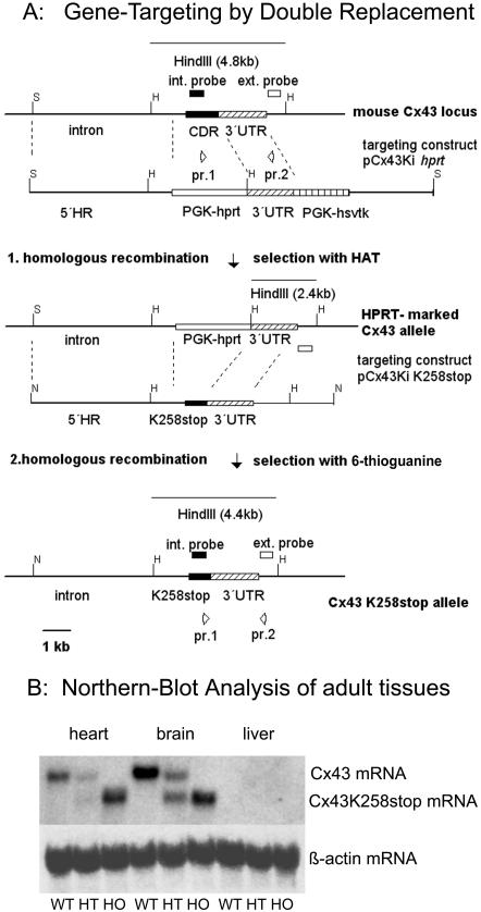
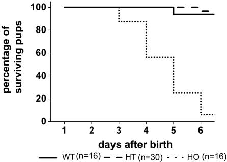
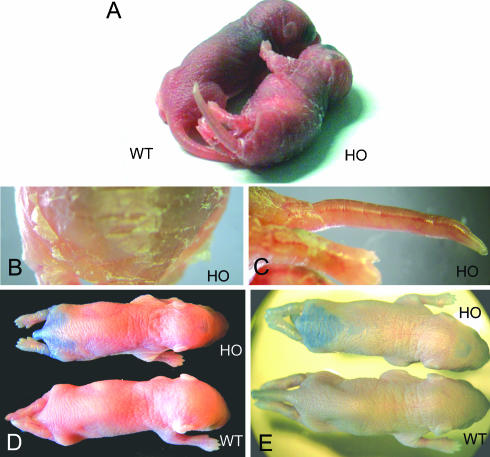
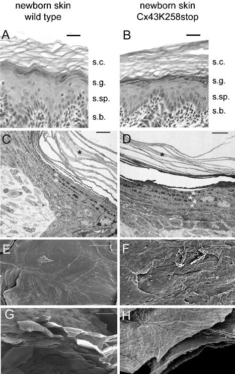
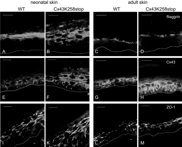
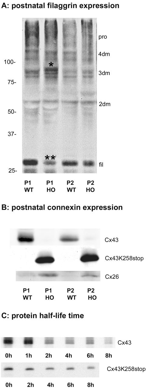
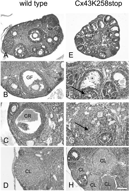
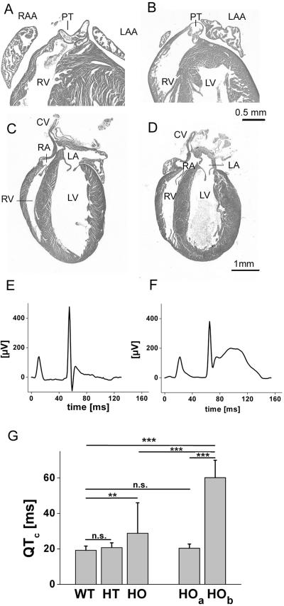
More than 97% of mice in which the C-terminal region of connexin43 (Cx43) was removed (designated as Cx43K258stop) die shortly after birth due to a defect of the epidermal barrier. The abnormal expression of Cx43K258stop protein in the uppermost layers of the epidermis seems to perturb terminal differentiation of keratinocytes. In contrast to Cx43-deficient mice, neonatal Cx43K258stop hearts show no lethal obstruction of the right ventricular outflow tract, but signs of dilatation. Electrocardiographies of neonatal hearts reveal repolarization abnormalities in 20% of homozygous Cx43K258stop animals. The very rare adult Cx43K258stop mice show a compensation of the epidermal barrier defect but persisting impairment of cardiac function in echocardiography. Female Cx43K258stop mice are infertile due to impaired folliculogenesis. Our results indicate that the C-terminally truncated Cx43K258stop mice lack essential functions of Cx43, although the truncated Cx43 protein can form open gap junctional channels.


Figures

Figure 1.
Figure 1.
Gene targeting and transcription analysis of the cx43K258stop allele. In a first homologous recombination in hprt-deficient embryonic stem cells, the cx43 coding region was replaced by a PGK-hprt-cassette (Plum et al., 2000). In the second homologous recombination, the PGK-hprt-cassette was replaced by the truncated cx43 coding region. Recombined clones were enriched by 6-thioguanine selection, validated by PCR analysis and Southern blot by using a 3′ external and a cx43 internal probe. S, SacI; H, HindIII; N, NotI; int, internal; ext., external; CDR, coding region; UTR, untranslated region; HR, homologous region; PGK-hprt, phosphoglycerate kinase promoter-hypoxanthine-guanine phosphoribosyl transferase-minigene; PGK-hsvtk, phosphoglycerate kinase promoterhepes simplex virus thymidine kinase cassette; Ki, knockin. (B) Total RNA blot of heart, brain and liver of WT, HT, and HO adult animals was probed with a radiolabeled DNA fragment corresponding to cx43 5′-UTR. The 2.6-kb cx43K258stop transcript was detected proportional to gene dosis. No 3.0-kb cx43 wild-type transcript was detected in homozygous mutant animals. Equal amounts of loaded total RNA were verified by reprobing with a β-actin probe.
Figure 2.
Figure 2.
Homozygous Cx43K258stop mice show high postnatal lethality. Ten heterozygous matings litters (62 pups) were subjected to a close-up observation. Homozygous animals were born at the expected Mendelian ratio of 25% (16 animals) but showed high postnatal lethality with only ∼2.5% (1 animal) surviving the first 5 d.
Figure 3.
Figure 3.
Homozygous Cx43K258stop pups show epidermal abnormalities and defective epidermal permeability barrier. (A–C) Comparison between littermates at postnatal day 4 revealed abnormal appearance of homozygous mutant epidermis with fissures and squames on back (A and B) and around joints (A). Constriction rings around tails were often found (C). (D and E) Penetration assays with toluidine blue revealed a defective epidermal permeability barrier of homozygous Cx43K258stop epidermis at postnatal 1 (D) and even more intensive at day 3 (E).
Figure 4.
Figure 4.
Comparison of wild-type and homozygous mutant epidermis by histology, transmission, and scanning electron microscopy. HE-stained paraffin sections (A and B), thin sections (C and D), and SEM of whole mount skin (E–H) revealed significant differences between morphology of s. corneum (s.c.) and s. granulosum (s.g.) of wild-type (A, C, E, and G) and homozygously mutated animals (B, D, F, and H). Characteristic of Cx43K258stop epidermis was the formation of more but thinner layers of corneocytes (C and D, black asterisks) and the premature formation of keratohyaline granules in s. spinosum (s.sp.) (C and D, white asterisks). In contrast to the even surface of wild-type epidermis (E) mutant skin showed frequent fissures (F). Bars, 20 μm (A, B, and G); 10 μm (C–F, and H). s.b., s. basale.
Figure 5.
Figure 5.
Immunofluorescence analysis of filaggrin, ZO-1, and Cx43 in epidermis. Cryosections of neonatal and adult epidermis of wild-type (A, C, E, G, I, and L) and homozygous Cx43K258stop animals (B, D, F, H, K, and M) were immunolabeled and the epidermis–dermis boundary was indicated by a white line. (A–D) Filaggrin was additionally detected in s. spinosum of neonatal homozygous Cx43K258stop animals (B). (E–H) In comparison with Cx43 (E and G), Cx43K258stop protein was abundantly expressed throughout neonatal homozygous mutant epidermis (K). In adult Cx43K258stop epidermis, the expression was elevated and detected mainly in the suprabasal s. spinosum (M). (I–M) ZO-1 was slightly elevated in homozygous mutant epidermis (K and M). Bars, 20 μm.
Figure 6.
Figure 6.
Immunoblot analyses of postnatal epidermis and Cx43K258stop protein turnover time in cultured HeLa cells. Immunoblot analysis (A and B) was carried out at postnatal days P1 (lanes 1 and 2) and P2 (lanes 3 and 4) of WT and HO epidermis. Amount of monumeric filaggrin was reduced in P1 homozygous epidermis (**), and an additional signal of higher molecular mass than the intermediate with three filaggrin domains (*) was found. Pro, profilaggrin; 4dm-2dm, intermediates with four, three, or two filaggrin domains; fil, monumeric filaggrin. (B) Membrane subsequently probed with primary antibodies to the cytoplasmic loop of Cx43 or to Cx26. Cx43K258stop and Cx26 amounts were elevated in homozygous epidermis. (C) Pulse-chase analyses on HeLa-Cx43WT and HeLa-Cx43K258stop cells revealed that the half-life time of Cx43K258stop was more than doubled to 4.2 h in comparison with 2.0 h for Cx43.
Figure 7.
Figure 7.
Histology of adult Cx43K258stop ovaries. (A–D) Normal folliculogenesis in wild-type ovaries (A and B), with ovulation indicated by formation of corpus rubrum (C) and subsequent development of corpora lutea (D). (E–H) In Cx43K258stop mice, follicular stages up to young Graafian follicles were observed, but no mature Graafian follicles developed (E). Preovulatory follicles showed morphological abnormalities (F, arrows) and degenerating oocytes (F, asterisk). Inappropriate luteinization (G) and corpora lutea formation without ovulation were observed (H). S, secondary follicle; GF, Graafian follicle; CR, corpus rubrum; CL, corpus luteum. Bars, 250 μm (A); 150 μm (B, C, D, F, and G); 375 μm (E); and 100 μm (H).
Figure 8.
Figure 8.
Histology and electrocardiography of neonatal Cx43K258stop hearts. (A–D) HE-stained sections of neonatal wild-type (A and C) and homozygous Cx43K258stop (B and D) hearts. The subpulmonary outlet of the Cx43K258stop heart (B) is normally open as that of the wild type (A). Cx43K258stop hearts (D) have a bulb shape with a dilated left ventricle and blunt apex. CV, caval vein; RA, right atrium; RV, right ventricle; LA, left atrium; LV, left ventricle; PT, pulmonary truncus; RAA, right atrial appendage; LAA, left atrial appendage. Bars, 500 μm (A and B); and 1 mm (C and D). (E and F) Signal-averaged ECG recordings of two homozygously mutated pups on postnatal day 2; the animal in E showed an unobtrusive ECG, whereas the animal in F displayed ST-elevation, QT-prolongation, and enlarged T-wave. (G) Frequency-corrected QTc-intervals demonstrated a significant longer repolarization in homozygous Cx43K258stop mice. The high SD in QTc of homozygous mice is explained by a subgroup of seven mice (HOb) demonstrating a significantly higher QTc compared with wild type (E, 60 ± 10 ms; p < 0.001). Remaining (n = 33) pups in this group (Hoa) demonstrated no differences to WT or HT. n.s., p > 0.05; **p < 0.01; ***p < 0.001.
Figure 9.
Figure 9.
Immunofluorescence analysis of hearts. Cryosections of neonatal (A, B, E, and F) and adult hearts (C, D, G, and H) were immunolabeled and costained for nuclei. (A–D) Compared with neonatal wild type (A), Cx43K258stop immunoreactive spots in the epicardial region of neonatal homozygous left ventricle (B) were increased in size. In adults, Cx43 and Cx43K258stop were detectable in intercalated discs in both genotypes (C and D). (E–H) ZO-1 expression was slightly elevated in homozygous mutant Cx43K258stop hearts (F and H). Bars, 20 μm.
Figure 10.
Figure 10.
Cx43K258stop gene dosage influences epidermal phenotype and postnatal survival. Post-weaning offspring was grouped according to genotypes. (A) Only 311 of 415 born animals from heterozygous Cx43WT/Cx43K258stop matings survived to adulthood. According to the expected Mendelian ratio of 50% heterozygous and both 25% wild-type and homozygous animals, percentages of survival were multiplied by 2 or 4, respectively. Less than 3% of all expected homozygous animals survived to adulthood. (B) Animals (105 of 131) from Cx43WT/Cx43K258stop × Cx43K258stop/Cx43KO matings survived to adulthood. According to the Mendelian ratio of each 25% Cx43/Cx43, Cx43WT/Cx43K258stop, Cx43K258stop/Cx43KO, and Cx43K258stop/Cx43KO animals, percentages of survival were multiplied by 4 to determin general survival percentage of the different groups. Cx43/Cx43, Cx43/Cx43K258stop, and Cx43/Cx43KO displayed survival in the range between 75 and 100%. Whereas Cx43K258stop/Cx43KO animals still displayed a reduced survival, >16 times as many animals of this genotype reached adulthood compared with homozygous Cx43K258stop animals. (C–E) In contrast to homozygous neonatal Cx43K258stop animals (C), toluidine dye penetration assay revealed no staining due to a defective barrier in Cx43K258stop/Cx43KO (D) and Cx43/Cx43KO (E) animals.

Similar articles

Cited by

References

    1. Ackert, C.L., Gittens, J.E., O'Brien, M.J., Eppig, J.J., and Kidder, G.M. (2001). Intercellular communication via connexin43 gap junctions is required for ovarian folliculogenesis in the mouse. Dev. Biol. 233, 258-270. - PubMed
    1. Bakirtzis, G., et al. (2003). Targeted epidermal expression of mutant Connexin 26(D66H) mimics true Vohwinkel syndrome and provides a model for the pathogenesis of dominant connexin disorders. Hum. Mol. Genet. 12, 1737-1744. - PubMed
    1. Barker, R.J., Price, R.L., and Gourdie, R.G. (2002). Increased association of ZO-1 with connexin43 during remodeling of cardiac gap junctions. Circ. Res. 90, 317-324. - PubMed
    1. Brissette, J.L., Kumar, N.M., Gilula, N.B., Hall, J.E., and Dotto, G.P. (1994). Switch in gap junction protein expression is associated with selective changes in junctional permeability during keratinocyte differentiation. Proc. Natl. Acad. Sci. USA 91, 6453-6457. - PMC - PubMed
    1. Cartlidge, P. (2000). The epidermal barrier. Semin. Neonatol. 5, 273-280. - PubMed

Publication types

MeSH terms