. 2004 Oct;15(10):4568-83.
doi: 10.1091/mbc.e04-04-0330. Epub 2004 Jul 28.

Protein-protein interactions governing septin heteropentamer assembly and septin filament organization in Saccharomyces cerevisiae

Affiliations

Protein-protein interactions governing septin heteropentamer assembly and septin filament organization in Saccharomyces cerevisiae

Matthias Versele et al. Mol Biol Cell. 2004 Oct.

Abstract

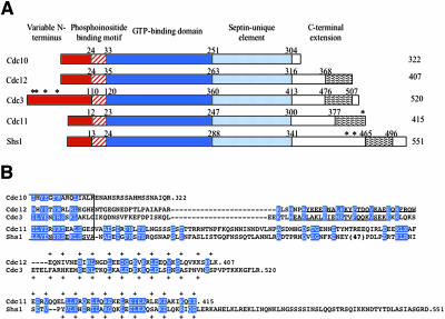
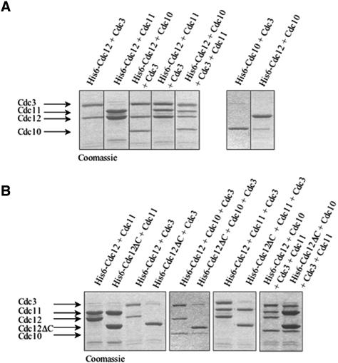
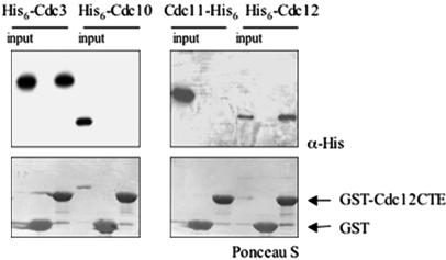
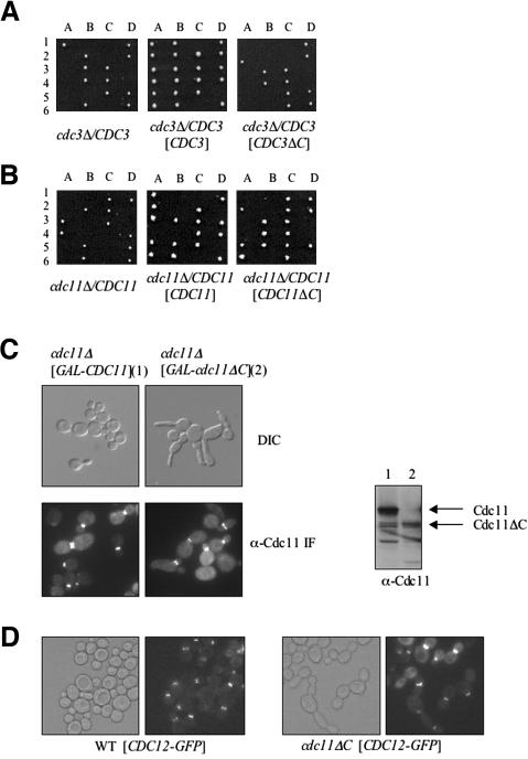
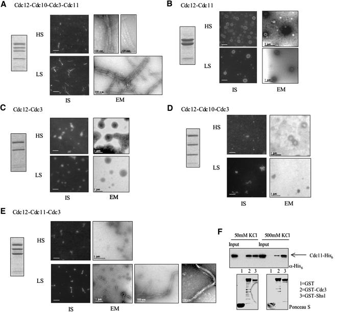
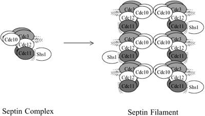
Mitotic yeast (Saccharomyces cerevisiae) cells express five related septins (Cdc3, Cdc10, Cdc11, Cdc12, and Shs1) that form a cortical filamentous collar at the mother-bud neck necessary for normal morphogenesis and cytokinesis. All five possess an N-terminal GTPase domain and, except for Cdc10, a C-terminal extension (CTE) containing a predicted coiled coil. Here, we show that the CTEs of Cdc3 and Cdc12 are essential for their association and for the function of both septins in vivo. Cdc10 interacts with a Cdc3-Cdc12 complex independently of the CTE of either protein. In contrast to Cdc3 and Cdc12, the Cdc11 CTE, which recruits the nonessential septin Shs1, is dispensable for its function in vivo. In addition, Cdc11 forms a stoichiometric complex with Cdc12, independent of its CTE. Reconstitution of various multiseptin complexes and electron microscopic analysis reveal that Cdc3, Cdc11, and Cdc12 are all necessary and sufficient for septin filament formation, and presence of Cdc10 causes filament pairing. These data provide novel insights about the connectivity among the five individual septins in functional septin heteropentamers and the organization of septin filaments.


Figures

Figure 1.
Figure 1.
Primary structure of S. cerevisiae mitotic septins. (A) Schematic depiction of the domain structure of yeast septins. Characteristic features include N termini of variable length and sequence (red box), tract of basic and hydrophobic residues implicated in septin-phosphoinositide interaction (hatched box), globular GTPase domain (dark blue box), a sequence element diagnostic of septins (light blue box), and the variable C-terminal extensions (white box) containing, where indicated, a predicted coiled coil segment (wavy box). The number of residues in each protein is indicated to the right, and the residue numbers above demarcate the boundaries of the indicated domains. Asterisks denote known sites of covalent attachment of Smt3 (yeast SUMO). (B) Amino acid sequence of the C-termini beyond the last 15 residues of the septin-unique element (boxed). In each of the indicated septins, the CTEs (number of residues) are as follows: Cdc10 (18), Cdc12 (91), Cdc3 (107), Cdc11 (115), and Shs1 (210). Identities and conservative substitutions shared by all five septins, or by the indicated septin pairs, are shown as white letters on blue boxes. Period indicates the C-terminal end of each polypeptide; dashes indicate gaps introduced to maximize the alignment, or to position the predicted coiled coil forming segments in the same approximate register. Strongly predicted α-helical segments (in Cdc3 and Cdc12 only) are underlined; residues constituting the diagnostic 4-3 repeat of primarily hydrophobic residues characteristic of coiled coils are indicated by the plus sign. Number in parentheses indicates a large insert present in Shs1.
Figure 2.
Figure 2.
The essential function of Cdc12 requires its C-terminal extension. (A) A cdc12Δ/CDC12 heterozygous diploid (Y21935) was transformed with a CEN plasmid carrying either wild-type CDC12 (pMVB45), CDC12ΔC (pMVB48), CDC12(S43V)ΔC (pMVB52), or CDC12(T48N)ΔC (pMVB53), sporulated and individual tetrads (1–6) were dissected and the resulting spore clones (A–D) were germinated on YPD plates. Samples (30 μg of total protein) of extracts were prepared and analyzed by SDS-PAGE and immunoblotting with an anti-Cdc12ΔC antibody [lane 1, Y21935 transformed with a CEN plasmid expressing wild-type CDC12; lane 2, Y21935 transformed with the same CEN vector expressing CDC12ΔC (right-most panel)]. (B) Wild-type cells (BY4741) were transformed with a CEN plasmid expressing either CDC12-GFP (pLP17) or CDC12ΔC-GFP (pMVB33), grown at 26°C to midexponential phase on SCGlc(-Leu), and samples of each culture were analyzed by differential interference contrast (DIC) optics (left) or fluorescence microscopy (right). Samples (30 μg of total protein) of extracts were prepared from the same cultures and analyzed by SDS-PAGE and immunoblotting with an anti-GFP monoclonal antibody (lane 1, BY4741 expressing CDC12-GFP; lane 2, BY4741 expressing CDC12ΔC-GFP; right-most panel). (C) Cdc12ΔC interferes with septin collar assembly. A wild-type strain (BJ2168) was transformed with an empty vector or with either an integrating vector (YMVB17) or a multi-copy (2 μm DNA) vector expressing CDC12ΔC, and samples of each were examined by bright field (left) or stained with DAPI to reveal the location of nuclei and examined by fluorescence microscopy (right). Samples (30 μg of total protein) of extracts were prepared from the same cultures and analyzed by SDS-PAGE and immunoblotting with polyclonal anti-Cdc12ΔC antibody (lane 1, BJ2168 transformed with empty vector; lane 2, BJ2168 transformed with the integrating vector expressing CDC12ΔC; lane 3, BJ2168 transformed with the multi-copy vector expressing CDC12ΔC; right-most panel). (D) A wild-type strain (BY4741) expressing either GAL1-CDC12-GFP (pMVB2) (left column) or GAL1-CDC12ΔC-GFP (pMVB160) (right column), or the same strains coexpressing either GAL1-CDC12 (pMVB162) or GAL1-CDC12ΔC (pMVB164) along with each of the indicated plasmids [CDC3-GFP, CDC10-GFP (pLA10) or CDC11-GFP (pSB5)], were grown to midexponential phase on SCGlc(-Leu,-Ura) at 30°C, and samples were analyzed by DIC optics (left) or fluorescence microscopy to visualize GFP-tagged septins (right).
Figure 7.
Figure 7.
Cdc10 is essential for normal septin collar organization at the bud neck. (A) Wild-type strain (BY4741) and a cdc10Δ mutant (YMVB5) were transformed with plasmids expressing Cdc12-GFP (pLP17) or Cdc12ΔC-GFP (pMVB33), grown at 26°C to midexponential phase on SCGlc(-Leu), and samples of each culture were viewed by differential interference contrast optics (left) or fluorescence (right) microscopy (GFP). Samples (30 μg of total protein) of extracts prepared from the same cultures was analyzed by SDS-PAGE and immunoblotting by using an anti-GFP monoclonal antibody. The blot was stained with Ponceau S as a loading control. (B) Wild-type strain (BY4741) and cdc10Δ mutant (YMVB5) were transformed with either a CEN plasmid (pLP17) or a multi-copy (2 μm DNA) vector (pMVB62) expressing Cdc12-GFP, grown to midexponential phase at 26°C on SCGlc(-Leu), and samples of each culture were incubated with calcofluor white to stain chitin and then viewed by fluorescence microscopy, by using appropriate cut-off filters to view GFP and chitin staining, as indicated.
Figure 3.
Figure 3.
Role of septin CTEs in pairwise septin–septin interactions. (A) Equivalent amounts of the indicated GST-septin fusions [Cdc3, Cdc3(Δ427–521), Cdc10, Cdc11, Cdc11(Δ357–416), Cdc12, and Cdc12(Δ339–407)] were immobilized on glutathione-agarose beads, incubated with either Cdc12(His)6 or Cdc12ΔC(His)6, and washed extensively. Bound proteins were eluted with SDS-PAGE sample buffer, resolved by SDS-PAGE, and analyzed either by Coomassie Blue (to verify equal loading of the GST-fusion proteins) or by immunoblotting by using anti-His(C-term) antibody to detect the input and bound (His)6-tagged proteins. Input, 20% of the amount of Cdc12-His6 or Cdc12ΔC-His6 added in each binding reaction. (B) The same GST-septin fusions as in A, immobilized on glutathione-agarose, were incubated with His6-Cdc3, His6-Cdc10, and Cdc11-His6, as indicated, and analyzed as described in A, except either anti-His antibody or anti-His(C-term) was used, as appropriate. C. GST-Shs1 and GST-Shs1(Δ418–551) were immobilized on glutathione-agarose and incubated with the indicated His6-tagged septins and analyzed as described in B.
Figure 4.
Figure 4.
Reconstitution of stoichiometric septin complexes requires the CTE of Cdc12. (A) His6-Cdc12 or His6-Cdc10, as indicated, was coexpressed in E. coli with the indicated untagged septin(s), and any resulting complexes were purified using Ni2+-NTA-agarose. After washing, bound proteins were eluted with imidazole, resolved by SDS-PAGE, and stained with Coomassie Blue. (B) His6-Cdc12 or His6-Cdc12ΔC, as indicated, was coexpressed with the other indicated untagged septin(s), and analyzed as described in A.
Figure 5.
Figure 5.
The Cdc12 CTE is sufficient for interaction with Cdc3. GST-Cdc12(339–407) [GST-CT-ECdc12] expressed in bacteria from pMVB172 was immobilized on glutathione-agarose and incubated with the indicated purified His6-tagged septins. After washing, bound proteins were eluted, resolved by SDS-PAGE, and analyzed either by Ponceau S dye (to verify equal loading of the GST-fusion proteins) or by immunoblotting by using anti-His monoclonal antibody to detect the input and bound (His)6-tagged proteins. Input, 20% of the total amount of the indicated His6-tagged protein that was added in each binding reaction.
Figure 6.
Figure 6.
The Cdc3 CTE is essential, but the Cdc11 CTE is not. (A) A cdc3Δ/CDC3 heterozygous diploid (Y25223) was transformed with empty vector, or a CEN plasmid carrying either CDC3 (pMVB100) or CDC3ΔC (pMVB102), sporulated and the meiotic products analyzed described in the legend of Figure 2A. (B) A cdc11Δ/CDC11 heterozygous diploid (YMVB32) was transformed with empty vector, or a CEN plasmid expressing CDC11 (pSB1) or CDC11ΔC (pSB2), and analyzed as in A. C. A cdc11Δ mutant expressing either CDC11 or CDC11ΔC from the GAL1 promoter on CEN vectors (YCpUG-CDC11 and YCpUG-CDC11ΔC, respectively) were grown to midexponential phase and analyzed by differential interference contrast (top) or indirect immunofluorescence by using rabbit polyclonal anti-Cdc11 as the primary antibody (bottom). Samples (30 μg total protein) of extracts prepared from the same cultures were analyzed by SDS-PAGE and immunoblotting by using the anti-Cdc11 antibody. D. A cdc11Δ mutant expressing either CDC11 (WT) from pSB1 or CDC11ΔC (cdc11ΔC) from pSB2 was transformed with a plasmid (pLP17) expressing Cdc12-GFP, grown to midexponential phase and viewed by DIC (left) or fluorescence (right) microscopy (GFP).
Figure 8.
Figure 8.
Ultrastructural analysis of reconstituted septin filaments. (A–E) The ability of the indicated purified, recombinant, heteromeric septin complexes (left panel) to form filaments in the high-salt elution buffer used for purification (HS) or after dialysis against 50 mM KCl (LS) were analyzed, as indicate, either by immunostaining using anti-Cdc12ΔC antibodies (IS, bars represent 2% μm) or by EM after negative staining (bar represents scale indicated). (F) The interaction between Cdc3 and Cdc11 is weakened at high salt concentration. Binding assay was performed as described in the legend to Figure 3 at the salt concentrations indicated.
Figure 9.
Figure 9.
Model for septin heteropentamer organization and assembly into filaments. Together, the results obtained in this study indicate that septin heteropentamers are assembled with the interseptin contacts indicated and suggest that filaments are formed by the end-to-end polymerization of Cdc3–Cdc12–Cdc11 complexes, with Cdc10 serving as a bridge to bundle the polymers into paired filaments.

Similar articles

Cited by

References

    1. Bardwell, L., Cook, J.G., Zhu-Shimoni, J.X., Voora, D., and Thorner, J. (1998). Differential regulation of transcription: repression by unactivated mitogen-activated protein kinase kss1 requires the dig1 and dig2 proteins. Proc. Natl. Acad. Sci. USA 95, 15400-15405. - PMC - PubMed
    1. Barral, Y., Mermali, V., Mooseker, M.S., and Snyder, M. (2000). Compartmentalization of the cell cortex by septins is required for maintenance of cell polarity in yeast. Mol. Cell 5, 841-851. - PubMed
    1. Barral, Y., Parra, M., Bidlingmaier, S., and Snyder, M. (1999). Nim1-related kinases coordinate cell-cycle progression with the organization of the peripheral cytoskeleton in yeast. Genes Dev. 13, 176-187. - PMC - PubMed
    1. Beites, C.L., Xie, H., Bowser, R., and Trimble, W.S. (1999). The septin CDCrel-1 binds syntaxin and inhibits exocytosis. Nat. Neurosci. 2, 434-439. - PubMed
    1. Brachmann, C.B., Davies, A., Cost, G.J., Caputo, E., Li, J., Hieter, P., and Boeke, J.D. (1998). Designer deletion strains derived from Saccharomyces cerevisiae S288C: a useful set of strains and plasmids for PCR-mediated gene disruption and other applications. Yeast 14, 115-132. - PubMed

Publication types

MeSH terms