. 2004 Aug 18;23(16):3346-55.
doi: 10.1038/sj.emboj.7600341. Epub 2004 Jul 29.

Visualization of RNA-protein interactions in living cells: FMRP and IMP1 interact on mRNAs

Affiliations

Visualization of RNA-protein interactions in living cells: FMRP and IMP1 interact on mRNAs

Oliver Rackham et al. EMBO J. .

Abstract

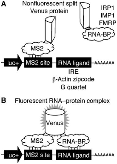
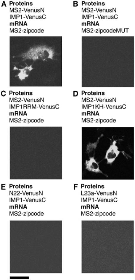
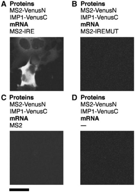
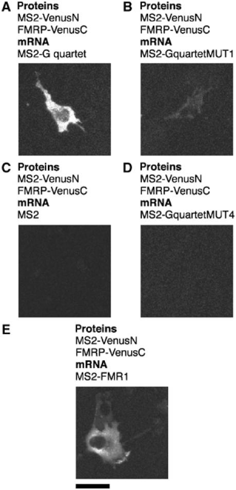
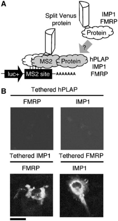
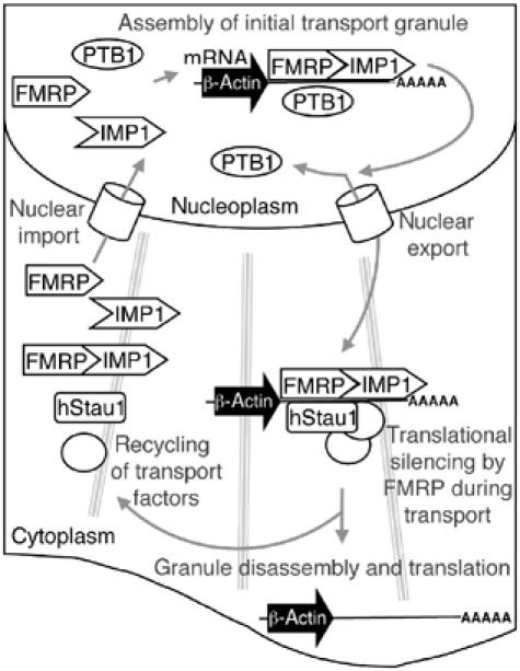
Protein expression depends significantly on the stability, translation efficiency and localization of mRNA. These qualities are largely dictated by the RNA-binding proteins associated with an mRNA. Here, we report a method to visualize and localize RNA-protein interactions in living mammalian cells. Using this method, we found that the fragile X mental retardation protein (FMRP) isoform 18 and the human zipcode-binding protein 1 ortholog IMP1, an RNA transport factor, were present on common mRNAs. These interactions occurred predominantly in the cytoplasm, in granular structures. In addition, FMRP and IMP1 interacted independently of RNA. Tethering of FMRP to an mRNA caused IMP1 to be recruited to the same mRNA and resulted in granule formation. The intimate association of FMRP and IMP1 suggests a link between mRNA transport and translational repression in mammalian cells.


Figures

Figure 1
Figure 1
A TriFC method to study RNA–protein interactions in living cells. A portion of the Venus fluorescent protein is attached to a reporter mRNA by the bacteriophage MS2 coat protein–RNA interaction. The complementing portion of Venus is fused to an RNA-binding protein (A). If the RNA-binding protein interacts with a sequence of interest within the reporter mRNA, the two portions of Venus are brought into close proximity to form a fluorescent product (B).
Figure 2
Figure 2
Detection of specific IMP1–zipcode interactions in living cells. Cells were transfected with plasmids expressing protein fusions to either the N- or C-terminal complementing portions of Venus (VenusN or VenusC, respectively) and reporter mRNAs as indicated. Scale bar, 20 μm.
Figure 3
Figure 3
Detection of specific IRP1–IRE interactions in living cells. Cells were transfected with plasmids expressing the MS2 coat protein fusion to the N-terminal complementing portion of Venus protein (MS2-VenusN) and IRP1 fused to the C-terminal complementing portion of Venus (IRP1-VenusC), and reporter mRNAs as indicated. Scale bar, 20 μm.
Figure 4
Figure 4
Detection of specific FMRP–RNA interactions in living cells. Cells were transfected with plasmids expressing the MS2 coat protein fusion to the N-terminal complementing portion of Venus protein (MS2-VenusN) and FMRP isoform 18 fused to the C-terminal complementing portion of Venus (VenusC), and reporter mRNAs containing the MS2 operator and additional sequences as indicated. Scale bar, 20 μm.
Figure 5
Figure 5
IMP1, FMRP, hStau and PTB associate with multiple mRNA sequences in vivo. Cells were transfected with plasmids expressing the MS2 coat protein fused to the N-terminal complementing portion of Venus, an RNA-binding protein (IMP1, FMRP, hStau or PTB) fused to the C-terminal complementing portion of Venus, and a reporter mRNA containing an MS2 coat protein recognition site and RNA sequence of interest as indicated. Scale bar, 20 μm.
Figure 6
Figure 6
IMP1 and FMRP associate with zipcode and FMR1 3′UTR RNA sequences. Immunoprecipitation confirms the association of IMP1 and FMRP with β-actin zipcode- and FMR1 3′UTR-containing mRNAs. An EGFP fusion to the MS2 coat protein allowed the immunoprecipitation of reporter mRNAs containing an MS2 coat protein recognition site and an RNA sequence of interest. Immunoprecipitations were performed using anti-GFP antibodies. FMRP and IMP1 were coexpressed as mRFP1 and FLAG-tagged fusion proteins, respectively. The theoretical molecular weights of FMRP-mRFP1 and FLAG-IMP1 are 91 and 66 kDa, respectively. The efficiency of immunoprecipitation was shown by immunoblotting with anti-GFP antibodies (lower panel). The theoretical molecular weight of MS2-EGFP is 40 kDa. The association of FMRP and IMP1 with reporter mRNAs was determined by immunoblotting with anti-FLAG and anti-FMRP antibodies, respectively.
Figure 7
Figure 7
FMRP and IMP1 interact in the absence of reporter mRNA. Expression of protein fusions to complementing portions of Venus demonstrated homomeric and heteromeric interactions between FMRP and IMP1. The unrelated IRP1 and L23a RNA-binding proteins do not interact with either FMRP or IMP1.
Figure 8
Figure 8
FMRP and IMP1 associate via protein–protein interactions. (A) Cells were transfected with plasmids expressing FMRP fused to VenusC and FLAG-tagged IMP1. FMRP-VenusC was immunoprecipitated with an anti-GFP antibody. To test for nonspecific immunoprecipitation, an irrelevant antibody, anti-COX IV, was used in a control reaction. Immunoblotting with anti-GFP antibodies was used to confirm the immunoprecipitation of FMRP-VenusC (theoretical molecular weight of 93 kDa). The association of FLAG-IMP1 (theoretical molecular weight of 66 kDa) was determined by immunoblotting with anti-FLAG antibodies. A reaction in which RNase A was used in place of RNasin determined the RNA-independent nature of the interaction. (B) Cells were transfected with plasmids expressing an IMP1-Venus fusion protein and mRFP1 tagged FMRP (both of theoretical molecular weight 91 kDa). Immunoprecipitations were performed as in (A). The association of FMRP with IMP1 was determined by immunoblotting with anti-FMRP antibodies. In a control reaction, lysates expressing only FMRP-mRFP1 were subjected to immunoprecipitation.
Figure 9
Figure 9
Tethered IMP1 recruits FMRP to an mRNA, and tethered FMRP can recruit IMP1 to an mRNA. (A) Schematic representation of how TriFC can be used to analyze the recruitment of specific protein to an mRNP complex. (B) Cells were transfected with plasmids expressing reporter mRNA containing the MS2 coat protein recognition site and the MS2 coat protein fused to the N-terminal complementing portion of Venus. A soluble form of hPLAP, IMP1 or FMRP was tethered to the reporter mRNA by fusion to the MS2 coat protein sequence. Recruitment of IMP1 or FMRP to reporter mRNAs was determined by coexpression of fusions to the C-terminal portion of Venus.
Figure 10
Figure 10
Model of the mRNA localization process involving IMP1 and FMRP. The localization process begins in the nucleus, with IMP1 and FMRP associating with the RNA at the transcription site. In the example shown, the target is β-actin mRNA. IMP1 and FMRP may bind sequentially, or attach as a preformed complex. The binding of PTB in the nucleus may in some way facilitate the granule assembly or nuclear export processes. The hStau1 protein could attach in the nucleus or the cytoplasm along with other general mRNA localization components, such as molecular motors. The granule then travels to its final location at the cell periphery. While in transit, attached FMRP silences the translation of transported mRNAs. Once anchored at its final location, the granule complex is disassembled. Removal of FMRP leaves mRNAs competent for translation. The free transport factors then return to take part in further rounds of mRNA localization.

References

    1. Antar LN, Bassell GJ (2003) Sunrise at the synapse: the FMRP mRNP shaping the synaptic interface. Neuron 37: 555–558 - PubMed
    1. Arn EA, Cha BJ, Theurkauf WE, Macdonald PM (2003) Recognition of a bicoid mRNA localization signal by a protein complex containing Swallow, Nod, and RNA binding proteins. Dev Cell 4: 41–51 - PubMed
    1. Bassell GJ, Zhang H, Byrd AL, Femino AM, Singer RH, Taneja KL, Lifshitz LM, Herman IM, Kosik KS (1998) Sorting of beta-actin mRNA and protein to neurites and growth cones in culture. J Neurosci 18: 251–265 - PMC - PubMed
    1. Brown V, Jin P, Ceman S, Darnell JC, O'Donnell WT, Tenenbaum SA, Jin X, Feng Y, Wilkinson KD, Keene JD, Darnell RB, Warren ST (2001) Microarray identification of FMRP-associated brain mRNAs and altered mRNA translational profiles in fragile X syndrome. Cell 107: 477–487 - PubMed
    1. Brown V, Small K, Lakkis L, Feng Y, Gunter C, Wilkinson KD, Warren ST (1998) Purified recombinant Fmrp exhibits selective RNA binding as an intrinsic property of the fragile X mental retardation protein. J Biol Chem 273: 15521–15527 - PubMed

Publication types