. 2004 Aug 18;23(16):3270-81.
doi: 10.1038/sj.emboj.7600342. Epub 2004 Jul 29.

LRIG1 restricts growth factor signaling by enhancing receptor ubiquitylation and degradation

Affiliations

LRIG1 restricts growth factor signaling by enhancing receptor ubiquitylation and degradation

Gal Gur et al. EMBO J. .

Abstract

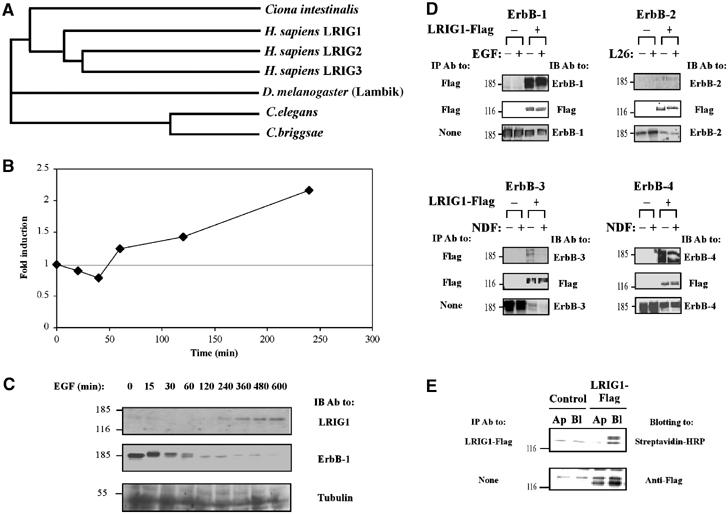
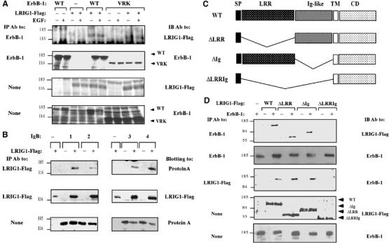
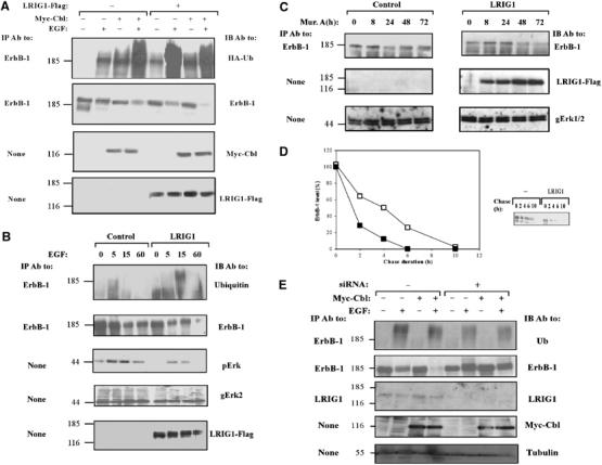
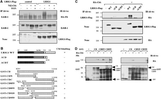
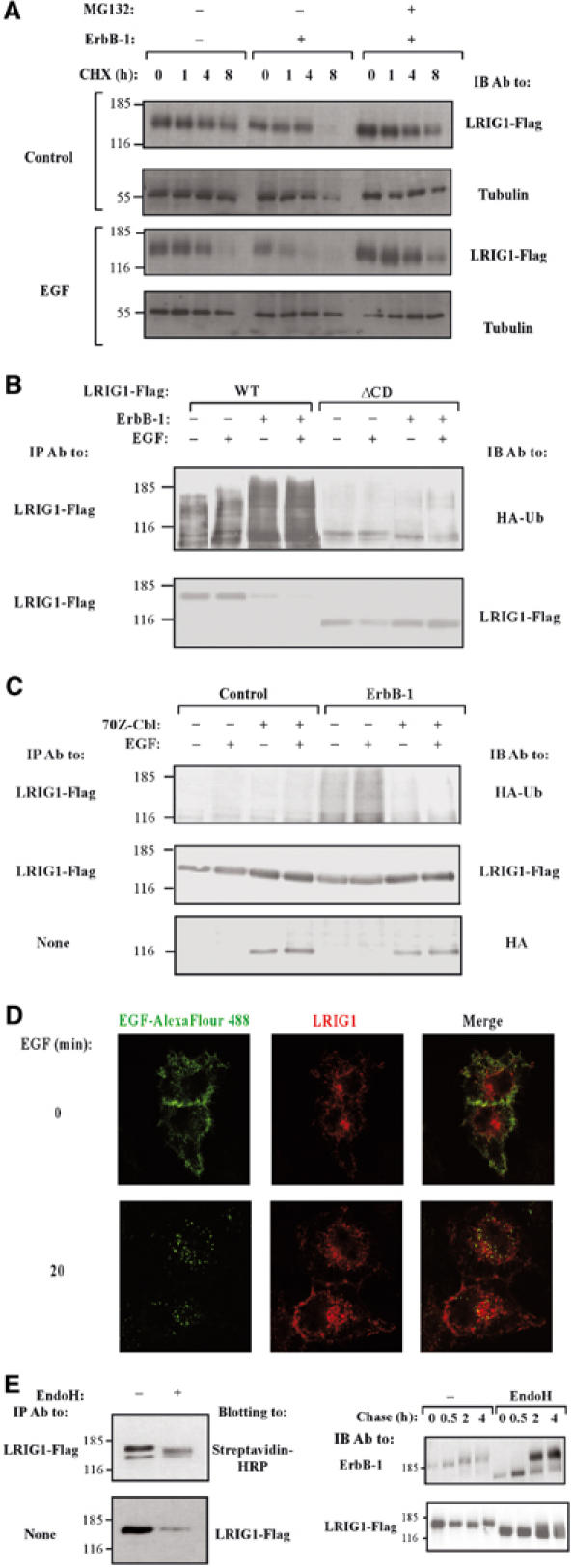
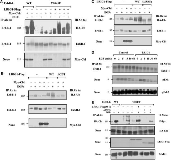
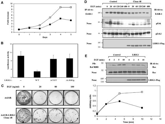
Kekkon proteins negatively regulate the epidermal growth factor receptor (EGFR) during oogenesis in Drosophila. Their structural relative in mammals, LRIG1, is a transmembrane protein whose inactivation in rodents promotes skin hyperplasia, suggesting involvement in EGFR regulation. We report upregulation of LRIG1 transcript and protein upon EGF stimulation, and physical association of the encoded protein with the four EGFR orthologs of mammals. Upregulation of LRIG1 is followed by enhanced ubiquitylation and degradation of EGFR. The underlying mechanism involves recruitment of c-Cbl, an E3 ubiquitin ligase that simultaneously ubiquitylates EGFR and LRIG1 and sorts them for degradation. We conclude that LRIG1 evolved in mammals as a feedback negative attenuator of signaling by receptor tyrosine kinases.


Figures

Figure 1
Figure 1
LRIG1 mRNA is induced by EGF and the encoded protein physically associates with the EGF receptor and other ErbB proteins. (A) LRIG proteins form a distinct, conserved LRR-Ig subfamily in metazoans. LRIG orthologs assembled from predicted proteomes of completed genomes were used for multi-alignment using ClustalW, gap trimming and tree construction. The following proteins were assembled: human LRIG1 (gene accession AB018349), LRIG2 (AY358288) and LRIG3 (AY505340); Ciona intestinalis gene 77.18.1; D. melanogaster gene CG8434 (Lambik); C. briggsae predicted protein P6018; and C. elegans predicted protein T21D12.9. (B) Subconfluent HeLa cells (∼1.7 × 10⁶ cells/plate) were serum starved for 24 h, followed by treatment with EGF (20 ng/ml) for the indicated time intervals. Cells were then extracted, and total RNA prepared, followed by reverse transcription with random hexamer primers. Real-time PCR was carried out with primers specific to LRIG1. The level of gene expression was quantified in comparison to a standard curve created by serial dilutions of the template cDNA. The experiment was repeated thrice. (C) HeLa cells were treated with EGF for the indicated intervals as in (B). Whole-cell extracts were resolved by electrophoresis and analyzed by IB with the indicated antibodies. (D) HEK-293T cells co-expressing a Flag-tagged LRIG1 and the indicated ErbB proteins were incubated for 15 min at 37°C with the respective ligands, or with a mAb to ErbB-2 (L26), prior to cell lysis. Cleared cell extracts were subjected to IP and IB, as indicated. (E) Filter-grown MDCK cells stably expressing a Flag peptide-tagged LRIG1 were labeled on the apical (Ap) or the basolateral (Bl) surface with biotin. Whole-cell extracts were prepared and subjected to blotting either directly (lower panel) or after IP. For control we used the parental, untransfected MDCK cells.
Figure 2
Figure 2
Recognition between LRIG1 and ErbB-1 occurs via the respective ectodomains. (A) HEK-293T cells co-expressing a Flag peptide-tagged LRIG1 and WT-ErbB-1, or a mutant lacking the whole cytoplasmic domain (VRK), were analyzed. Monoclonal antibodies to either Flag or the ectodomain of ErbB-1 were used to probe immunoprecipitates or whole-cell extracts. (B) Whole extracts derived from HEK-293T cells co-expressing LRIG1-Flag and the indicated Fc–ErbB fusion proteins (IgBs) were analyzed by using the indicated antibodies. (C) Schematic diagram depicting the domain structure of LRIG1, including a signal peptide (SP), a LRR, three Ig-like domains, a transmembrane domain (TM) and a cytoplasmic domain (CD). Also shown are deletion mutants. (D) HEK-293T cells co-expressing ErbB-1 and the indicated forms of LRIG1-Flag were extracted and analyzed either directly or following IP with the indicated antibodies.
Figure 3
Figure 3
LRIG1 enhances ubiquitylation and degradation of ErbB-1. (A) HEK-293T cells co-expressing ErbB-1 and HA-tagged ubiquitin, along with Myc-Cbl and LRIG1-Flag, as indicated, were stimulated with EGF (100 ng/ml) for 10 min. Cell extracts were analyzed using the indicated antibodies. (B) HEK-293 cells stably expressing pIND-LRIG1-Flag, or a control vector, were transfected with a plasmid encoding ErbB-1, and 24 h later they were treated with Muristerone A (0.1 μM) for another 24 h. Thereafter, cultures were treated at 37°C with EGF (100 ng/ml) for the indicated intervals. Cell extracts were analyzed either directly (None), or after IP, and the immunoprecipitates were analyzed by using the indicated antibodies, including antibodies to Erk2 (gErk2) and the doubly phosphorylated form of Erks (pErk). (C) HEK-293 cells stably expressing the ecdysone receptor were stably transfected with a plasmid expressing LRIG1-Flag from the ecdysone-inducible promoter (pIND-LRIG1-Flag). For control we used an empty pIND plasmid. Cells were incubated at 37°C without or with Muristerone A (2 μM) for the indicated time intervals. Thereafter, cell extracts were prepared and the endogenous ErbB-1 immunoprecipitated. Immunoprecipitates and whole-cell extracts were analyzed with antibodies to ErbB-1 or to the Erk1 and Erk2 proteins. (D) CHO cells expressing ErbB-1, either alone (open symbols) or together with LRIG1-Flag (closed symbols), were metabolically labeled for 16 h with 35S-labeled amino acids. Thereafter, cells were chased at 37°C in fresh medium containing EGF (100 ng/ml). An autoradiogram of the immunoprecipitated ErbB-1 is shown, along with the respective quantification of the ErbB-1 signals. (E) HeLa cells were transfected with siRNA-encoding plasmids and the indicated vectors, together with a pBabe-puro vector for Puromycin selection. At 24 h post transfection, cells were re-plated in the presence of 2 μg/ml Puromycin. After 48 h, cells were stimulated with EGF (100 ng/ml) for 10 min and cell extracts analyzed either directly or following IP with the indicated antibodies.
Figure 4
Figure 4
The juxtamembrane domain of LRIG1 interacts with c-Cbl. (A) HEK-293T cells co-expressing ErbB-1 and HA-tagged ubiquitin, along with a dominant-negative form of c-Cbl, 70Z-Cbl and LRIG1-Flag, as indicated, were stimulated with EGF (100 ng/ml) for 10 min. Cell extracts were analyzed using the indicated antibodies. (B) Schematic diagrams of WT-LRIG1 and mutants lacking portions of the cytoplasmic domain. Also shown are diagrams of GST–LRIG1 fusion proteins containing either the full cytoplasmic domain (GST-CD), or the indicated regions. Binding of c-Cbl to individual forms of LRIG1 is indicated in the right column. (C) HEK-293T cells co-expressing HA-c-Cbl, or a control vector, and the indicated forms of LRIG1-Flag were extracted and co-immunoprecipitation analyzed with the indicated antibodies. (D) The indicated glutathione immobilized GST-LRIG1 proteins (1 μg; see (B)) were incubated with extracts derived from HEK-293T cells transfected with a control or a HA-c-Cbl plasmid. Proteins pulled down (PD) were washed extensively, resolved by electrophoresis and detected using the indicated antibodies. The lower panels show the various GST fusion proteins (arrowheads).
Figure 5
Figure 5
Subcellular localization of LRIG1, ubiquitylation by c-Cbl and proteasomal degradation. (A) CHO cells transiently expressing LRIG1, either alone or together with ErbB-1, were treated with cycloheximide (CHX, 10 μg/ml) for the indicated intervals in the absence or presence of EGF (100 ng/ml) and MG132 (10 μM). Cells were then extracted, and analyzed directly with the indicated antibodies. (B) HEK-293T cells co-expressing the indicated forms of LRIG1-Flag and HA-ubiquitin, in the absence or presence of ErbB-1, were stimulated with EGF (100 ng/ml) for 10 min. Cells were extracted using SDS (1%) containing buffer, and analyzed with the indicated antibodies. (C) HEK-293T cells co-expressing LRIG1-Flag and HA-tagged ubiquitin, along with ErbB-1 and the dominant-negative form of c-Cbl, 70Z-Cbl, as indicated, were stimulated with EGF (100 ng/ml) for 10 min. Cell extracts were analyzed using the indicated antibodies. (D) HeLa cells co-expressing ectopic ErbB-1 and Flag-tagged LRIG1 were pre-incubated for 40 min at 4°C with an EGF conjugated to Alexa Fluor 488. Thereafter, cells were either fixed (0 min), or incubated for 20 min at 37°C. This was followed by fixation, permeabilization, staining with an anti-Flag antibody and detection using a Cy3-conjugated secondary antibody. Confocal micrographs reflecting the distribution of fluorescent EGF and Flag-LRIG1 are shown, along with merge panels depicting both signals. (E) Left panels: The surface of LRIG1-expressing CHO cells was labeled with biotin, followed by IP of LRIG1 and treatment with EndoH (New England Biolabs, Beverly, MA, USA), as indicated. Right panels: CHO cells transiently co-expressing ErbB-1 or LRIG1 were subjected to a 20 min-long metabolic labeling with [35S]methionine (pulse), followed by a variable length chase. Thereafter, ErbB-1 and LRIG1 immunoprecipitates were untreated or treated with EndoH, resolved by electrophoresis and detected using autoradiograpy.
Figure 6
Figure 6
LRIG1-induced ubiquitylation of ErbB-1 is independent of direct docking of c-Cbl at ErbB-1. (A) HEK-293T cells co-expressing ErbB-1, either WT or Y1045F, along with HA-tagged ubiquitin, without or with Myc-Cbl and LRIG1-Flag, were stimulated with EGF (100 ng/ml) for 10 min. Cell extracts were analyzed using the indicated antibodies. (B, C) HEK-293T cells co-expressing Y1045F-ErbB-1 and HA-tagged ubiquitin, along with Myc-Cbl and the indicated forms of LRIG1, were stimulated with EGF (100 ng/ml) for 10 min and cell extracts analyzed using the indicated antibodies. (D) CHO cells expressing Y1045F-ErbB-1, either alone or with LRIG1-Flag, were stimulated with EGF (100 ng/ml) for the indicated time intervals. Cell extracts were analyzed either directly, or after immunoprecipitation with antibodies to ErbB-1, Erk2, and the phosphorylated form of Erk proteins (pErk). (E) CHO cells co-expressing ErbB-1, either WT or Y1045F, along with HA-Cbl and either WT or ΔCDT-LRIG1-Flag, were stimulated with EGF (100 ng/ml) for 10 min. Tyrosine phosphorylation of c-Cbl was analyzed using the indicated antibodies.
Figure 7
Figure 7
LRIG1 negates EGF-induced signals. (A) HEK-293 cells expressing the ecdysone receptor were stably transfected with a plasmid expressing LRIG1-Flag from an ecdysone-inducible promoter (pIND-LRIG1-Flag). For control, we used an empty pIND plasmid. Parental cells (open symbols) and a mixture of stable clones expressing Flag-tagged LRIG1 (closed symbols) were plated in 96-well plates (5000 cells/well) in the presence of Muristerone A (2 μM) and EGF (20 ng/ml). Cell proliferation was monitored for 5 days by using MTT. (B) COS-7 cells co-expressing a fos-luciferase reporter gene, and the indicated forms of LRIG1-Flag were treated without or with EGF (20 ng/ml) for 12 h. The luminescence signal was determined and presented as the mean±s.d. of six cultures, relative to unstimulated cells. (C) A clone of A431R cells stably expressing a Flag-tagged LRIG1 (clone 48) was plated at a density of 200 cells/cm2, in the presence of the indicated concentrations of EGF. At 14 days after plating, cells were fixed using methanol, stained with Giemsa, and photographed. The parental A431R cells were used as control. (D) A431R and A431R-LRIG1 (clone 48) cells were stimulated in the presence of EGF (50 ng/ml) for the indicated time intervals. Thereafter, cell extracts were analyzed with the indicated antibodies. (E) A431R and A431R-LRIG1 (clone 48) cells were untreated (open symbols) or treated with EGF (20 ng/ml; closed symbols) for 2–10 min and cell extracts subjected to a Raf1 RBD pull-down assay. Immunoblotting with anti-Ras antibodies is shown, along with quantification of Ras signals.

References

    1. Alvarado D, Rice AH, Duffy JB (2004) Bipartite inhibition of Drosophila epidermal growth factor receptor by the extracellular and transmembrane domains of Kekkon 1. Genetics 167: 187–202 - PMC - PubMed
    1. Brummelkamp TR, Bernards R, Agami R (2002) Stable suppression of tumorigenicity by virus-mediated RNA interference. Cancer Cell 2: 243–247 - PubMed
    1. Chen X, Levkowitz G, Tzahar E, Karunagaran D, Lavi S, Ben Baruch N, Leitner O, Ratzkin BJ, Bacus SS, Yarden Y (1996) An immunological approach reveals biological differences between the two NDF/heregulin receptors, ErbB-3 and ErbB-4. J Biol Chem 271: 7620–7629 - PubMed
    1. de Melker AA, van der Horst G, Calafat J, Jansen H, Borst J (2001) c-Cbl ubiquitinates the EGF receptor at the plasma membrane and remains receptor associated throughout the endocytic route. J Cell Sci 114: 2167–2178 - PubMed
    1. de Rooij J, Bos JL (1997) Minimal Ras-binding domain of Raf1 can be used as an activation-specific probe for Ras. Oncogene 14: 623–625 - PubMed

Publication types

MeSH terms