

Amygdaloid inputs define a caudal component of the ventral striatum in primates
- PMID: 15282709
- PMCID: PMC2424286
- DOI: 10.1002/cne.20228
Amygdaloid inputs define a caudal component of the ventral striatum in primates
Abstract
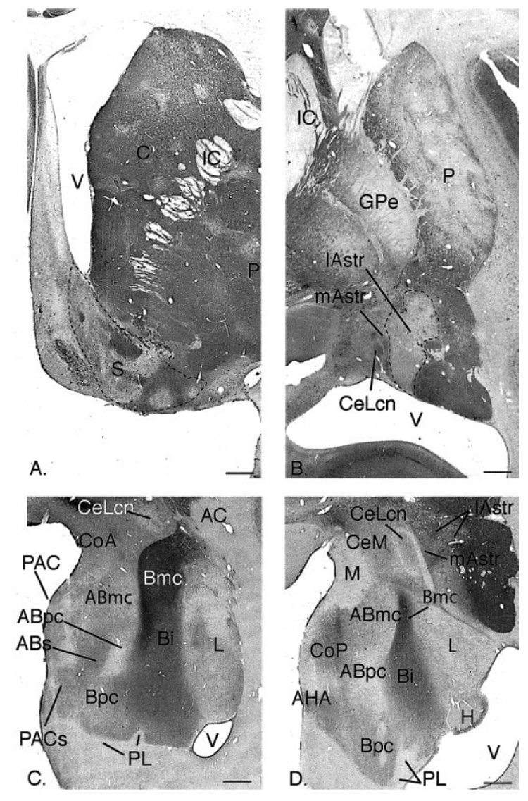
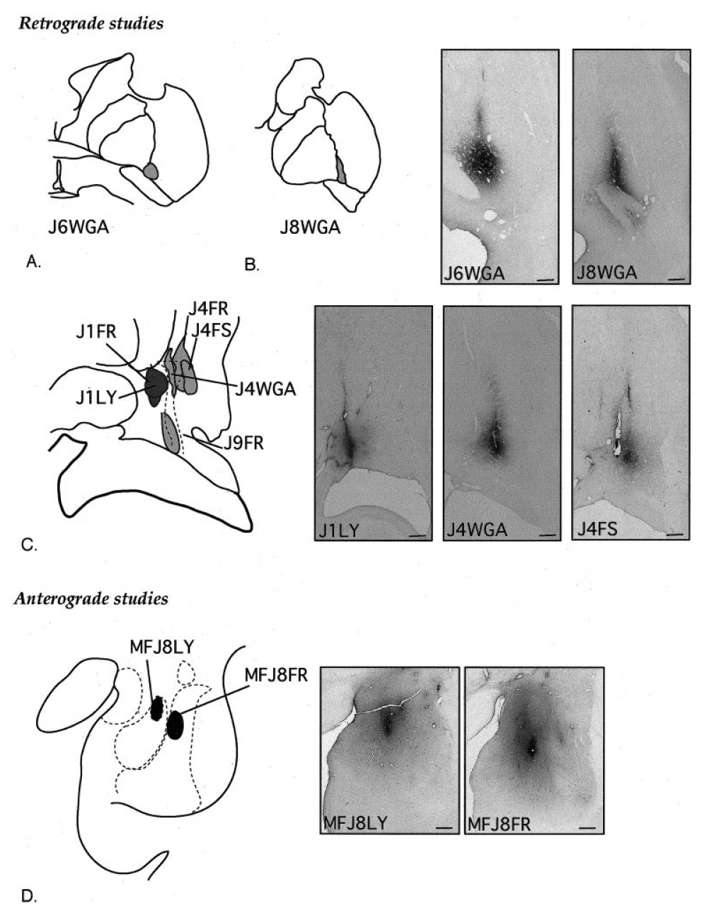
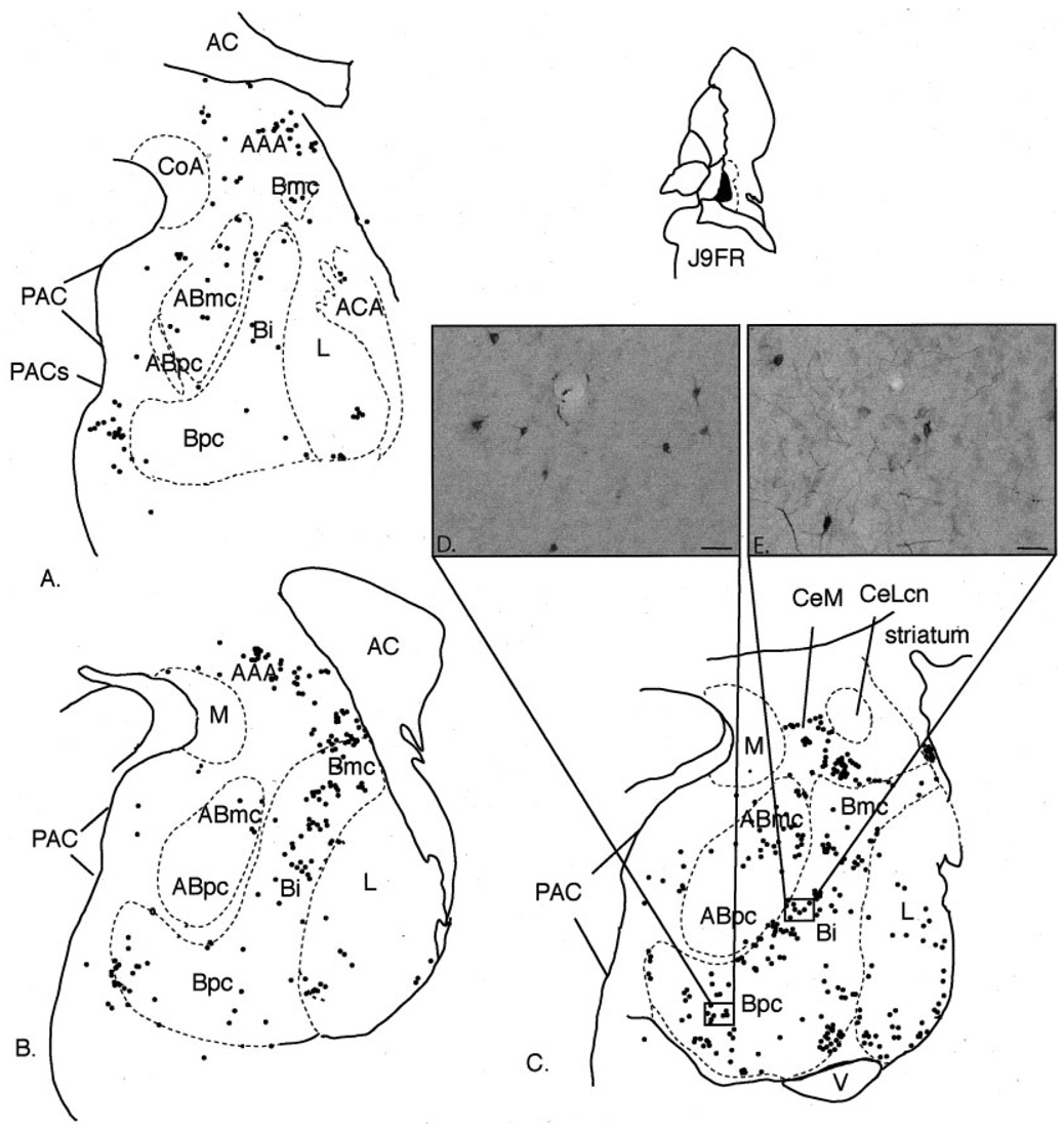
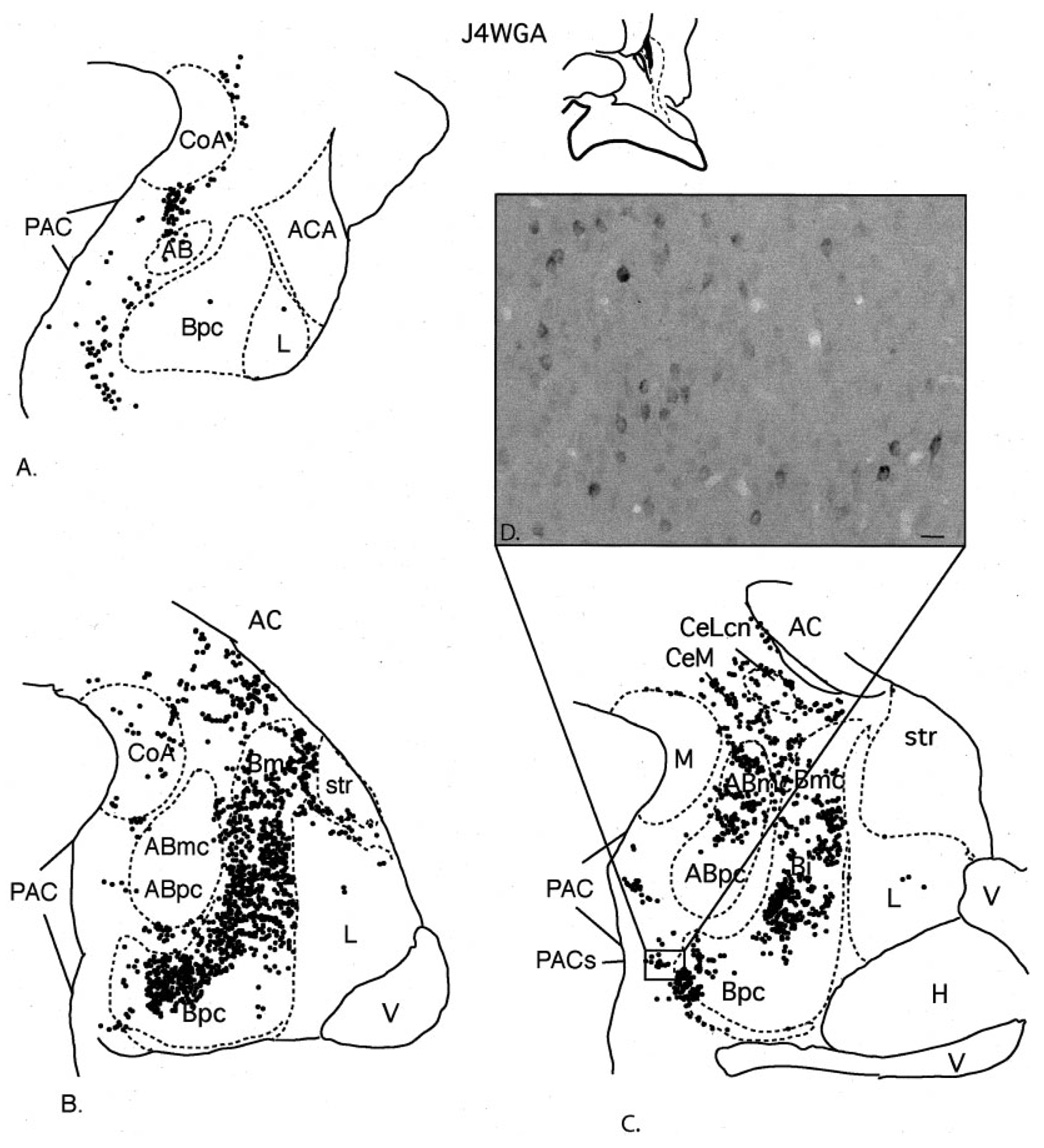
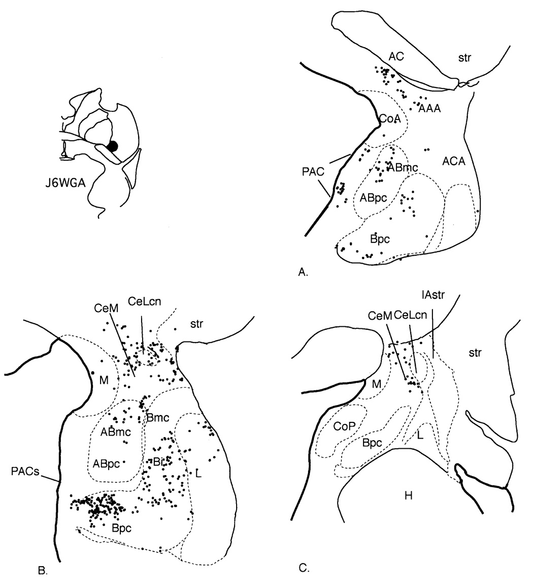
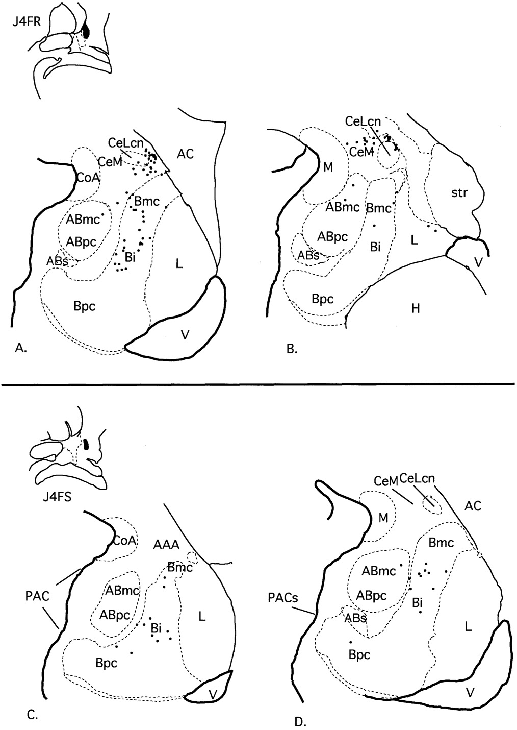
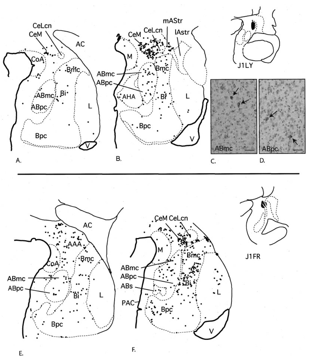
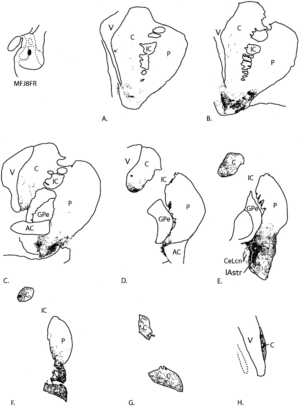
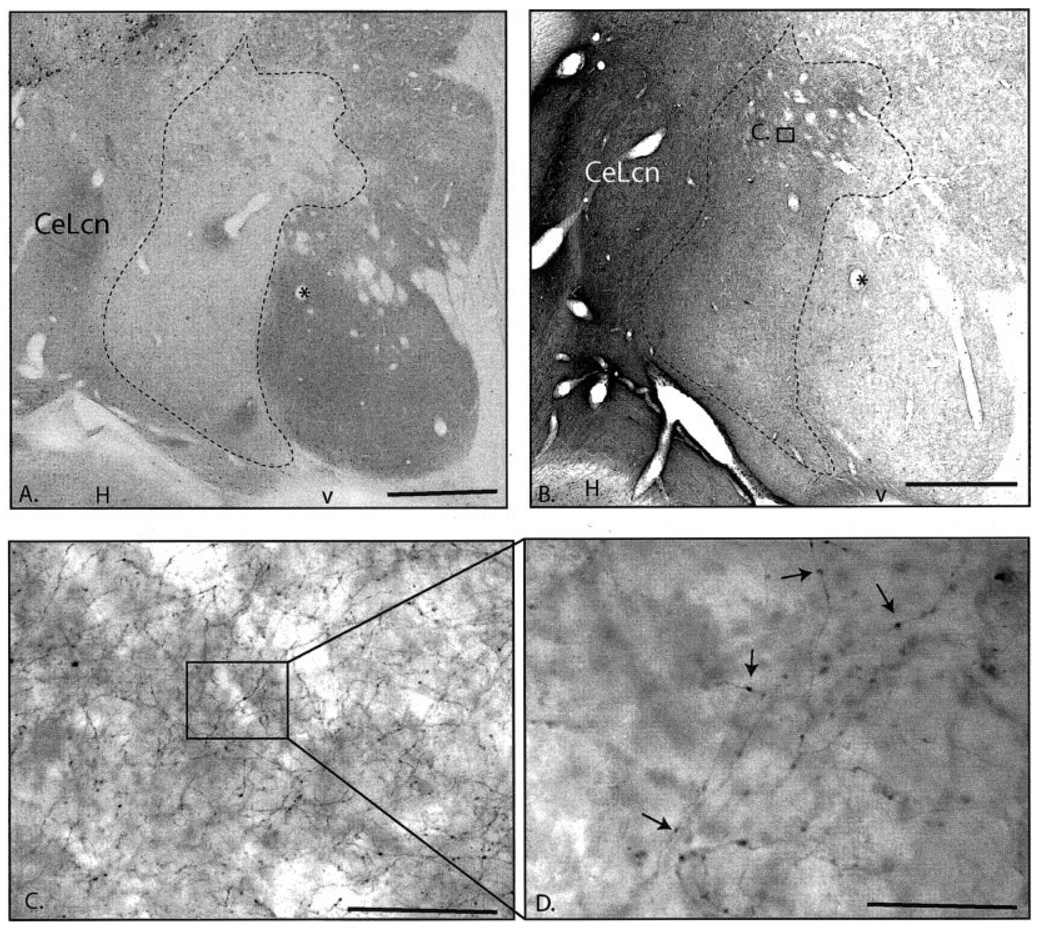
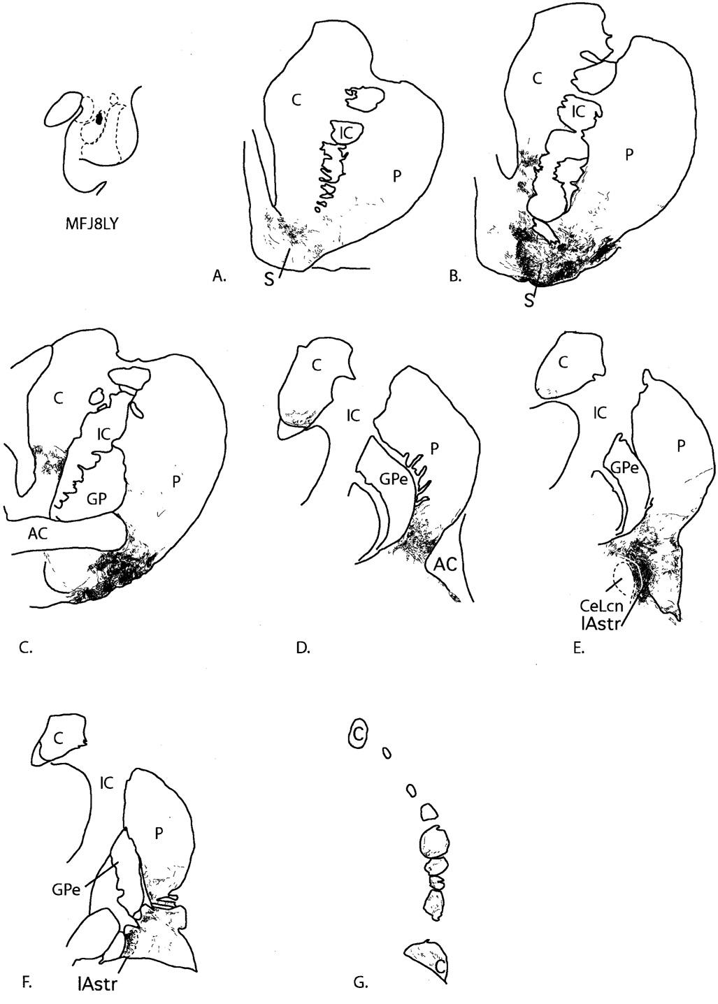
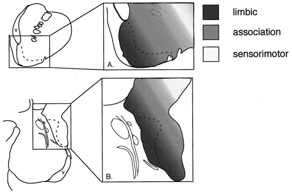
The ventral striatum mediates goal-directed behavior through limbic afferents. One well-established afferent to the ventral striatum is the amygdaloid complex, which projects throughout the shell and core of the nucleus accumbens, the rostral ventromedial caudate nucleus, and rostral ventromedial putamen. However, striatal regions caudal to the anterior commissure also receive inputs from the amygdala. These caudal areas contain histochemical and cytoarchitectural features that resemble the shell and core, based on our recent studies. Specifically, there is a calcium binding protein (CaBP)-poor region in the lateral amygdalostriatal area that resembles the "shell." To examine the idea that the caudal ventral striatum is part of the "classic" ventral striatum, we placed small injections of retrograde tracers throughout the caudal ventral striatum/amygdalostriatal area and charted the distribution of specific amygdaloid inputs. Amygdaloid inputs to the CaBP-poor zone in the lateral amygdalostriatal area arise from the basal nucleus, the magnocellular subdivision of the accessory basal nucleus, the periamygdaloid cortex, and the medial subdivision of the central nucleus, resembling that of the shell of the ventral striatum found in our previous studies. There are also amygdaloid inputs to CaBP-positive areas outside the shell, which originate mainly in the basal nucleus. Taken together, the "limbic-related" striatum forms a continuum from the rostral ventral striatum through the caudal ventral striatum/lateral amygdalostriatal area based on histochemical and cellular similarities, as well as inputs from the amygdala.
Figures
Similar articles
-
Amygdaloid projections to ventromedial striatal subterritories in the primate.Neuroscience. 2002;110(2):257-75. doi: 10.1016/s0306-4522(01)00546-2. Neuroscience. 2002. PMID: 11958868
-
Insular and gustatory inputs to the caudal ventral striatum in primates.J Comp Neurol. 2005 Sep 19;490(2):101-18. doi: 10.1002/cne.20660. J Comp Neurol. 2005. PMID: 16052493 Free PMC article.
-
Defining the caudal ventral striatum in primates: cellular and histochemical features.J Neurosci. 2002 Dec 1;22(23):10078-82. doi: 10.1523/JNEUROSCI.22-23-10078.2002. J Neurosci. 2002. PMID: 12451107 Free PMC article.
-
Amygdalostriatal projections in the neurocircuitry for motivation: a neuroanatomical thread through the career of Ann Kelley.Neurosci Biobehav Rev. 2013 Nov;37(9 Pt A):1932-45. doi: 10.1016/j.neubiorev.2012.11.019. Epub 2012 Dec 7. Neurosci Biobehav Rev. 2013. PMID: 23220696 Free PMC article. Review.
-
Functional anatomy of the basal ganglia: limbic aspects.Rev Neurol (Paris). 2012 Aug-Sep;168(8-9):569-75. doi: 10.1016/j.neurol.2012.06.015. Epub 2012 Aug 14. Rev Neurol (Paris). 2012. PMID: 22902172 Review.
Cited by
-
Striatum on the anxiety map: Small detours into adolescence.Brain Res. 2017 Jan 1;1654(Pt B):177-184. doi: 10.1016/j.brainres.2016.06.006. Epub 2016 Jun 6. Brain Res. 2017. PMID: 27276526 Free PMC article. Review.
-
Amygdala activity for the modulation of goal-directed behavior in emotional contexts.PLoS Biol. 2018 Jun 5;16(6):e2005339. doi: 10.1371/journal.pbio.2005339. eCollection 2018 Jun. PLoS Biol. 2018. PMID: 29870524 Free PMC article.
-
Cortical Granularity Shapes the Organization of Afferent Paths to the Amygdala and Its Striatal Targets in Nonhuman Primate.J Neurosci. 2022 Feb 23;42(8):1436-1453. doi: 10.1523/JNEUROSCI.0970-21.2021. Epub 2021 Dec 28. J Neurosci. 2022. PMID: 34965977 Free PMC article.
-
Neuropeptide Y input to the rat basolateral amygdala complex and modulation by conditioned fear.J Comp Neurol. 2016 Aug 15;524(12):2418-39. doi: 10.1002/cne.23960. Epub 2016 Feb 2. J Comp Neurol. 2016. PMID: 26779765 Free PMC article.
-
Heterogeneous dopamine populations project to specific subregions of the primate amygdala.Neuroscience. 2010 Feb 17;165(4):1501-18. doi: 10.1016/j.neuroscience.2009.11.004. Epub 2009 Nov 13. Neuroscience. 2010. PMID: 19914353 Free PMC article.
References
-
- Aggleton JP. A description of intra-amygdaloid connections in old world monkeys. Exp Brain Res. 1985;57:390–399. - PubMed
-
- Alheid GF, Heimer L. New perspectives in basal forebrain organization of special relevance for neuropsychiatric disorders: the striatopallidal, amygdaloid, and corticopetal components of substantia innominata. Neuroscience. 1988;27:1–39. - PubMed
-
- Alheid GF, Shammah-Lagnado SJ, Beltramino CA. The interstitial nucleus of the posterior limb of the anterior commissure: a novel layer of the central division of extended amygdala. Ann NY Acad Sci. 1999;877:645–654. - PubMed
-
- Amaral DG, Bassett JL. Cholinergic innervation of the monkey amygdala: an immunohistochemical analysis with antisera to choline acetyltransferase. J Comp Neurol. 1989;281:337–361. - PubMed
Publication types
MeSH terms
Substances
Grants and funding
LinkOut - more resources
Full Text Sources
Research Materials
